
Main menu

  • Home
  • Content
    • Current
      • JNMT Supplement
    • Ahead of print
    • Past Issues
    • Continuing Education
    • JNMT Podcast
    • SNMMI Annual Meeting Abstracts
  • Subscriptions
    • Subscribers
    • Rates
    • Journal Claims
    • Institutional and Non-member
  • Authors
    • Submit to JNMT
    • Information for Authors
    • Assignment of Copyright
    • AQARA Requirements
  • Info
    • Reviewers
    • Permissions
    • Advertisers
    • Corporate & Special Sales
  • About
    • About Us
    • Editorial Board
    • Contact Information
  • More
    • Alerts
    • Feedback
    • Help
    • SNMMI Journals
  • SNMMI
    • JNMT
    • JNM
    • SNMMI Journals
    • SNMMI

User menu

  • Subscribe
  • My alerts
  • Log in
  • My Cart

Search

  • Advanced search
Journal of Nuclear Medicine Technology
  • SNMMI
    • JNMT
    • JNM
    • SNMMI Journals
    • SNMMI
  • Subscribe
  • My alerts
  • Log in
  • My Cart
Journal of Nuclear Medicine Technology

Advanced Search

  • Home
  • Content
    • Current
    • Ahead of print
    • Past Issues
    • Continuing Education
    • JNMT Podcast
    • SNMMI Annual Meeting Abstracts
  • Subscriptions
    • Subscribers
    • Rates
    • Journal Claims
    • Institutional and Non-member
  • Authors
    • Submit to JNMT
    • Information for Authors
    • Assignment of Copyright
    • AQARA Requirements
  • Info
    • Reviewers
    • Permissions
    • Advertisers
    • Corporate & Special Sales
  • About
    • About Us
    • Editorial Board
    • Contact Information
  • More
    • Alerts
    • Feedback
    • Help
    • SNMMI Journals
  • Watch or Listen to JNMT Podcast
  • Visit SNMMI on Facebook
  • Join SNMMI on LinkedIn
  • Follow SNMMI on Twitter
  • Subscribe to JNMT RSS feeds
Review ArticleTeaching Case Studies

18F-FDG PET Assessment of Lewy Body Dementia with Cerebellar Diaschisis

Zaid Al-faham, Rami K. Zein and Ching-yee Oliver Wong
Journal of Nuclear Medicine Technology December 2014, 42 (4) 306-307; DOI: https://doi.org/10.2967/jnmt.114.139295
Zaid Al-faham
Department of Diagnostic Radiology and Molecular Imaging, William Beaumont School of Medicine and Health System, Oakland University, Royal Oak, Michigan
  • Find this author on Google Scholar
  • Find this author on PubMed
  • Search for this author on this site
Rami K. Zein
Department of Diagnostic Radiology and Molecular Imaging, William Beaumont School of Medicine and Health System, Oakland University, Royal Oak, Michigan
  • Find this author on Google Scholar
  • Find this author on PubMed
  • Search for this author on this site
Ching-yee Oliver Wong
Department of Diagnostic Radiology and Molecular Imaging, William Beaumont School of Medicine and Health System, Oakland University, Royal Oak, Michigan
  • Find this author on Google Scholar
  • Find this author on PubMed
  • Search for this author on this site
  • Article
  • Figures & Data
  • Info & Metrics
  • PDF
Loading

Abstract

Alzheimer disease and Lewy body dementia are the 2 most common causes of dementia. Each disease has distinctive regional metabolic reduction patterns on 18F-FDG PET. In this report, we present a rare case of an elderly man with dementia whereby 18F-FDG PET clearly showed Lewy body disease with crossed cerebellar diaschisis.

  • Lewy body dementia
  • crossed cerebellar diaschisis
  • 18F-FDG PET

Lewy body disease and Alzheimer dementia may coexist in the same patient and may be difficult to separate clinically or by imaging. Recognition of additional occipital cortex involvement favors the diagnosis of Lewy body disease. In this report, we present a rare case of an elderly man with dementia whereby 18F-FDG PET clearly showed asymmetric Lewy body disease with crossed cerebellar diaschisis.

CASE REPORT

A 71-y-old man with parkinsonism presented with progressively worsening short-term memory loss since 2011. The patient was complaining of motor tics and had been experiencing a decline in multiple domains, including executive functioning, visuospatial functioning, memory, language, attention, and concentration. There was no history of stroke or trauma. The patient had a mini–mental state examination score of 6/30. PET with 18F-FDG was ordered to evaluate for the etiology of the dementia.

After intravenous administration of 540.2 MBq (14.6 mCi) of 18F-FDG, multiple tomographic slices of the brain were obtained. The 18F-FDG PET scan (in quantitative scale with each color step representing 1 SD from the mean [−5.0 to +5.0]) demonstrated decreased radiotracer activity in the left parietal lobe, right parietal lobe, left temporal lobe, and occipital lobes (left greater than right), which is compatible with Lewy body dementia (Figs. 1 and 2). Also, there was decreased radiotracer uptake in the right cerebellum secondary to crossed cerebellar diaschisis (Fig. 3).

FIGURE 1.
  • Download figure
  • Open in new tab
  • Download powerpoint
FIGURE 1.

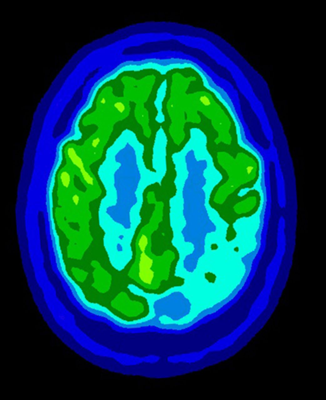
18F-FDG PET scan demonstrating severe decreased radiotracer activity in left parietal lobe and moderately decreased metabolism in right parietal lobe.

FIGURE 2.
  • Download figure
  • Open in new tab
  • Download powerpoint
FIGURE 2.

18F-FDG PET scan demonstrating severe decreased radiotracer activity in left temporal lobe and occipital lobes (left greater than right).

FIGURE 3.
  • Download figure
  • Open in new tab
  • Download powerpoint
FIGURE 3.

18F-FDG PET scan demonstrating crossed cerebellar diaschisis. This image shows mildly reduced metabolism in right cerebellum secondary to crossed cerebellar diaschisis.

DISCUSSION

Lewy body dementia is the second most common cause of dementia after Alzheimer disease. Lewy body dementia manifests as widespread neuronal degeneration. The pathologic etiology centers on deposition of Lewy bodies and Lewy neurites, which contain α synuclein as a major filamentous component (1). The diagnosis of Lewy body dementia is clinical and is based on consensus guidelines (2). In some cases, it is difficult to distinguish between Alzheimer disease and Lewy body dementia clinically, and in these cases the use of noninvasive nuclear imaging such as SPECT or 18F-FDG PET is useful (2). Lewy body dementia and Alzheimer disease have a similar hypometabolic cerebral distribution with marked decreases in association cortices and relative sparing of subcortical structures and primary somatomotor cortex. However, the metabolic reduction in the occipital cortex is the key feature to support the diagnosis of Lewy body dementia (3). The reduced metabolism in the right cerebellum is consistent with diaschisis, which is defined as depression of regional neuronal metabolism and cerebral blood flow caused by dysfunction in anatomically separate but functionally related neuronal regions (4). The mechanism appears to be an interruption of the corticopontine–cerebellar pathway (5). This phenomenon has been associated with Lewy body dementia (2) and with other cortical pathologies, specifically tumor, stroke, or trauma (5).

CONCLUSION

Alzheimer disease and Lewy body dementia are the two most common causes of dementia. Each disease has distinctive regional metabolic reduction patterns on 18F-FDG PET. Therefore, knowing these patterns is important to support the likely diagnosis.

DISCLOSURE

No potential conflict of interest relevant to this article was reported.

Footnotes

  • Published online Sep. 4, 2014.

REFERENCES

  1. 1.↵
    1. Galvin JE,
    2. Uryu K,
    3. Lee VM,
    4. Trojanowski JQ
    . Axon pathology in Parkinson’s disease and Lewy body dementia hippocampus contains alpha-, beta-, and gamma-synuclein. Proc Natl Acad Sci USA. 1999;96:13450–13455.
    OpenUrlAbstract/FREE Full Text
  2. 2.↵
    1. Mirzaei S,
    2. Knoll P,
    3. Koehn H,
    4. Bruecke T
    . Assessment of diffuse Lewy body disease by 2–[18F]fluoro-2-deoxy-D-glucose positron emission tomography (FDG PET). BMC Nucl Med. 2003;20;3:1.
  3. 3.↵
    1. Ishii K
    . PET approaches for diagnosis of dementia. AJNR Am J Neuroradiol. August 14, 2013 (Epub ahead of print).
  4. 4.↵
    1. Brunberg JA,
    2. Frey KA,
    3. Horton JA,
    4. Kuhl DE
    . Crossed cerebellar diaschisis occurrence and resolution demonstrated with PET during carotid temporary balloon occlusion. AJNR Am J Neuroradiol. 1992;13:58–61.
    OpenUrlFREE Full Text
  5. 5.↵
    Mettler FA, Guiberteau, J Milton. Essentials of Nuclear Medicine Imaging. Philadelphia, PA: Saunders/Elsevier; 2006:388–389.
  • Received for publication February 19, 2014.
  • Accepted for publication April 8, 2014.
PreviousNext
Back to top

In this issue

Journal of Nuclear Medicine Technology: 42 (4)
Journal of Nuclear Medicine Technology
Vol. 42, Issue 4
December 1, 2014
  • Table of Contents
  • About the Cover
  • Index by author
Print
Download PDF
Article Alerts
Sign In to Email Alerts with your Email Address
Email Article

Thank you for your interest in spreading the word on Journal of Nuclear Medicine Technology.

NOTE: We only request your email address so that the person you are recommending the page to knows that you wanted them to see it, and that it is not junk mail. We do not capture any email address.

Enter multiple addresses on separate lines or separate them with commas.
18F-FDG PET Assessment of Lewy Body Dementia with Cerebellar Diaschisis
(Your Name) has sent you a message from Journal of Nuclear Medicine Technology
(Your Name) thought you would like to see the Journal of Nuclear Medicine Technology web site.
Citation Tools
18F-FDG PET Assessment of Lewy Body Dementia with Cerebellar Diaschisis
Zaid Al-faham, Rami K. Zein, Ching-yee Oliver Wong
Journal of Nuclear Medicine Technology Dec 2014, 42 (4) 306-307; DOI: 10.2967/jnmt.114.139295

Citation Manager Formats

  • BibTeX
  • Bookends
  • EasyBib
  • EndNote (tagged)
  • EndNote 8 (xml)
  • Medlars
  • Mendeley
  • Papers
  • RefWorks Tagged
  • Ref Manager
  • RIS
  • Zotero
Share
18F-FDG PET Assessment of Lewy Body Dementia with Cerebellar Diaschisis
Zaid Al-faham, Rami K. Zein, Ching-yee Oliver Wong
Journal of Nuclear Medicine Technology Dec 2014, 42 (4) 306-307; DOI: 10.2967/jnmt.114.139295
Twitter logo Facebook logo LinkedIn logo Mendeley logo
  • Tweet Widget
  • Facebook Like
  • Google Plus One
Bookmark this article

Jump to section

  • Article
    • Abstract
    • CASE REPORT
    • DISCUSSION
    • CONCLUSION
    • DISCLOSURE
    • Footnotes
    • REFERENCES
  • Figures & Data
  • Info & Metrics
  • PDF

Related Articles

  • No related articles found.
  • PubMed
  • Google Scholar

Cited By...

  • Prognostic Implications of Total Hemispheric Glucose Metabolism Ratio in Cerebrocerebellar Diaschisis
  • Google Scholar

More in this TOC Section

  • High-Sensitivity Troponin Elevation in a Young Woman with Typical Chest Pain: The Heart of the Matter
  • Pulmonary Adenocarcinoma Revealed by Parathyroid Scintigraphy: An Incidental Case to Remember
  • Prominent Right Ventricular Tracer Uptake: A Harbinger of Multivessel Coronary Artery Disease
Show more Teaching Case Studies

Similar Articles

Keywords

  • Lewy body dementia
  • crossed cerebellar diaschisis
  • 18F-FDG PET
SNMMI

© 2025 SNMMI

Powered by HighWire