


Article Figures & Data
Figures
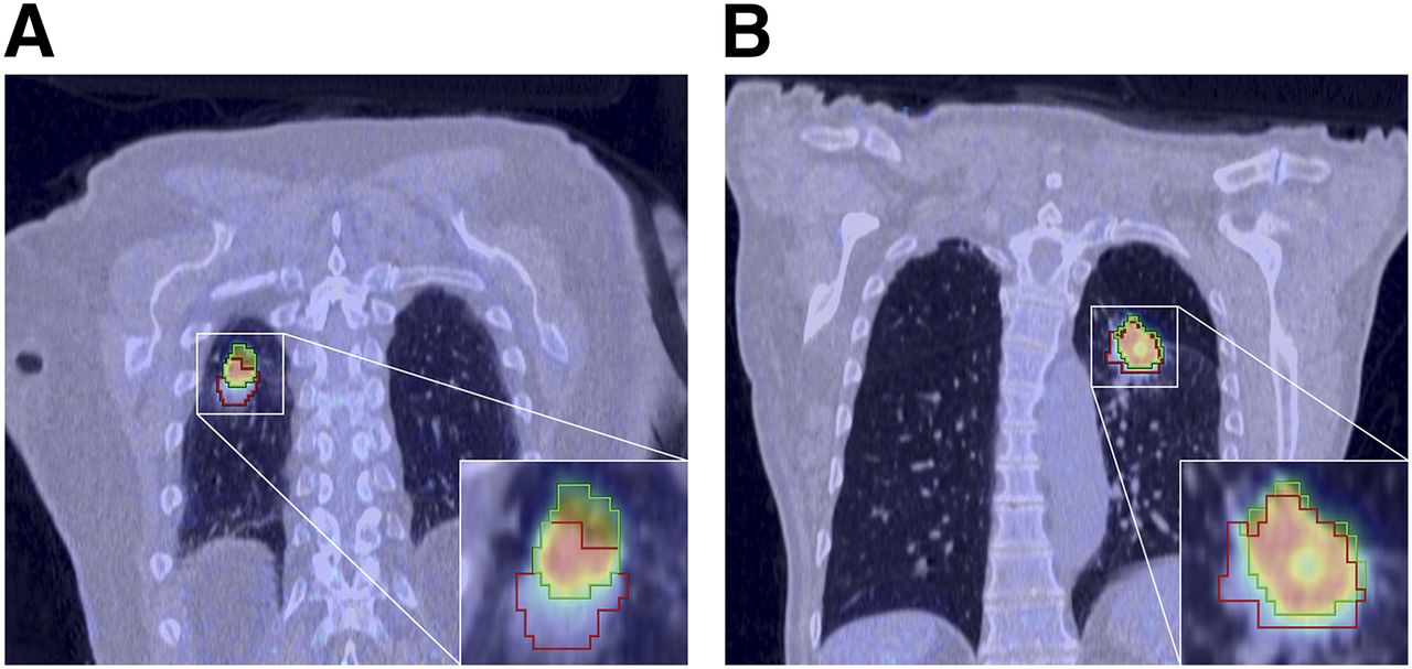
- FIGURE 1.
Coronal respiratory-gated PET images. (A) Patient with squamous cell carcinoma in FB group. Lesion delineation using PET (green) and CT (red). Difference between centroids of PET and CT images is 10.5 mm. JSC is 0.29. (B) Patient with adenocarcinoma in BH group. PET lesion delineation is green, and CT delineation is red. Difference between centroids of PET and CT images is 4.0 mm; JSC is 0.50.
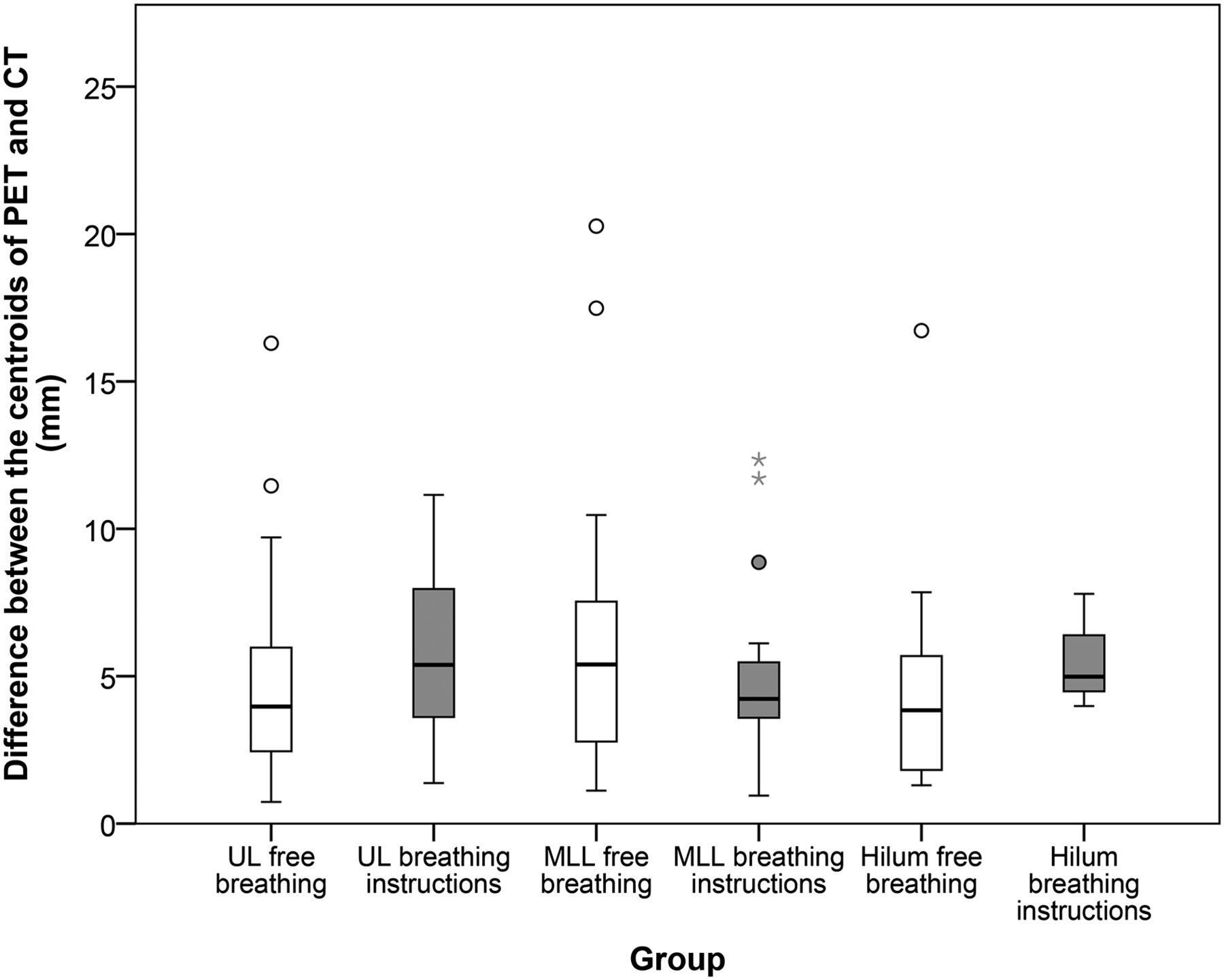
- FIGURE 2.
Distribution between difference of centroids between PET and CT scan. There are several outliers, depicted as ○, which are values that do not fall in inner fences, and even extreme outliers (*), which are more than 3 times height of boxes. UL = upper lobes; MLL = middle and lower lobes; hilum = central lesions, connected to hilum of lung.
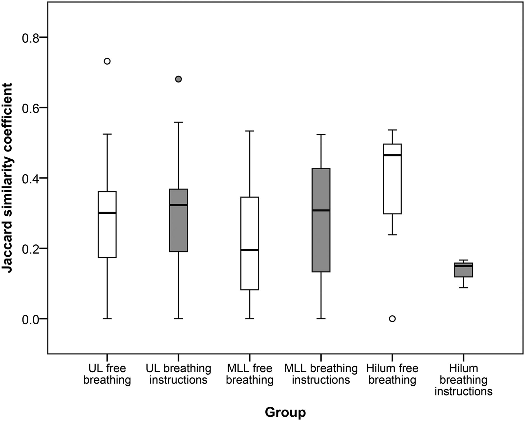
- FIGURE 3.
Distribution of JSC for both FB and BH groups for 3 localizations. There are several outliers (values that do not fall in inner fences). UL = upper lobes; MLL = middle and lower lobes; hilum = central lesions, connected to hilum of lung.


Tables
Characteristic FB group BH group Average age in y 66.7 (9.5) 70.9 (9.8) Average weight in kg 77.7 (12.1) 70.6 (11.9) Sex Female 12 6 Male 24 10 Confirmed malignancy Primary lung cancer 25 10 Metastasis 3 3 Other and unconfirmed 8 3 Lesion size on CT in mm3 8,261.6 (22,914.4) 8,890.2 (21,872.8) Average 18F-FDG dose in MBq 247.7 (40.2) 225.8 (41.5) Minimum SUVmax in g/cm3 0.67 1.03 Data in parentheses are SDs.
SUVmax = maximum SUV.
Anatomic location FB group BH group Upper lobes 45 17 Middle and lower lobes 30 19 Central 11 3 Total 86 39 Location FB group (mm) BH group (mm) P Upper lobes 4.7 ± 3.1 6.0 ± 3.0 0.11 Middle and lower lobes 5.8 ± 4.3 5.1 ± 2.9 0.70 Central 4.8 ± 4.6 5.6 ± 2.0 0.24 Total 5.1 ± 3.8 5.5 ± 2.9 0.16 Location FB group BH group P Upper lobes 0.28 ± 0.17 0.28 ± 0.19 0.83 Middle and lower lobes 0.22 ± 0.16 0.28 ± 0.18 0.20 Central 0.39 ± 0.17 0.13 ± 0.04 0.04 Total 0.27 ± 0.17 0.27 ± 0.18 0.95


Jump to section
Related Articles
Cited By...
- Improvement of Anatomic Alignment and Image Quality Using a Respiratory Motion Reduction Block in Oncologic PET/CT
- Improving the Spatial Alignment in PET/CT Using Amplitude-Based Respiration-Gated PET and Patient-Specific Breathing-Instructed CT
- Improving the Spatial Alignment in PET/CT Using Amplitude-Based Respiration-Gated PET and Respiration-Triggered CT