


Abstract
For phantom studies involving 90Y PET/CT, homogeneous solutions of 90Y, for example, 90Y citrate, are commonly used. However, the microsphere biodistribution of a postradioembolization liver is never homogeneous; therefore, such phantoms are physiologically unrealistic for simulating clinical scenarios. The aim of this work was to develop a safe and practical phantom capable of simulating the heterogeneous microsphere biodistribution of a postradioembolization liver. Methods: Gelatin (5%) was used to suspend 90Y resin microspheres, poured into plastic containers to simulate a liver with 2 tumors. Microspheres were added while the gelatin was maintained in a liquid state on a hot plate and continuously stirred with magnetic stir bars. The liquid microsphere mixture was then rapidly cooled in an ice bath while being stirred, resulting in a heterogeneous suspension of microspheres. The completed phantom was serially scanned by 90Y PET/CT over 2 wk. Results: All scans demonstrated a heterogeneous microsphere distribution throughout the liver and tumor inserts. Serendipitously, magnetic stir bars left inside the phantom produced CT artifacts similar to those caused by embolization coils, whereas pockets of air trapped within the gelatin during its preparation mimicked gas within hollow viscus. The microspheres and tumor inserts remained fixed and suspended within the gelatin, with no evidence of breakdown or leakage. Conclusion: A gelatin phantom realistically simulating the physiologic microsphere biodistribution of a postradioembolization liver is feasible to construct in a radiopharmacy.
Radioembolization of liver malignancies with 90Y is brachytherapy delivered by arterially injected β−-emitting microspheres, which may be made of resin (SIR-Spheres; Sirtex Medical Limited) or glass (TheraSphere; BTG). There has been recent interest in 90Y PET with concomitant low-dose CT. Coincidence imaging of 90Y is possible because of a minor decay branch to the 0+ first excited state of 90Zr, followed by β−β+ internal pair production, at a low branching ratio of 31.86 ± 0.47 × 10⁻⁶ (1,2). Despite background noise in the reconstructed images due to naturally occurring 176Lu within the lutetium-based crystal of today’s time-of-flight PET scanners, recent studies have shown 90Y PET quantification to be feasible and accurate (3–8).
For 90Y PET/CT phantom studies, homogeneous solutions of 90Y, for example, 90Y-citrate, are commonly used. However, the microsphere biodistribution of a postradioembolization liver is never homogeneous (9); therefore, such phantoms are physiologically unrealistic for simulating clinical scenarios. The aim of this work was to develop a safe and practical phantom capable of simulating the heterogeneous microsphere biodistribution of a postradioembolization liver throughout both tumor and nontumor liver compartments with a physiologically realistic tumor–to–normal liver ratio.
MATERIALS AND METHODS
Phantom Construction
Institutional Review Board approval was not required for the conduct and publication of this phantom study, which did not involve any human subjects. The phantom design was inspired by a gel-based phantom used by Goedicke et al. to overcome the problem of microsphere sedimentation (7). For this phantom, the medium used to suspend the microspheres was gelatin. A 5%-by-weight solution of gelatin was prepared by slowly dissolving 75 g of gelatin into 1,500 mL of distilled sterile water (for injection/irrigation). The solution was kept at 45°C on a hot plate and magnetically stirred at 300 rpm to ensure that the gelatin would not set. The liver phantom was a 1,330-mL rectangular homeware container made of clear, rigid plastic. The 2 tumor inserts were round containers made of similar plastic material, measuring 27 and 58 mL by volume. A magnetic stir bar (Spinbar; Sigma-Aldrich) was placed into each of the 3 inserts, and then the inserts were filled to maximum capacity with gelatin. The inserts were kept on a hot plate with the stir bar at 300 rpm to ensure proper distribution of heat and to prevent setting.
90Y resin microspheres from a full vial with a total activity of 3.144 GBq were carefully dispensed into each of the 3 inserts to achieve a realistic tumor–to–normal liver ratio of approximately 5 for hepatocellular carcinoma (10). The total vial activity was determined from calibration factors provided by the manufacturer, to an uncertainty of ±10%. For this vial, the derived total activity of 3.144 ± 0.31 GBq was in good agreement with the measured activity of 3.139 GBq by the dose calibrator (CRC-35; Capintec) using the 90Y setting of 480 × 10. Individual activities were 281 ± 28 MBq in 27 mL (10.4 MBq/mL) and 573 ± 57 MBq in 58 mL (9.9 MBq/mL) for the 2 tumor inserts and 2,290 ± 229 MBq in 1,245 mL (1.8 MBq/mL) for the liver insert. All 90Y activities were determined by volume from the original vial of well-suspended microspheres. All microspheres were completely dispensed into the phantom, with negligible residual vial activity.
After the microspheres were added, the hot plate was turned off and the gelatin was rapidly cooled in an ice-water bath while still being stirred. This stirring process resulted in a gelatin-based suspension of heterogeneously distributed microspheres throughout both tumor and nontumor liver compartments. The magnetic stir bars were intentionally left within the inserts to simulate the metallic coils used for prophylactic coil embolization. The plastic lids of all 3 inserts were secured with adhesive tape. The 2 tumor inserts were placed into the liver insert to complete the liver phantom. Therefore, the whole-liver total activity was 3.144 ± 0.31 GBq distributed throughout 1,330 mL of gelatin, with a mean radioconcentration of 2.36 MBq/mL. To simulate the attenuation within a patient, the liver phantom was fixed using adhesive strips near the bottom of a 5-liter plastic body phantom and further weighed down using saline bags placed on its lid. The body phantom was then completely filled with water and sealed with adhesive tape.
90Y PET/CT Protocol
90Y PET/CT scans were obtained on a Biograph mCT-S(64)4R (Siemens Medical Solutions), which uses lutetium oxyorthosilicate crystals and has time-of-flight capability. Because 90Y was not available on this system as a radionuclide choice at the time of this study, all studies were acquired using 86Y settings, with a half-life of 14.74 h and a branching ratio of 0.33. PET was acquired at 15 min per bed position and reconstructed using the vendor-supplied TrueX algorithm plus time-of-flight reconstruction (UltraHD-PET; Siemens) with 1 iteration, 21 subsets, and a filter of 8 mm, decay-corrected to the start of the scan. CT was performed at 120 kVp, 50 mAs (CARE Dose 4D; Siemens), a Z-coverage per rotation of 64 × 0.6 mm, 5-mm slices, and a pitch of 0.75. All phantom scans were obtained using a single bed position. All reconstructed 90Y PET/CT images were qualitatively reviewed; 90Y PET quantification of activity was not performed.
Phantom Physical Density
The main constituent of gelatin is collagen. Because our liver phantom was prepared in a nonsterile manner, environmental bacteria or fungi may have been inadvertently introduced into the phantom, with the potential to cause gelatin hydrolysis. Furthermore, the effect of 90Y β radiation on the structural integrity of gelatin has not been well described. For our study, the physical density (g/cm3) of the liver phantom was empirically used as a surrogate measure of possible gelatin breakdown or leakage occurring during the study. We postulated that as gelatin degrades, its density may change due to the upward migration of air pockets trapped during preparation of the liver phantom.
To avoid radiation exposure to the operator, the physical density of gelatin was indirectly measured based on the CT component of the PET/CT scan. By assuming a linear relationship between the CT number (Hounsfield unit) and the physical density of a water-based object of interest (11), we calculated the physical density of the liver phantom as (mean CT number + 1,000)/1,000. CT analysis was performed using OsiriX software (version 5.6; Pixmeo). The mean CT number of the liver phantom was determined from a cylindric volume of interest with a 4-cm diameter and 4.5-cm length, placed within a region of gelatin free from large air pockets or tumor inserts. This volume of interest was repeated across all phantom scans to obtain its mean physical density and compared with an expected result of 1.05 g/cm3. As a control, the physical density of water within the body phantom was similarly measured and compared with an expected result of 1 g/cm3.
RESULTS
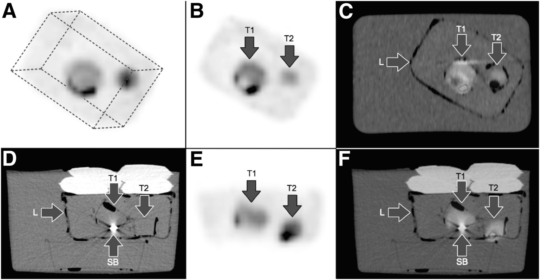
Twelve phantom scans were acquired over 13 d. All scans demonstrated a heterogeneous distribution of 90Y microspheres throughout the liver and tumor inserts, fixed and suspended within the gelatin (Fig. 1). Barring the expected deterioration in perceived image quality due to increasing noise as 90Y decayed over the course of the study, the overall visual activity distribution within the phantom was generally unchanged.

Gelatin-based liver phantom containing suspended 90Y resin microspheres. (A) Maximum-intensity projection of liver phantom in oblique view. Dotted lines outline liver phantom. (B and C) 90Y PET/CT scan in coronal plane demonstrates heterogeneous distribution of 90Y resin microspheres within 2 tumor inserts, T1 and T2, and throughout nontumorous liver, L. Small air pockets can be seen within and around tumor inserts and liver phantom. (D–F) 90Y PET/CT in transaxial view demonstrates CT artifacts caused by a magnetic stir-bar (SB) at bottom of T1 tumor insert. (A color version of this figure is available as a supplemental file online at http://tech.snmjournals.org/.)
Serendipitously, magnetic stir bars left inside the liver phantom produced CT artifacts similar to those caused by embolization coils, and pockets of air trapped inside the gelatin during its preparation mimicked gas within hollow viscus. Throughout the study, the 2 tumor inserts remained unchanged in position and there was no detectable activity along the bottom of the water-filled body phantom to suggest any sedimentation of leaked 90Y resin microspheres.
The physical density of the liver phantom was estimated as 1.011 ± 0.0012 g/cm3 (median, 1.011 g/cm3; 95% confidence interval, 1.010–1.011), which remained unchanged throughout the study, and was lower than the expected density of 1.05 g/cm3, likely due to pockets of air trapped within the gelatin. As a methodologic control, the physical density of water in the body phantom was estimated as 1.0017 ± 0.00066 g/cm3 (median, 1.0017 g/cm3; 95% confidence interval, 1.0013–1.0021), representing a low mean error of less than 0.2%, and provided quality assurance for this technique.
DISCUSSION
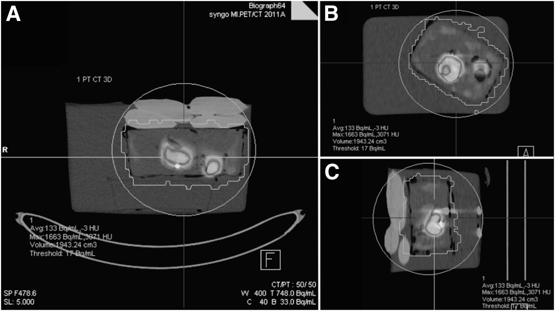
Although homogeneous solutions of 90Y (e.g., 90Y-citrate) are less affected by uncertainties in activity measurement and are convenient for use in phantom studies, they do not represent the physiologic reality of a postradioembolization liver. This study has achieved its objective of developing a phantom capable of simulating the heterogeneous distribution of microspheres in both tumor and nontumor liver compartments, with a realistic tumor–to–normal liver ratio. Such a phantom may have theoretic benefits in improving the qualitative and quantitative accuracy of 90Y PET/CT phantom scans when simulating postradioembolization clinical scenarios (Fig. 2).

Example of volume-of-interest analysis of 90Y PET/CT phantom scan generated using 1% volumetric isocontour threshold, which may be used for quantitative analysis. Images are shown in transaxial (A), coronal (B), and sagittal planes (C). (A color version of this figure is available as a supplemental file online at http://tech.snmjournals.org/.)
For this phantom design, the tumor inserts and 90Y resin microspheres remained fixed and suspended within the gelatin throughout a 2-wk period, with no evidence of breakdown or microsphere leakage. This phantom was made from materials readily available in a radiopharmacy and was feasible to construct.
There were several limitations to this study. First, 90Y PET activity quantification or voxel-based analyses were not adequately performed at the time of this report because technical issues related to our scan protocols were still being optimized. Second, our phantom was not anthropometrically realistic of the liver and abdomen and was also missing the lungs and skeleton. The impact of such anatomic discrepancies was not investigated in this study. Third, although we achieved a realistic tumor–to–normal liver ratio of approximately 5 for hepatocellular carcinoma, the heterogeneous distribution of microspheres would not be exactly reproducible at the microscopic level from phantom to phantom, which is an inherent limitation of our phantom construction technique. Last, although the magnetic spin bars left intentionally within the liver phantom produced CT artifacts visually similar to those of embolization coils, the physical shape and metallic composition of the spin bars (alloy of aluminum, nickel, iron, and cobalt) were different from those of embolization coils (platinum-based). The effects of these physical differences were not further investigated.
CONCLUSION
A gelatin-based liver phantom realistically simulating the physiologic biodistribution of 90Y resin microspheres in a postradioembolization liver is safe and feasible.
DISCLOSURE
No potential conflict of interest relevant to this article was reported.
Footnotes
Published online Nov. 11, 2014.
REFERENCES
- Received for publication July 3, 2014.
- Accepted for publication October 15, 2014.


Jump to section
Related Articles
Cited By...
- No citing articles found.