


Article Figures & Data
Figures
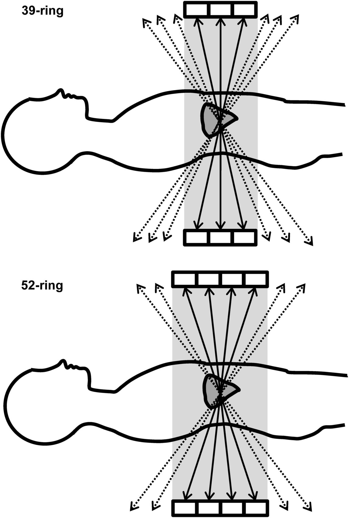
- FIGURE 1.
A 3D emission acquisition of 39- (upper) and 52-ring (lower) scanners. The 52-ring scanner has large acceptance angle. Thus, system sensitivity of 52-ring scanner is higher than that of 39-ring scanner.
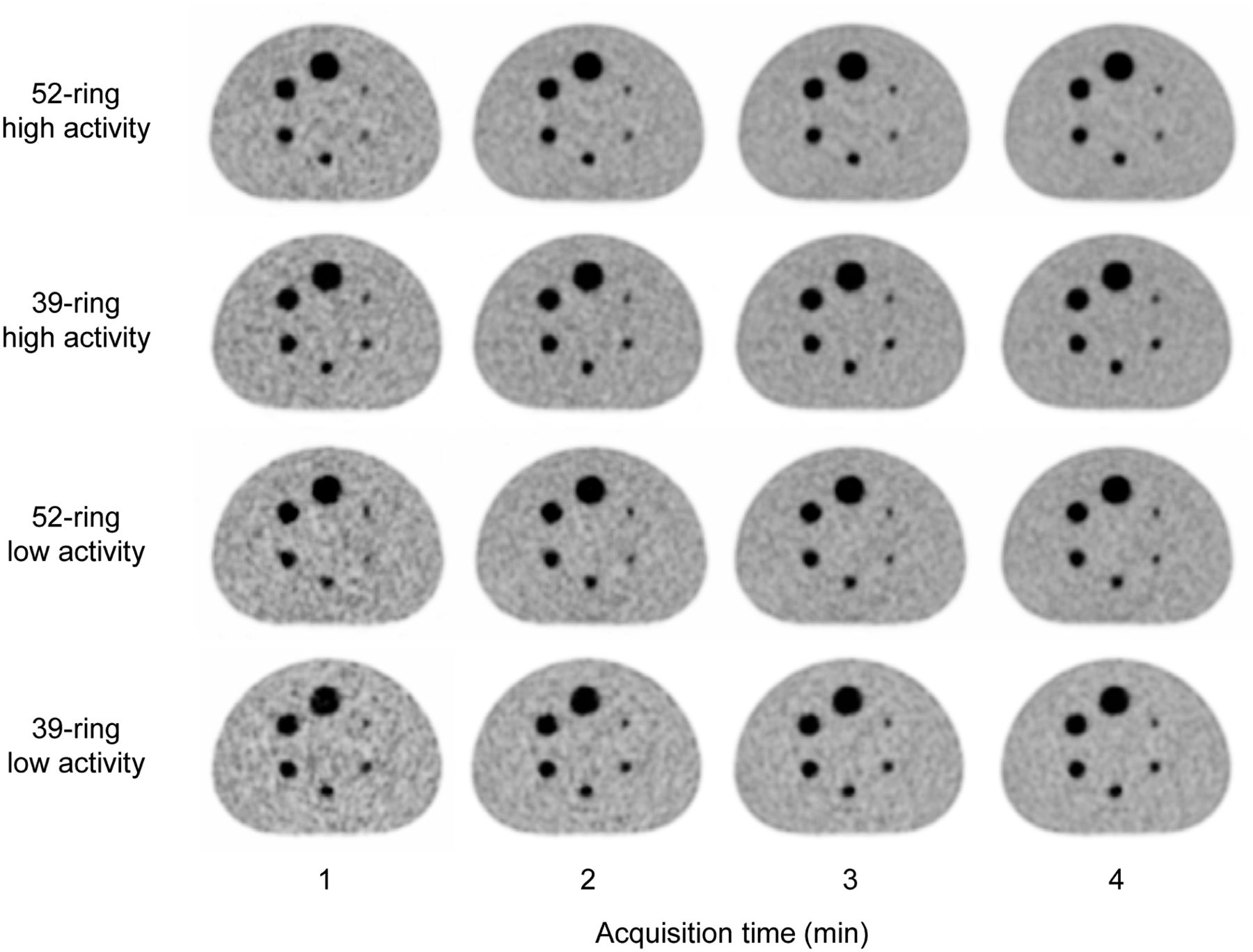
- FIGURE 2.
PET images reconstructed using OSEM + PSF + TOF with various acquisition times, different activity levels, and 2 types of detector ring. In terms of background uniformity, images acquired using 52-ring scanner were superior to those acquired using 39-ring scanner.
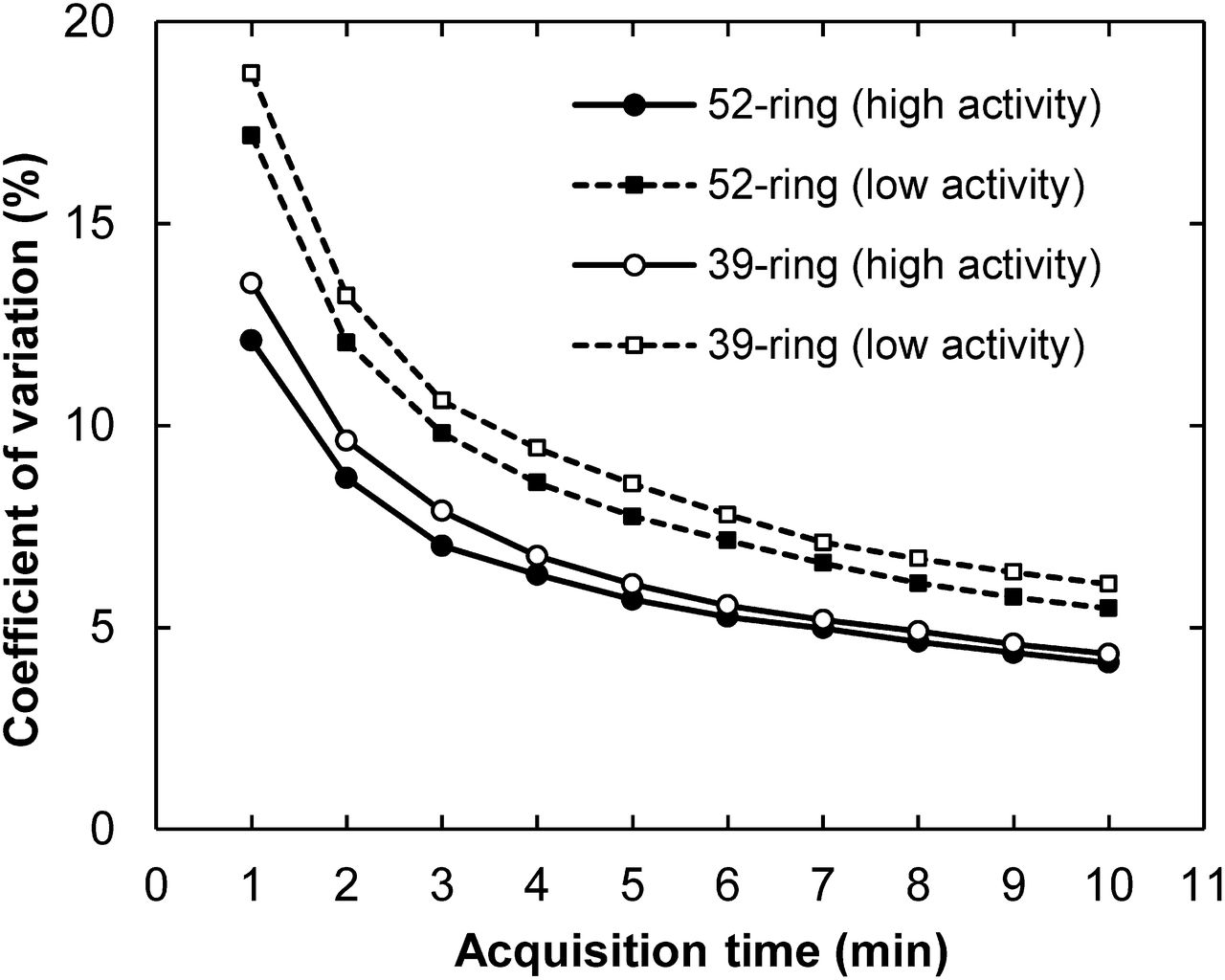
- FIGURE 3.
CV for OSEM + PSF + TOF images in relation to acquisition times. CV of 52-ring scanner was superior to that of 39-ring scanner by approximately 10%. CV of 52-ring scanner using 3-min acquisition was equivalent to that of 39-ring scanner using 4-min acquisition.
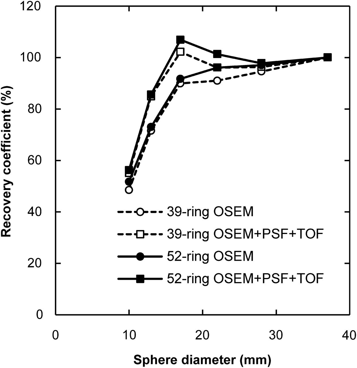
- FIGURE 4.
Relative RC curves at 10-min acquisition in relation to reconstruction methods and number of detector rings. Although PSF and TOF also increased RC, RC curves were nearly identical independent of number of detector rings.


Tables
Parameter D (cm) 39-ring system 52-ring system Sensitivity (kcps/MBq) 0 5.4 9.2 10 5.9 9.4 Total sensitivity (kcps/MBq) 5.6 9.3 D = radial distance in from center of FOV.
Parameter NECR (kcps) 52-ring high activity 115.9 52-ring low activity 59.0 39-ring high activity 72.0 39-ring low activity 37.8


