


Article Figures & Data
Figures
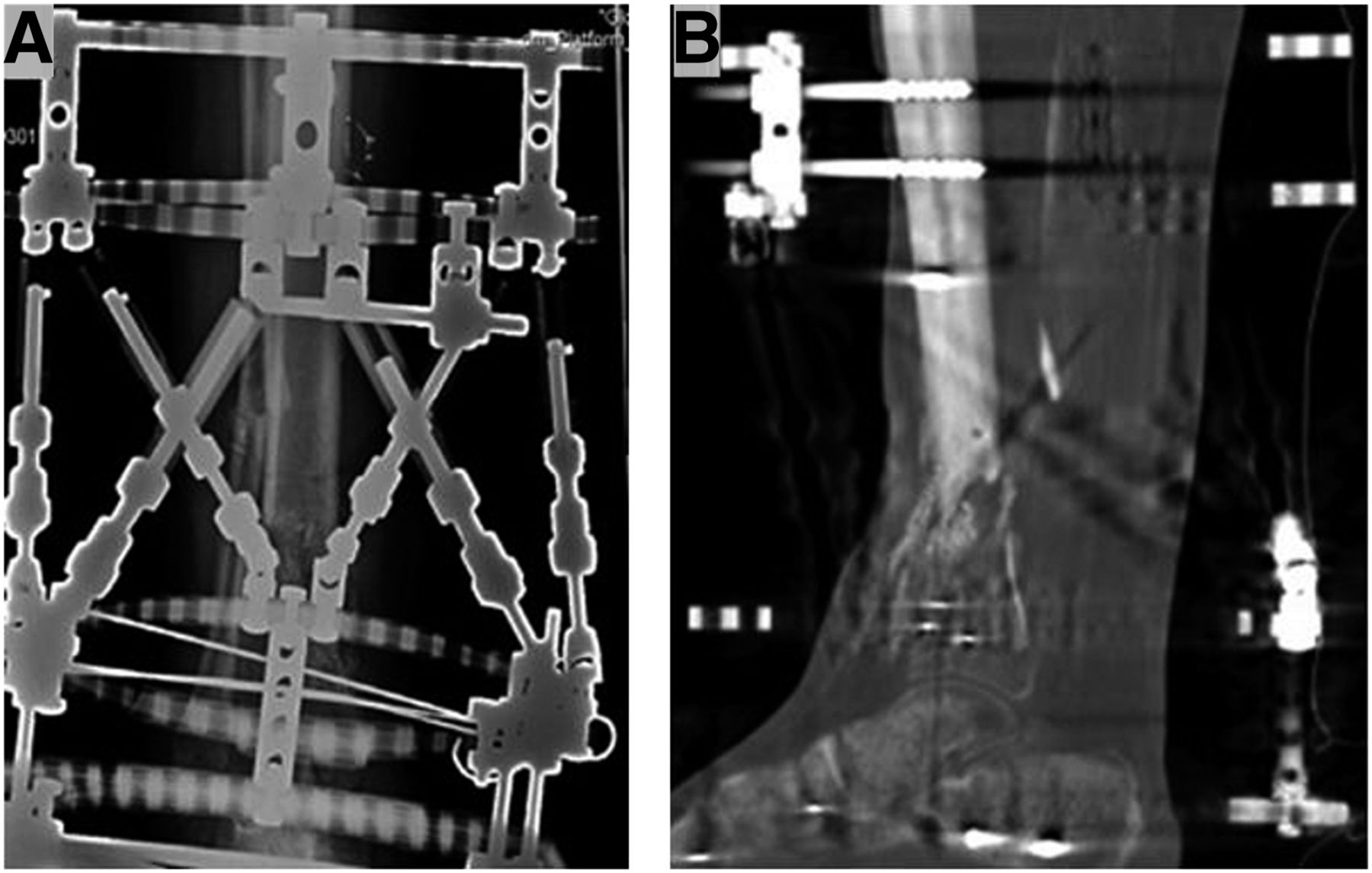
- FIGURE 1.
Typical planar radiograph (A) and CT scan (B) of patient with Taylor spatial frame.
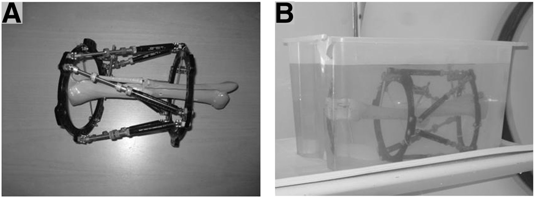
- FIGURE 2.
(A) Taylor spatial frame attached to plastic leg. (B) Phantom (in water bath) about to be inserted into PET/CT scanner.
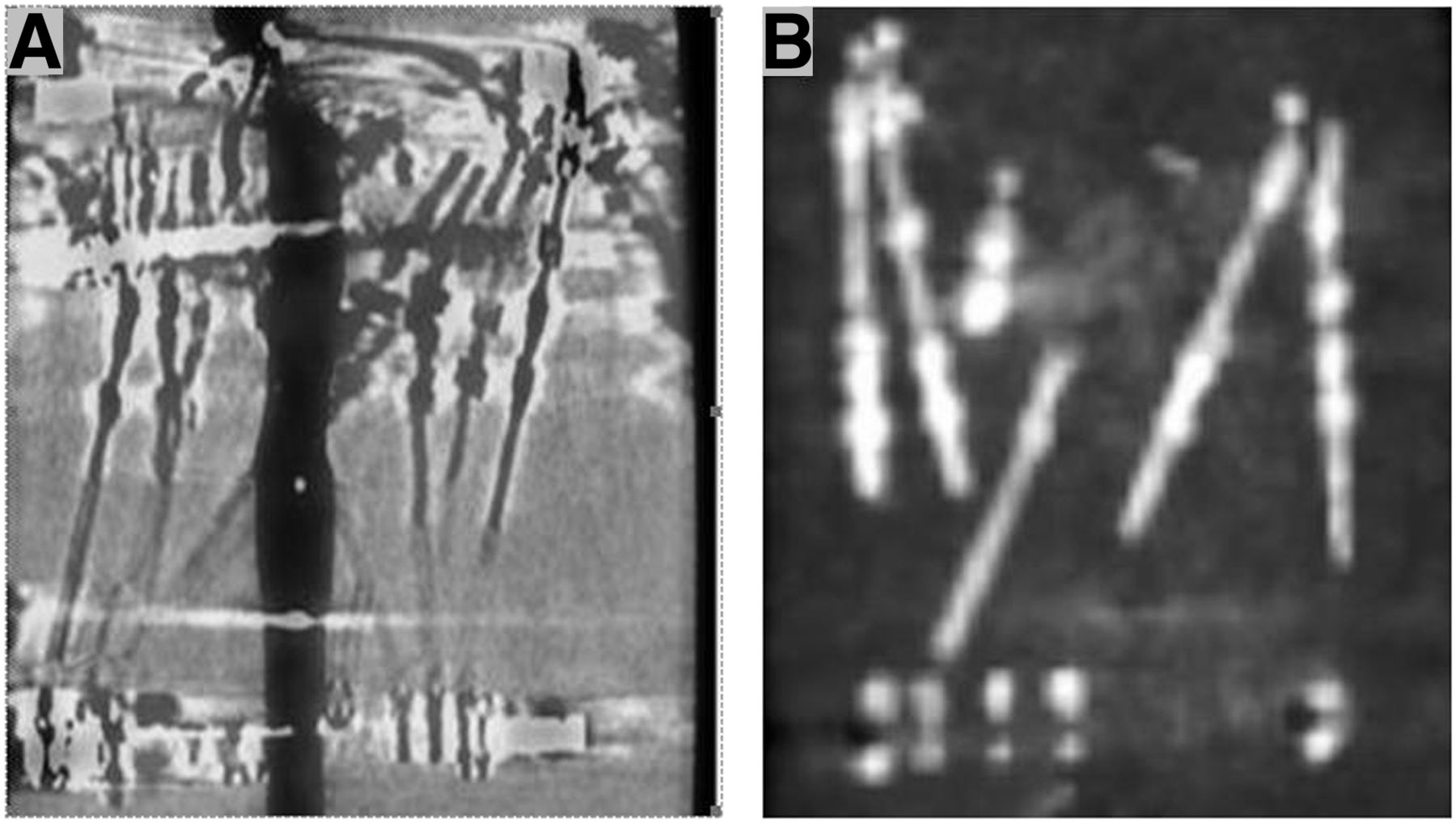
- FIGURE 3.
Reconstructed PET/CT scans of phantom: (A) When metal reduction algorithm is not used, no metal artifacts are seen in reconstruction of leg and no uptake of 18F is seen in Taylor spatial frame. (B) When metal reduction algorithm is used, uptake of 18F is seen in metal of Taylor spatial frame.
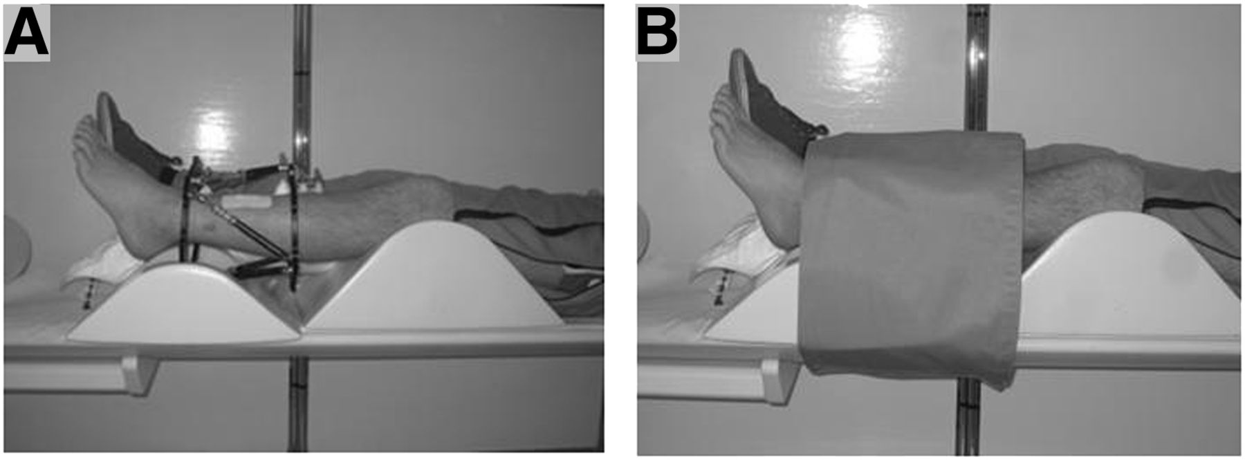
- FIGURE 4.
Initial (A) and final prescanning (B) positions of patient.
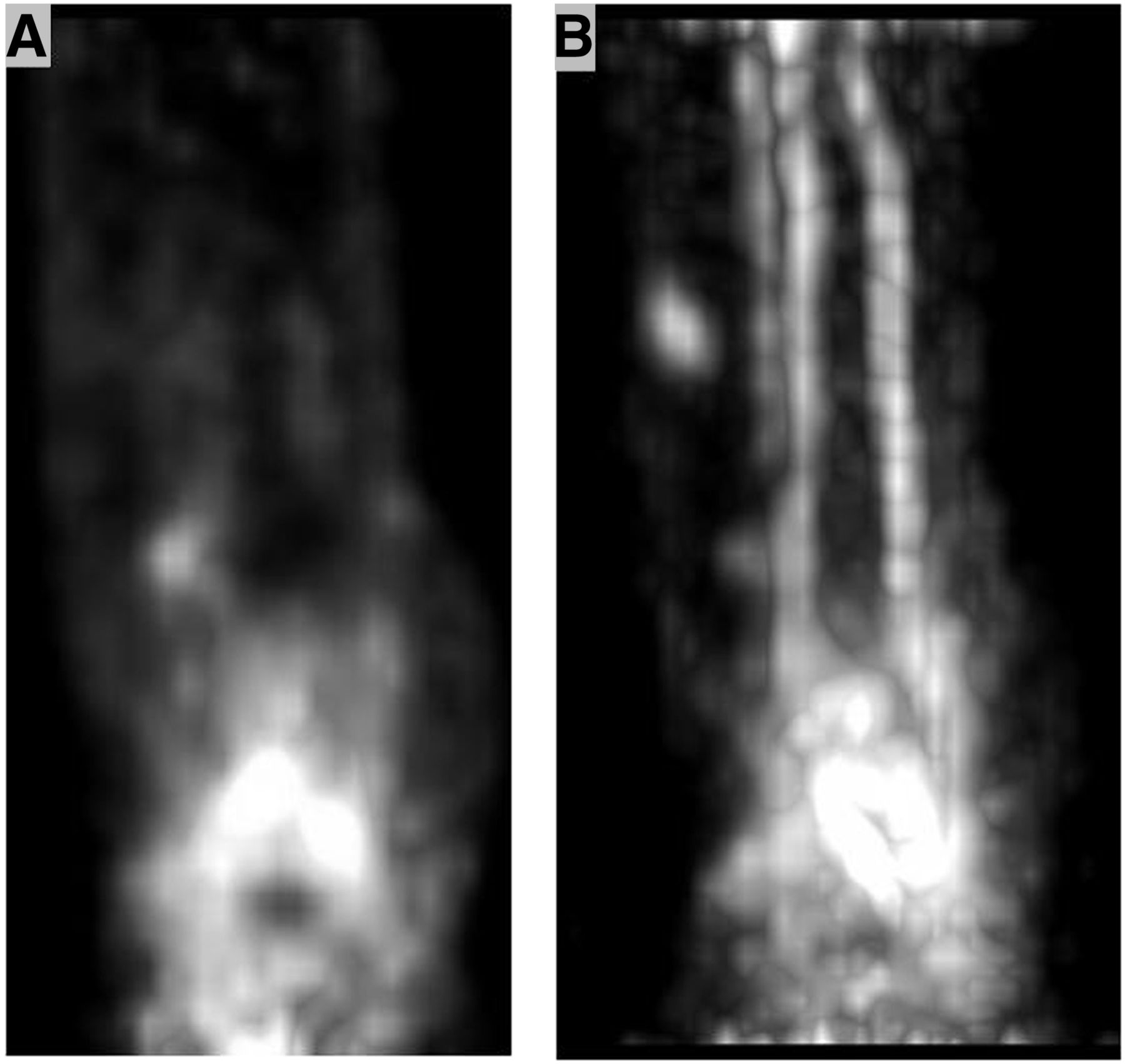
- FIGURE 5.
PET scans reconstructed from list mode with 5 frames of 1 min each (A) and 3 min each (B).
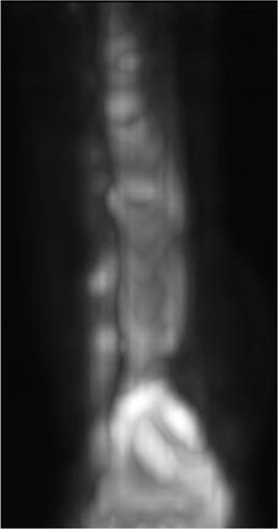
- FIGURE 6.
Initial 5-min static scan acquired after 60 min.
- FIGURE 7.
Three-dimensional reconstruction of axial view (A) and coronal view (B) of 5-min static PET scan fused with CT scan.


Jump to section
Related Articles
Cited By...
- No citing articles found.