


Article Figures & Data
Figures
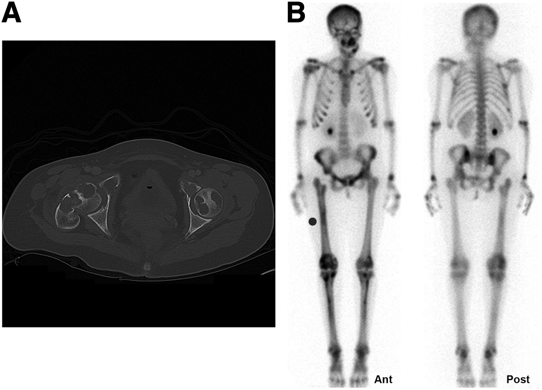
- FIGURE 1.
(A) Axial CT image of hips (iopamidol, 85 mL; 120 kVp; 210 mA; 380-mm field of view) shows pathologic fracture of right femoral neck with multiple osteolytic lesions involving femurs bilaterally and bony pelvis. (B) Whole-body bone scan with multifocal lesions involving mandible, maxilla, pelvis, and tibial bones.
- FIGURE 2.
(A and B) Axial maxillofacial CT images reveal expansile osteolytic lesions involving right and left maxilla and left mandible. (C) Axial maxillofacial CT image demonstrates interval sclerosis and regression of left maxillary lesion after parathyroidectomy.
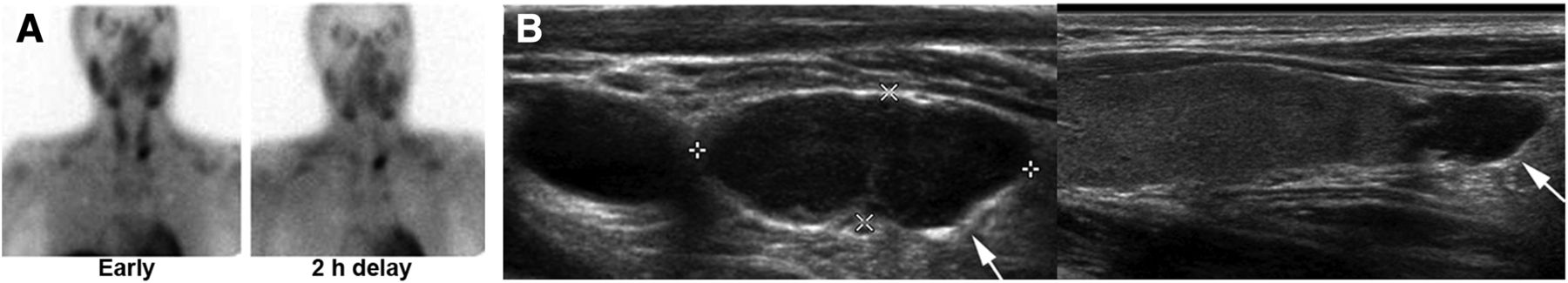
- FIGURE 3.
Planar 99mTc-sestamibi scan (A) and ultrasound (B) depict large left inferior parathyroid adenoma (arrows).
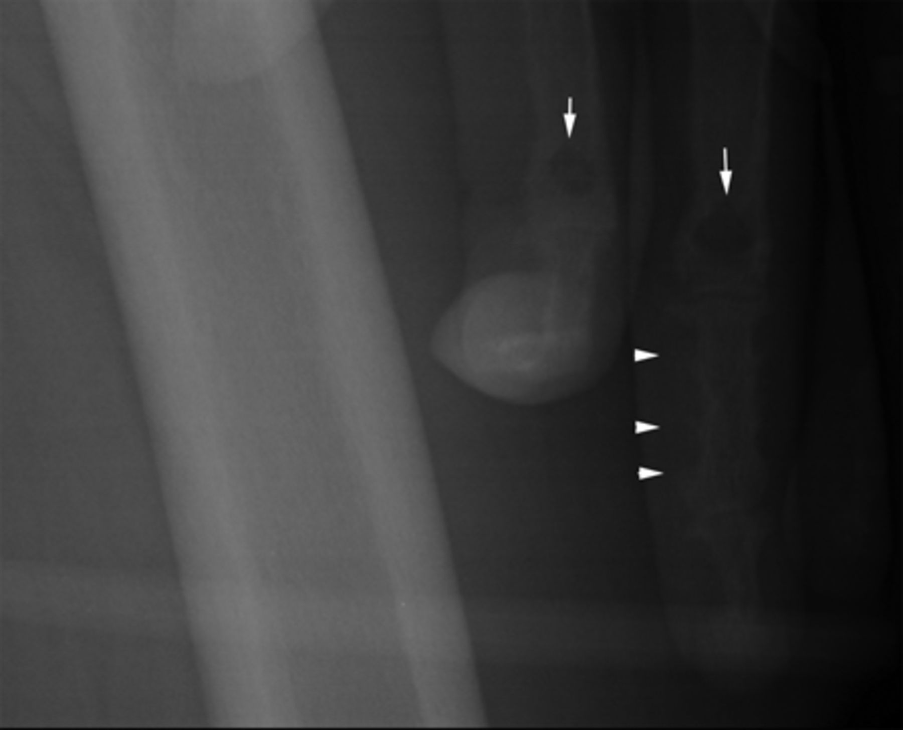
- FIGURE 4.
Frontal radiograph of left femur that incidentally included left hand depicts erosions and subperiosteal bone resorption on radial aspect of mid phalanx of middle finger (arrowheads) and brown tumors in proximal phalanges (arrows).


Jump to section
Related Articles
Cited By...
- No citing articles found.