
Main menu

  • Home
  • Content
    • Current
      • JNMT Supplement
    • Ahead of print
    • Past Issues
    • SNMMI Annual Meeting Abstracts
  • Subscriptions
    • Subscribers
    • Rates
    • Journal Claims
    • Institutional and Non-member
  • Authors
    • Submit to JNMT
    • Information for Authors
    • Assignment of Copyright
    • AQARA Requirements
  • Info
    • Continuing Education
    • Reviewers
    • Permissions
    • Advertisers
    • Corporate & Special Sales
  • About
    • About Us
    • Editorial Board
    • Editorial Contact
  • More
    • Alerts
    • Feedback
    • Help
    • SNMMI Journals
  • SNMMI
    • JNMT
    • JNM
    • SNMMI Journals
    • SNMMI

User menu

  • Subscribe
  • My alerts
  • Log in
  • My Cart

Search

  • Advanced search
Journal of Nuclear Medicine Technology
  • SNMMI
    • JNMT
    • JNM
    • SNMMI Journals
    • SNMMI
  • Subscribe
  • My alerts
  • Log in
  • My Cart
Journal of Nuclear Medicine Technology

Advanced Search

  • Home
  • Content
    • Current
    • Ahead of print
    • Past Issues
    • SNMMI Annual Meeting Abstracts
  • Subscriptions
    • Subscribers
    • Rates
    • Journal Claims
    • Institutional and Non-member
  • Authors
    • Submit to JNMT
    • Information for Authors
    • Assignment of Copyright
    • AQARA Requirements
  • Info
    • Continuing Education
    • Reviewers
    • Permissions
    • Advertisers
    • Corporate & Special Sales
  • About
    • About Us
    • Editorial Board
    • Editorial Contact
  • More
    • Alerts
    • Feedback
    • Help
    • SNMMI Journals
  • Follow SNMMI on Twitter
  • Visit SNMMI on Facebook
  • Join SNMMI on LinkedIn
  • Subscribe to JNMT RSS feeds
Case ReportCASE REPORTS

False-Positive Scalp Activity in 131I Imaging Associated with Hair Coloring

Di Yan, Mohan Doss, Ranee Mehra, Rosaleen B. Parsons, Barton N. Milestone and Jian Q. Yu
Journal of Nuclear Medicine Technology March 2013, 41 (1) 43-45; DOI: https://doi.org/10.2967/jnmt.112.117416
Di Yan
1Department of Medicine, Temple University School of Medicine, Philadelphia, Pennsylvania
  • Find this author on Google Scholar
  • Find this author on PubMed
  • Search for this author on this site
Mohan Doss
2Diagnostic Imaging, Fox Chase Cancer Center, Philadelphia, Pennsylvania; and
  • Find this author on Google Scholar
  • Find this author on PubMed
  • Search for this author on this site
Ranee Mehra
3Medical Oncology, Fox Chase Cancer Center, Philadelphia, Pennsylvania
  • Find this author on Google Scholar
  • Find this author on PubMed
  • Search for this author on this site
Rosaleen B. Parsons
2Diagnostic Imaging, Fox Chase Cancer Center, Philadelphia, Pennsylvania; and
  • Find this author on Google Scholar
  • Find this author on PubMed
  • Search for this author on this site
Barton N. Milestone
2Diagnostic Imaging, Fox Chase Cancer Center, Philadelphia, Pennsylvania; and
  • Find this author on Google Scholar
  • Find this author on PubMed
  • Search for this author on this site
Jian Q. Yu
2Diagnostic Imaging, Fox Chase Cancer Center, Philadelphia, Pennsylvania; and
  • Find this author on Google Scholar
  • Find this author on PubMed
  • Search for this author on this site
  • Article
  • Figures & Data
  • Info & Metrics
  • PDF
Loading

Article Figures & Data

Figures

  • FIGURE 1.
    • Download figure
    • Open in new tab
    • Download powerpoint
    FIGURE 1.

    Whole-body iodine scans before radioiodine ablation, in anterior and posterior projections. (A) Scan 1 mo before therapy. (B) Scan on day of radioiodine therapy. There is physiologic distribution of tracer in nasal mucosa, stomach, urinary bladder, and background.
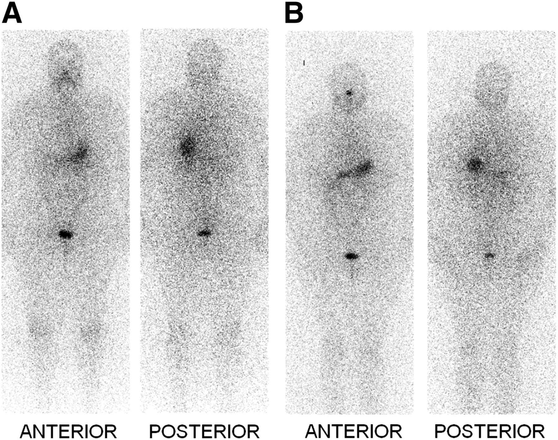
  • FIGURE 2.
    • Download figure
    • Open in new tab
    • Download powerpoint
    FIGURE 2.

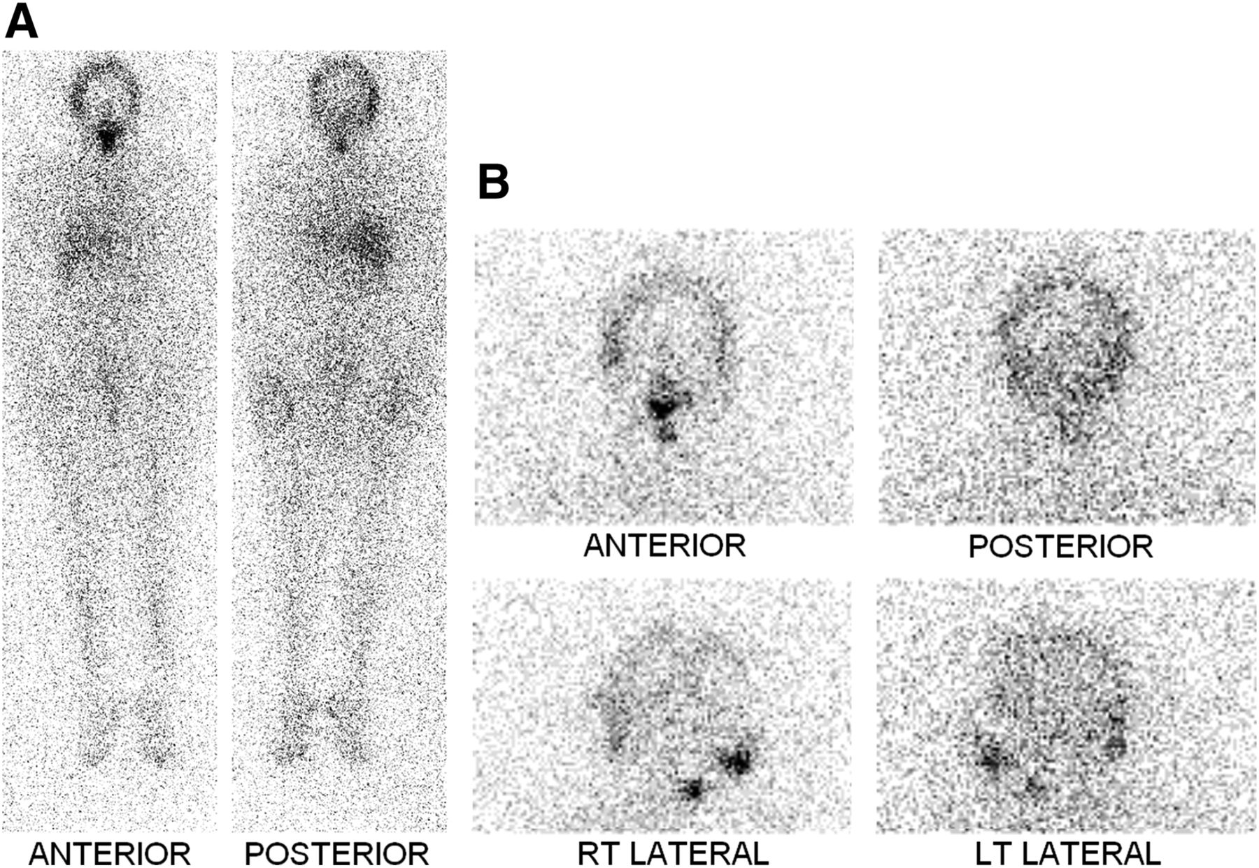
    (A) Whole-body iodine scan 1 wk after therapy. There is focal activity in lower neck consistent with iodine-avid tissue that had been treated with ablation dose. Liver activity is physiologic after therapy. There is diffuse helmet-shaped activity in head, likely in scalp or hair. (B) Images of head and neck in anterior, posterior, and lateral projections 10 d after therapy. Neck and nasal mucosal activity is present as expected. Activity in head is persistent and likely is in scalp, not in hair, because of patient’s reported good daily wash.

  • FIGURE 3.
    • Download figure
    • Open in new tab
    • Download powerpoint
    FIGURE 3.

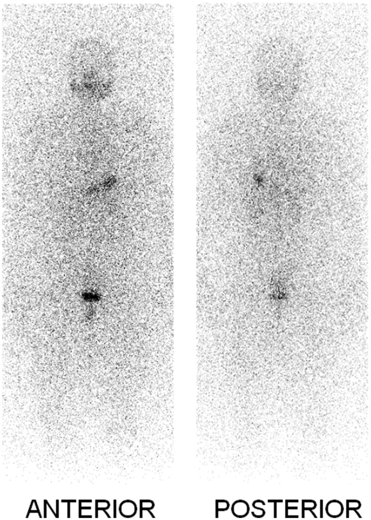
    Whole-body iodine scan for routine follow-up 15 mo after ablation. No abnormal activity is identified to suggest iodine-avid lesion. No focal activity in skull or scalp region is seen.

PreviousNext
Back to top

In this issue

Journal of Nuclear Medicine Technology: 41 (1)
Journal of Nuclear Medicine Technology
Vol. 41, Issue 1
March 1, 2013
  • Table of Contents
  • About the Cover
  • Index by author
Print
Download PDF
Article Alerts
Sign In to Email Alerts with your Email Address
Email Article

Thank you for your interest in spreading the word on Journal of Nuclear Medicine Technology.

NOTE: We only request your email address so that the person you are recommending the page to knows that you wanted them to see it, and that it is not junk mail. We do not capture any email address.

Enter multiple addresses on separate lines or separate them with commas.
False-Positive Scalp Activity in 131I Imaging Associated with Hair Coloring
(Your Name) has sent you a message from Journal of Nuclear Medicine Technology
(Your Name) thought you would like to see the Journal of Nuclear Medicine Technology web site.
Citation Tools
False-Positive Scalp Activity in 131I Imaging Associated with Hair Coloring
Di Yan, Mohan Doss, Ranee Mehra, Rosaleen B. Parsons, Barton N. Milestone, Jian Q. Yu
Journal of Nuclear Medicine Technology Mar 2013, 41 (1) 43-45; DOI: 10.2967/jnmt.112.117416

Citation Manager Formats

  • BibTeX
  • Bookends
  • EasyBib
  • EndNote (tagged)
  • EndNote 8 (xml)
  • Medlars
  • Mendeley
  • Papers
  • RefWorks Tagged
  • Ref Manager
  • RIS
  • Zotero
Share
False-Positive Scalp Activity in 131I Imaging Associated with Hair Coloring
Di Yan, Mohan Doss, Ranee Mehra, Rosaleen B. Parsons, Barton N. Milestone, Jian Q. Yu
Journal of Nuclear Medicine Technology Mar 2013, 41 (1) 43-45; DOI: 10.2967/jnmt.112.117416
Twitter logo Facebook logo LinkedIn logo Mendeley logo
  • Tweet Widget
  • Facebook Like
  • Google Plus One
Bookmark this article

Jump to section

  • Article
    • Abstract
    • CASE REPORT
    • DISCUSSION
    • CONCLUSION
    • DISCLOSURE
    • Footnotes
    • REFERENCES
  • Figures & Data
  • Info & Metrics
  • PDF

Related Articles

  • No related articles found.
  • PubMed
  • Google Scholar

Cited By...

  • No citing articles found.
  • Google Scholar

More in this TOC Section

  • Post-90Y Radioembolization PET/CT Scan with Respiratory Gating Using Time-of-Flight Reconstruction
  • Primary Hyperparathyroidism–Related Brown Tumors Mimicking Other Giant Cell–Containing Skeletal Tumors: Role of Correlative Imaging in Diagnosis
Show more Case Reports

Similar Articles

Keywords

  • 131I distribution
  • postablation scan
  • false-positive uptake
  • hair coloring
SNMMI

© 2023 Journal of Nuclear Medicine Technology

Powered by HighWire