


Article Figures & Data
Figures
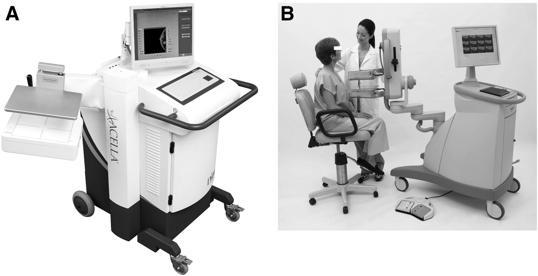
- FIGURE 1.
Breast imaging devices. (A) Single-photon imaging device from Dilon Diagnostics. (B) High-resolution PET scanner from Naviscan. In both systems, detectors are mounted on mammographic gantries, allowing for range of motion and positioning in either sitting or standing position. Each system also has small footprint.
- FIGURE 2.
Photographs showing positioning for craniocaudal (A), mediolateral oblique (B), and axillary (C) breast imaging views. System shown has single detector head and compression paddle. (Courtesy of Dilon Diagnostics.)
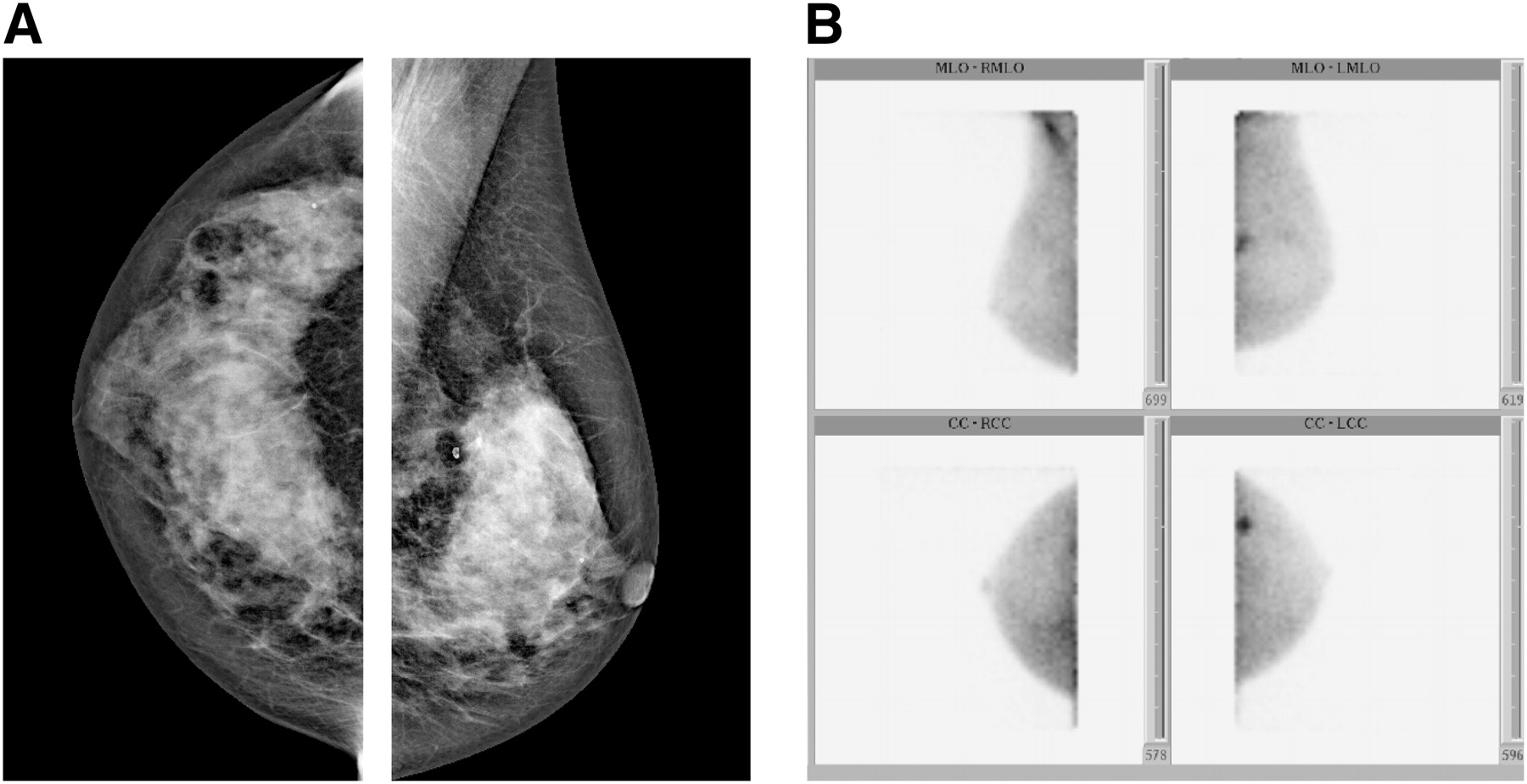
- FIGURE 3.
Breast cancer seen with 99mTc-sestamibi. (A) Craniocaudal and mediolateral oblique mammographic views of left breast in patient with dense breasts. Suggestive area corresponded to location of prior biopsy that documented benign lesion. (B) Nuclear images show uptake in area of prior biopsy, which was determined to be infiltrating lobular carcinoma. (Courtesy of Dilon Diagnostics.)
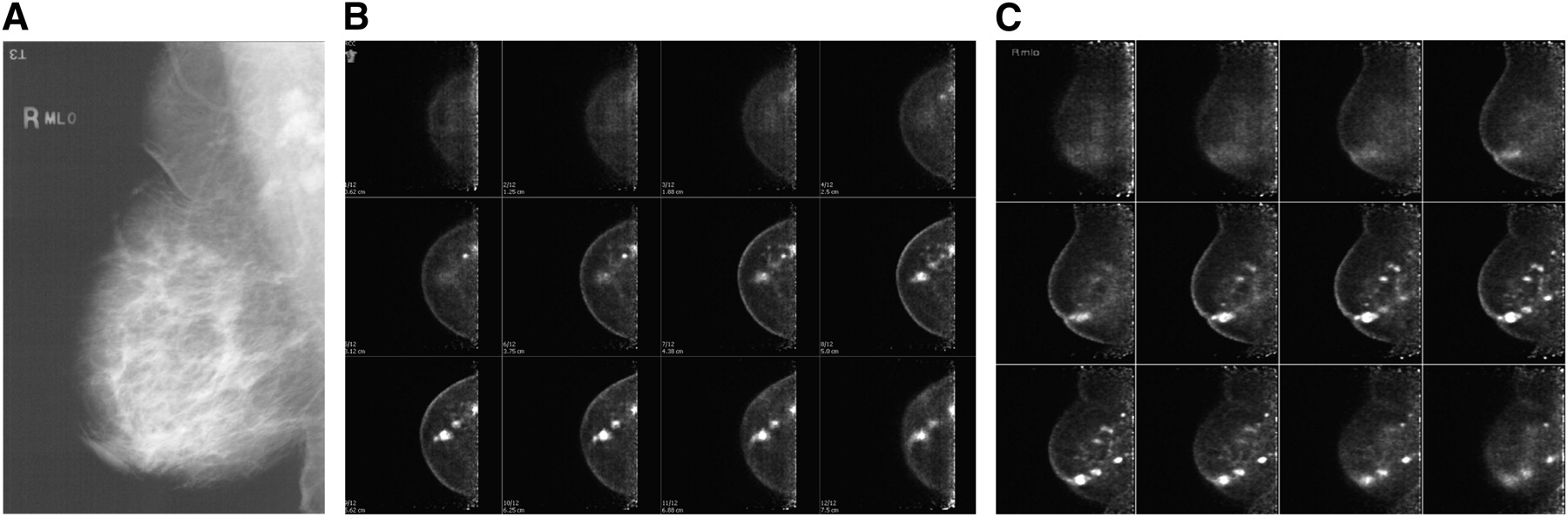
- FIGURE 4.
Tomographic slices from 18F-FDG breast study. Mediolateral oblique (B) and craniocaudal (C) views of multifocal cancer in right breast are shown, along with corresponding right mediolateral oblique mammogram (A). Slice thickness depends on distance between the 2 detectors, since this system always produces either 12-slice or 24-slice study. (Obtained from East Jefferson General Hospital, Metairie, Louisiana, and provided courtesy of Naviscan, Inc.)


Tables
Manufacturer Parameter Dilon Dilon GammaMedica Digirad GE Healthcare Naviscan Model 6800 6800 Acella LumaGem Ergo Discovery NM 530c High-resolution organ-specific PET scanner No. of heads 1 1 2 1 1 or 2 2 Detector material NaI(Tl) CsI(Tl) CZT CsI(Tl) CZT Lutetium yttrium oxyorthosilicate Detector size 3 × 3 × 6 mm 3 × 3 × 6 mm 1.6 × 1.6 × 5 mm 3 × 3 × 6 mm 2.6 × 2.6 mm 2 × 2 × 13 mm Photon/electron collection PSPMT p-type/intrinsic/n-type photodiode Application-specific integrated circuit Silicon photodiode Application-specific integrated circuit PSPMT Collimation γ-camera types γ-camera types Registered collimator γ-camera types, pinhole Registered collimator None Dead space 10 mm 10 mm 8 mm 12.7 mm “Minimal” 13 mm Imaging FOV 15 × 20 cm 20 × 25 cm 16 × 20 cm 40 × 30 cm 16 × 24 cm 17 × 24 cm Energy resolution 13.5% 7.9% <5% (140 keV) 7.9% (140 keV) 6.5% (140 keV) 13% Energy range 70–200 keV 50–170 keV Optimized for photons < 160 keV, capable of up to 511 keV 50–350 keV 80–200 keV 350–700 keV Spatial resolution 3.3 mm 3.3 mm 1.6 mm 3.3 mm 2.46 mm 2.4 mm Source for statistics Company’s specification sheet Company’s specification sheet Company’s specification sheet and written communication from James Hugg, Chief Technology Officer, GammaMedica, August 2011 Company’s specification sheet Company’s specification sheet Luo and MacDonald articles and written communication from Weidong Luo, Director of Imaging Physics for Naviscan, October 2011


