


Abstract
This study was done to examine the effect of asiaticoside on MCF-7 cell uptake of 99mTc-tetrofosmin (99mTc-Tfos) and 99mTc-sestamibi (99mTc-MIBI). Methods: The 3-(4,5-dimethylthiozol-2-yl)-2,5-diphenyltetrazolium bromide (MTT) assay was used to evaluate the effect of a 50% inhibitory concentration of asiaticoside on MCF-7 cell proliferation. MCF-7 cells were treated with 10, 20, 30, 40, and 50 μM asiaticoside for 48 h and then incubated with 59.2 MBq of either 99mTc-Tfos or 99mTc-MIBI tracer for 60 min. The uptake of the tracers was measured with a dose calibrator. Results: The 50% inhibitory concentration of asiaticoside for MCF-7 cells was determined with the MTT assay to be 40 μM. The uptake results were expressed as the mean ± SE radioactivity in MBq/mg of protein, and P values were also calculated (P values of 0.03 indicated significant differences). In the control (no asiaticoside) and at 10, 20, 30, 40, and 50 μM asiaticoside, the mean levels of 99mTc-Tfos uptake were 0.79 (SE, 0.059) (P = 0.14), 0.84 (SE, 0.057) (P = 0.60), 0.47 (SE, 0.034) (P = 0.03), 0.40 (SE, 0.050) (P = 0.03), 0.37 (SE, 0.050) (P = 0.03), and 0.15 (SE, 0.023) (P = 0.03), respectively; the mean levels of 99mTc-MIBI uptake were 0.95 (SE, 0.007) (P = 0.14), 0.81 (SE, 0.009) (P = 0.60), 0.79 (SE, 0.019) (P = 0.03), 0.63 (SE, 0.004) (P = 0.03), 0.13 (SE, 0.006) (P = 0.03), and 0.07 (SE, 0.008) (P = 0.03), respectively. Asiaticoside concentrations of 10, 20, 30, 40, and 50 μM revealed the uptake kinetics for both 99mTc-Tfos and 99mTc-MIBI in MCF-7 cells. 99mTc-Tfos and 99mTc-MIBI showed similar trends; the radioactivity uptake was dose dependent, and asiaticoside inhibited 16% and 47% of 99mTc-Tfos uptake and 99mTc-MIBI uptake in MCF-7 cells, respectively. Conclusion: This study showed that asiaticoside, acting as a biochemical modulator, may induce apoptosis and enhance antitumor activity in MCF-7 cells, as determined by 99mTc-Tfos and 99mTc-MIBI uptake. These findings are promising for cancer chemotherapy. Future studies should be performed to confirm our findings and to further delineate the clinical role of asiaticoside.
The cationic lipophilic agent 99mTc-hexakis-2-methoxyisobutylisonitrile, or 99mTc-sestamibi (99mTc-MIBI), is widely used for myocardial perfusion imaging (1). 99mTc-MIBI has been reported to accumulate within the mitochondria and cytoplasm of cells on the basis of transmembrane electrical potentials (2,3). 99mTc-tetrofosmin (99mTc-Tfos) is a monovalent cationic lipophilic agent (diphosphine group) (4,5) that was originally developed for myocardial perfusion imaging. It rapidly enters myocardial cells because of its lipophilic properties (3,6), although these properties alone may not be the sole determinants.
99mTc-Tfos has proved to be an excellent tumor-imaging agent, accumulating in thyroid and breast cancers and, recently, in a variety of tumors with a sensitivity of 95% or more and a specificity of 91% (7–9). Many in vitro and in vivo studies have reported that 99mTc-Tfos is a good tumor marker for many types of cancers, such as thyroid and brain cancers, malignant gliomas, and lung and breast cancers (10–12). Recently, several musculoskeletal sarcomas of the extremities or pelvis were examined (13). Malignant breast tumors were shown to have increased transmembrane potentials because of increased metabolic requirements, which induced the increased accumulation of 99mTc-MIBI in these tumors (3,6,14). 99mTc-Tfos was retained at high levels in malignant tumors and accumulated through a similar mechanism, which might be related to the tumor cell proliferation and lipophilicity of this tracer. One study reported that the uptake of both 99mTc-MIBI and 99mTc-Tfos through the cell membrane was related, in part, to the Na+/H+ antiporter system. Only a portion of the accumulated 99mTc-Tfos inside the cells entered the mitochondria, and most of the accumulated 99mTc-MIBI was related to mitochondrial uptake (15).
In the present study, human breast cancer cell line MCF-7 was used because it is a commonly available human breast cancer in vitro model. MCF-7 was previously studied with both 99mTc-Tfos and 99mTc-MIBI and shown to have high uptake in comparison with other types of cancer cell lines (16).
Cancer causes about 23.2% of all deaths. According to the American Cancer Society, more than 7.6 million people died from cancer worldwide in 2010 (17–19). Cancer chemoprevention is considered one of the most promising areas in current cancer research (20). Centella asiatica is a plant that is widely used in traditional Ayurvedic medicine for a variety of illnesses. Recent research has shown that components of C. asiatica, specifically, asiaticoside, show great promise in the prevention and treatment of cancer. A major advantage of using this plant is its relative lack of systemic toxicity. Few studies have investigated this effect (21,22). In the present study, we examined the chemopreventive and cytotoxic effects of asiaticoside on MCF-7 cell uptake of 99mTc-Tfos and 99mTc-MIBI.
MATERIALS AND METHODS
Materials
A tetrofosmin (Myoview) kit was purchased from Amersham International. 99mTc-MIBI (Cardiolite) was purchased from Bristol–Myers Squibb. Pertechnetate (99mTcO−4) was obtained from a molybdenum/technetium (99Mo/99mTc) generator (Amersham). Asiaticoside (molecular weight, 959.12) and all other reagents used in this study were supplied by Sigma.
Cells and Culture Media
All of the culture media and supplements were provided by BioWhittaker. MCF-7 is an established cell line derived from a breast tumor (American Type Culture Collection). MCF-7 cells were grown in advanced Dulbecco modified Eagle medium supplemented with 10% fetal calf serum, l-glutamine at 2 mmol/L, penicillin at 100 U/mL, and streptomycin at 100 mg/mL in a humidified atmosphere with 5% CO2 at 37°C. Unless otherwise stated, stock cultures of MCF-7 cells were seeded at a density of 2 × 10⁵ cells per milliliter in 25-cm2 flasks and allowed to multiply for 48–72 h. In chemotherapy experiments, the MCF-7 cells were drug-sensitive, wild-type cells that were allowed to grow exponentially to 70% confluence.
Preparation of 99mTc-Tfos
Fresh eluates of 99mTc were used each time to prepare 99mTc-Tfos in accordance with the Myoview kit instructions and recommendations. In brief, 1,110 MBq of 99mTc in 5 mL of saline (99mTcO−4) were added to a freeze-dried Myoview kit to produce 99mTc-Tfos, and the mixture was incubated for at least 15 min at room temperature. The quality of the prepared tracer was checked by thin-layer chromatography on silica plates (Silica Gel type G; Sigma) with a 35:65 (v/v) mixture of acetone and dichloromethane as the mobile phase in accordance with the manufacturer's instructions. The radiochemical purity was always greater than 95%, and the pH of prepared 99mTc-Tfos was 7.5.
Preparation of 99mTc-MIBI
Lyophilized 99mTc-MIBI vial products were reconstituted with 1,110 MBq of fresh 99mTcO−4. The vial was heated in a boiling water bath for 10 min. After the vial was cooled at room temperature, quality control procedures were performed with Whatman number 1 paper and a 75:25 solution of chloroform and methanol in accordance with the manufacturer's instructions. The radiochemical purity was greater than 95%.
99mTc-Tfos and 99mTc-MIBI Radiotracer Experiment and Uptake Determination
MCF-7 cells were cultured in 25-cm2 flasks, in triplicate, until they reached 70% confluence. After reaching 70% confluence, the MCF-7 cells were divided into 2 groups. The first group (control) consisted of MCF-7 cells that were not treated with asiaticoside (concentration of 0). The second group (asiaticoside treated) consisted of MCF-7 cells that were treated with various concentrations of asiaticoside (10, 20, 30, 40, and 50 μM).
Next, the cells were incubated in a humidified atmosphere with 5% CO2:95% air at 37°C for 48 h. After the 48-h incubation, 59.2 MBq of 99mTc-Tfos or 99mTc-MIBI were added to the cells. After 60 min, the uptake of 99mTc-Tfos or 99mTc-MIBI in the radioactive medium was measured with a dose calibrator (ATOMLAB 100; Biodex Medical Systems). The cells were washed 6 times with ice-cold phosphate-buffered saline (PBS) to eliminate the free tracer present in the extracellular spaces.
Next, the cells were incubated with a nonradioactive medium. The efflux of activity in the medium was measured. The cells were washed once with ice-cold PBS, trypsinized with 0.5 mL of trypsin for 3 min, neutralized with 0.5 mL of advanced Dulbecco modified Eagle medium, and centrifuged at 1,000 rpm for 2 min at 4°C. Activity in the medium was measured. Cell pellets were washed once with PBS to remove extracellular protein, and radioactivity uptake was measured. The last step was repeated 3 times.
Next, the cells were solubilized with 1% sodium dodecyl sulfate (SDS) in sodium borate (10 mmol/L; Sigma). The radioactivity in the cellular lysate was measured with the dose calibrator.
All of the experiments were performed in triplicate and repeated.
Cell Viability Assay
MCF-7 cells (106) were incubated in 25-cm2 flasks in triplicate. The flasks were set up for controls and various asiaticoside concentrations (0.0025, 0.01, 0.02, 0.04, 0.1, 0.2, 0.25, 0.3, 0.5, 1, 10, 20, 40, 50, 125, 250, and 500 μM) and then incubated in a humidified atmosphere with 5% CO2:95% air at 37°C for 24, 48, or 72 h. Cell viability was measured with the 3-(4,5-dimethylthiozol-2-yl)-2,5-diphenyltetrazolium bromide (MTT) assay, which is based on the conversion of MTT to MTT–formazan by mitochondria.
Also, in some experiments, the cells were seeded in flat-bottom, 96-well tissue culture plates in triplicate at a concentration of 105 cells per milliliter of medium in a volume of 100 μL per well and were allowed to grow to 70% confluence before asiaticoside was added. After the cells reached 70% confluence, various concentrations of asiaticoside were added separately, and the mixtures were incubated for 24, 48, and 72 h. Next, the medium was removed with a pipette, the cells were washed with PBS, and 100 μL of fresh medium as well as 20 μL of MTT (5 mg/mL) were added to each well. The plates were protected from light and incubated for 3 h, and the formazan crystals that formed were solubilized with 200 μL of dimethyl sulfoxide per well. The plates were kept in a shaker with gentle mixing for 20 min to dissolve the precipitate. The color that developed was measured with a 96-well plate scanner (Multiskan Spectrum; Thermo Electron Corp.) at dual filter wavelengths (540 and 690 nm). Cell viability was expressed as a percentage of the control value. This viability test was used to determine the optimum 50% inhibitory concentration (IC50) of asiaticoside for MCF-7 cells.
Protein Determination
Protein content was determined with a micro–bicinchoninic acid protein assay kit (Pierce).
Data Presentation and Statistical Analysis
All data, unless otherwise stated, were expressed as the mean (SE). The Student t test was used to determine statistical differences between 2 means, whereas Kruskal–Wallis nonparametric analysis of the 1-way ANOVA was used to evaluate differences between time points. Statistical analysis was performed with SPSS version 17.0 software (SPSS Inc.).
RESULTS
Cell Viability Assay
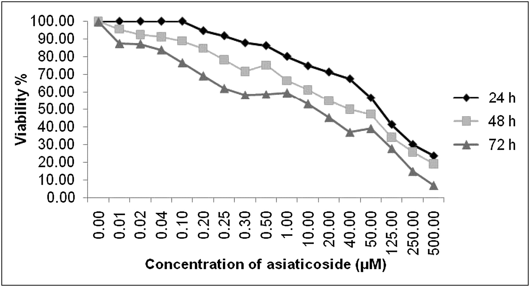
The IC50 of asiaticoside for MCF-7 cells was determined with the MTT assay to be 40 μM. Figure 1 shows the viability of MCF-7 cells treated with asiaticoside for 24, 48, and 72 h; viability was determined with the MTT assay and expressed as a percentage of the control value. Figure 2 shows the inhibition of MCF-7 cells treated with asiaticoside for 24, 48, and 72 h; inhibition was determined with the MTT assay and expressed as a percentage of the control value.

Cell viability, expressed as percentage of control value, for MCF-7 cells treated with asiaticoside for 24, 48, and 72 h. Viability was determined with MTT assay.

Cell inhibition, expressed as percentage of control value, for MCF-7 cells treated with asiaticoside for 24, 48, and 72 h. Inhibition was determined with MTT assay.
99mTc-Tfos and 99mTc-MIBI Radiotracer Experiment and Uptake Determination
The uptake results were expressed as the mean ± SE radioactivity in MBq/mg of protein, and P values were also calculated (P values of 0.03 indicated significant differences). Table 1 shows both 99mTc-Tfos uptake and 99mTc-MIBI uptake. In the control (no asiaticoside) and at 10, 20, 30, 40, and 50 μM asiaticoside, the mean levels of 99mTc-Tfos uptake were 0.79 (SE, 0.059) (P = 0.14), 0.84 (SE, 0.057) (P = 0.60), 0.47 (SE, 0.034) (P = 0.03), 0.40 (SE, 0.050) (P = 0.03), 0.37 (SE, 0.050) (P = 0.03), and 0.15 (SE, 0.023) (P = 0.03), respectively; the mean levels of 99mTc-MIBI uptake were 0.95 (SE, 0.007) (P = 0.14), 0.81 (SE, 0.009) (P = 0.60), 0.79 (SE, 0.019) (P = 0.03), 0.63 (SE, 0.004) (P = 0.03), 0.13 (SE, 0.006) (P = 0.03), and 0.07 (SE, 0.008) (P = 0.03), respectively. Asiaticoside concentrations of 10, 20, 30, 40, and 50 μM revealed the uptake kinetics for both 99mTc-Tfos and 99mTc-MIBI in MCF-7 cells. 99mTc-Tfos and 99mTc-MIBI showed similar trends; the radioactivity uptake was dose dependent, and asiaticoside inhibited 16% and 47% of 99mTc-Tfos uptake and 99mTc-MIBI uptake in MCF-7 cells, respectively. Both 99mTc-Tfos and 99mTc-MIBI showed significant reductions in MCF-7 cell uptake at asiaticoside concentrations of 20–50 μM, as determined with the Student paired t test (P = 0.03), relative to the results for their own controls. Also, there were no significant differences from the results for the controls at asiaticoside concentrations of less than or equal to 10 μM (P = 0.60).
Differences Between 99mTc-Tfos Uptake and 99mTc-MIBI Uptake in MCF-7 Cells After Treatment with Asiaticoside (n = 12)
DISCUSSION
In this study, the IC50 of asiaticoside for MCF-7 cells was determined with the MTT assay to be 40 μM. This finding is in agreement with the results of a study reporting that the IC50 of asiatic acid and asiaticoside ranged from 20.42 to 76.45 μM (23). Here, the nuclear medicine radiopharmaceuticals 99mTc-Tfos and 99mTc-MIBI were used to examine the potential protective effect of asiaticoside on MCF-7 cells. The results suggested that asiaticoside may have chemopreventive and antitoxic effects on MCF-7 cells, enhancing antitumor activity. These effects were demonstrated by reduced radiotracer uptake. Our results are in agreement with the suggestion of some studies investigating cell uptake that asiaticoside, acting as a biochemical modulator, may induce apoptosis or has a protective effect against β-amyloid neurotoxicity (22,24). Here, we demonstrated that the radiotracers 99mTc-Tfos and 99mTc-MIBI were dependent on asiaticoside concentrations to similar degrees. In fact, 99mTc-Tfos and 99mTc-MIBI were reported to be accurate and equally efficient for the detection of breast malignancies (13,16), and our results are in agreement with such reports.
Many studies have reported that the radiotracers 99mTc-MIBI and 99mTc-Tfos are trapped within the mitochondria because of a highly negative mitochondrial transmembrane potential that, in the normal physiologic state, is lower than that of the cytosol. Also, the accumulation of 99mTc-MIBI and 99mTc-Tfos by the mitochondria is related to the ability to transduce metabolic energy into an electronegative transmembrane potential (25,26). In addition, their uptake is related to increased energy-dependent metabolism and cell proliferation. 99mTc-MIBI and 99mTc-Tfos localize primarily in normally functioning mitochondria. Apoptosis is accompanied by a decrease in the mitochondrial transmembrane potential toward that of the cytosol, reducing the localization of these radiotracers (27). In this study, both 99mTc-MIBI uptake and 99mTc-Tfos uptake in MCF-7 cells were significantly reduced when asiaticoside concentrations were increased and when different concentrations of asiaticoside were used. These results may suggest that asiaticoside stimulates the process of programmed cell death, cell apoptosis, through a certain mechanism. Gurfinkel et al. reported that disruption of the cellular endoplasmic reticulum and alterations in calcium homeostasis were early events in asiaticoside-induced cell apoptosis (28). Other studies suggested that the administration of asiaticoside caused a disturbance in mitochondrial function (29) that was manifested as cell apoptosis (30).
Recent studies demonstrated that the cellular accumulation of 99mTc-MIBI and 99mTc-Tfos was reduced when multidrug resistance proteins were overexpressed (31). These results may suggest multidrug resistance proteins as a mechanism (22,32) for the effect of asiaticoside in this study. Many studies have reported a positive correlation between the administration of asiaticoside and antitumor activity (30), inhibition of DNA synthesis (33), therapeutic interventions for many human cancer types (34,35), and protection of the liver from damage. The mechanism has been suggested to be related to upregulation of mitochondrial voltage-dependent anion channels and inhibition of the process of mitochondrial permeability transition (36).
CONCLUSION
This study showed that asiaticoside, acting as a biochemical modulator, may induce apoptosis and enhance antitumor activity in MCF-7 cells, as determined by 99mTc-Tfos and 99mTc-MIBI uptake. These findings are promising for cancer chemotherapy. Future studies should be performed to confirm our findings and to further delineate the clinical role of asiaticoside.
Acknowledgments
I would like to thank Research Core Facility Projects GM 01/01 and GM 01/05 for valuable technical support and Kuwait University Research Administration for funding (Research Grant No. MN 01/09). No other potential conflict of interest relevant to this article was reported.
Footnotes
Published online Nov. 11, 2011.
REFERENCES
- 1.
- 2.
- 3.
- 4.
- 5.
- 6.
- 7.
- 8.
- 9.
- 10.
- 11.
- 12.
- 13.
- 14.
- 15.
- 16.
- 17.
- 18.
- 19.
- 20.
- 21.
- 22.
- 23.
- 24.
- 25.
- 26.
- 27.
- 28.
- 29.
- 30.
- 31.
- 32.
- 33.
- 34.
- 35.
- 36.
- Received for publication May 11, 2011.
- Accepted for publication August 9, 2011.


