Table of Contents
Cover image

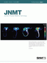
ON THE COVER
Maximum-intensity-projection 18F-FDG microPET images of mouse tails all scaled to the same display window of 50% injected dose per gram. The first image on the left is a mouse injected intraperitoneally. The next three images from left to right are mice injected intravenously via lateral tail veins and classified qualitatively as good, intermediate, and poor injections.
See page 264.