


Article Figures & Data
Figures
- FIGURE 1.
Horizontal slices of 123I-PE2I template with manually delineated striatum (yellow and cyan), putamen (green and blue), caudate nucleus (pink and red), and reference (orange) ROIs. (A color version of this figure is available as a supplemental file online at http://tech.snmjournals.org/.)
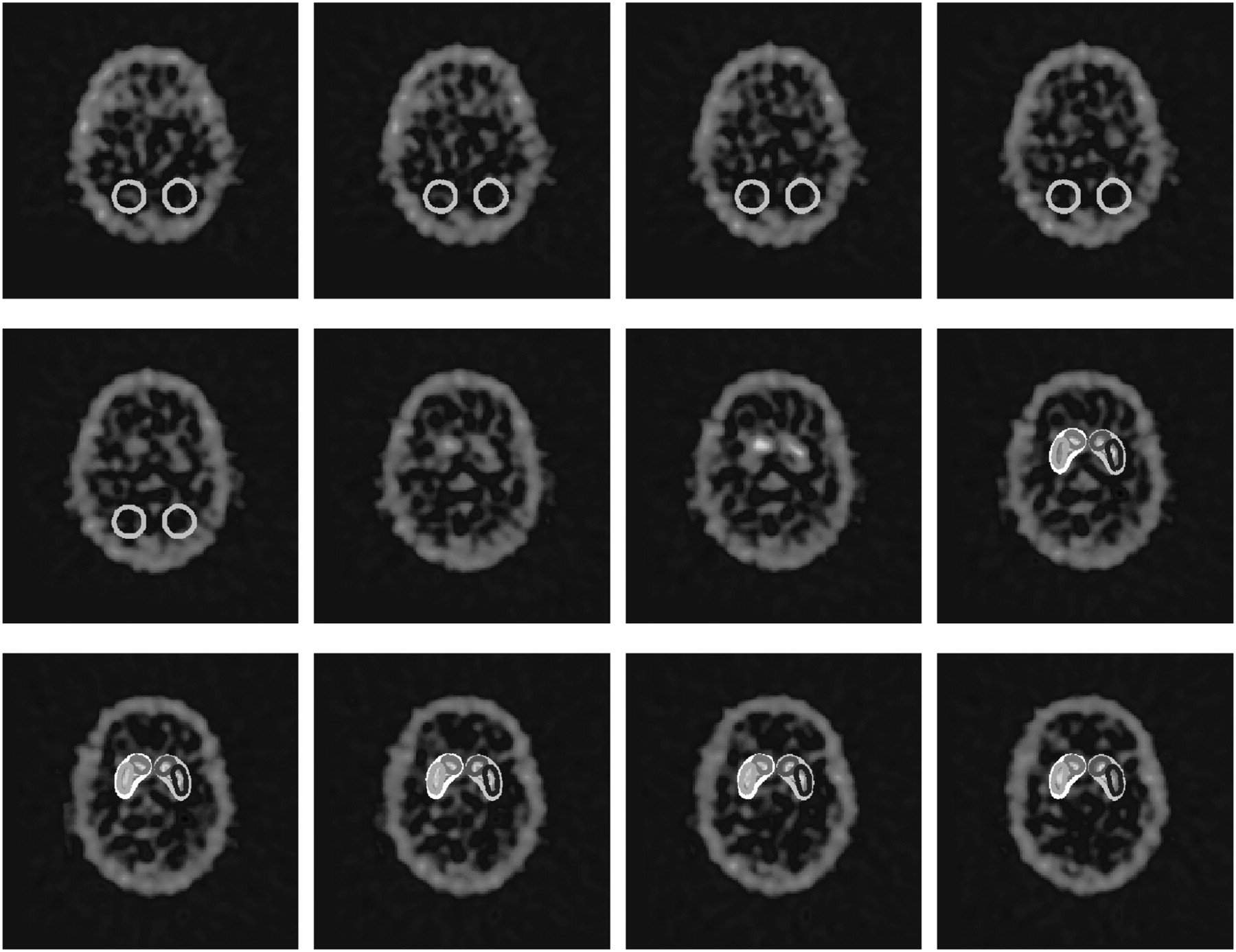
- FIGURE 2.
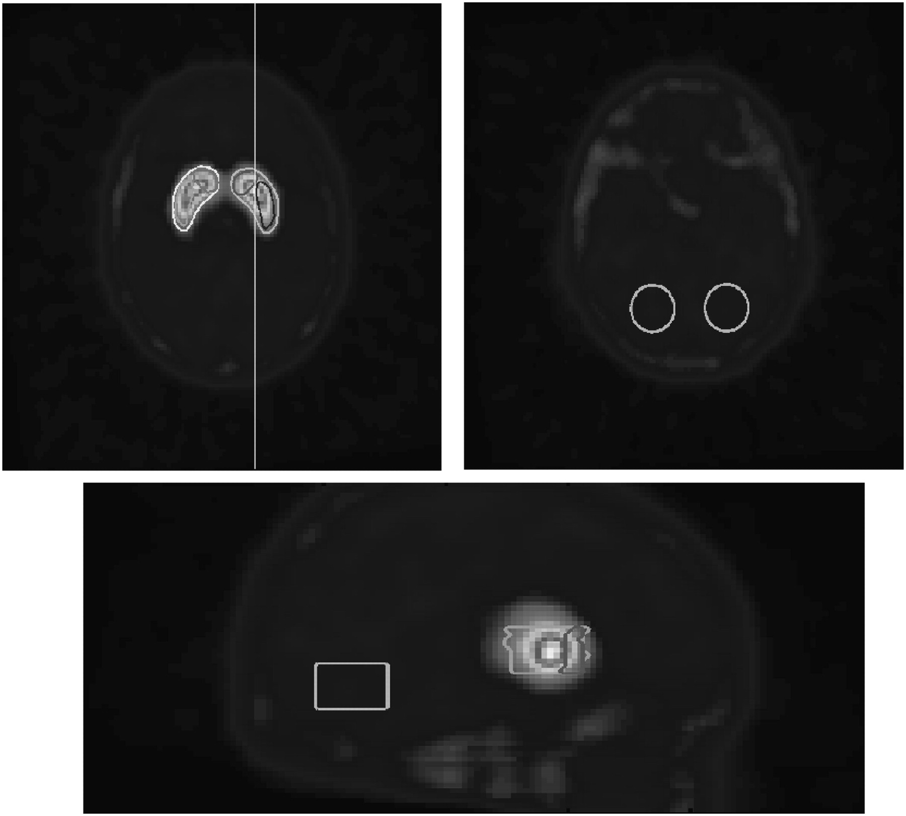
Two horizontal slices (upper row) and 1 sagittal slice (lower row) of 123I-PE2I template illustrating exact positions and configurations of manually defined striatal and reference ROIs. Vertical line in first horizontal slice (upper row, left) illustrates location of sagittal slice. (A color version of this figure is available as a supplemental file online at http://tech.snmjournals.org/.)
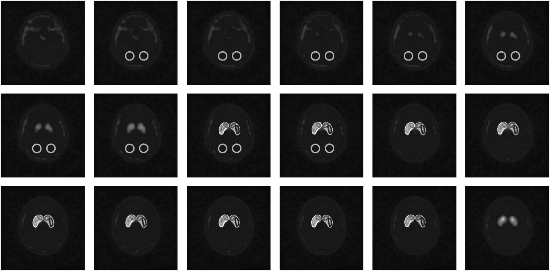
- FIGURE 3.
Example of part of patient's normalized 123I-PE2I image in template space with automatic DATquan-delineated ROIs. Each ROI covers 5 consecutive slices, and there is a gap of 2 slices between reference and striatal ROIs. (A color version of this figure is available as a supplemental file online at http://tech.snmjournals.org/.)
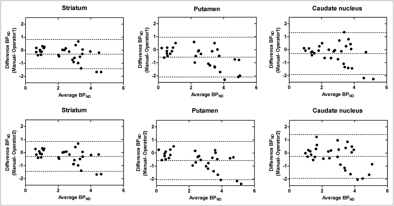
- FIGURE 4.
Bland–Altman plots of difference between ROI-specific BPND estimates obtained manually (by experienced physician) and semiautomatically (with DATquan) against mean of estimates. Top and bottom rows show plots for first and second DATquan operators, respectively. For each ROI-specific plot, mean error and 95% limits of agreements are indicated by dashed lines.
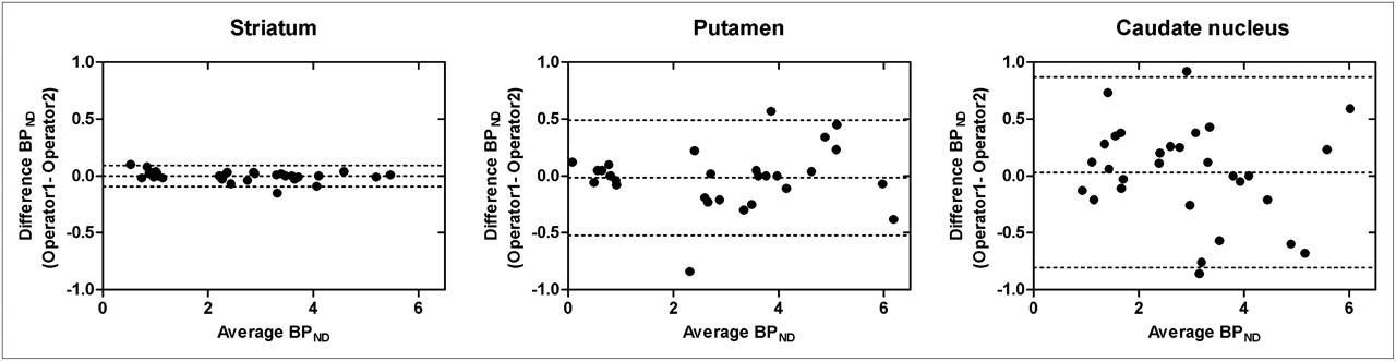
- FIGURE 5.
Bland–Altman plots of difference between ROI-specific BPND estimates obtained by 2 DATquan operators against mean of estimates. For each ROI-specific plot, mean difference and 95% limits of agreements are indicated by dashed lines.
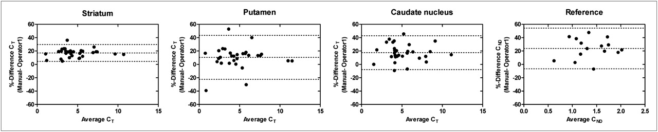
- FIGURE 6.
Bland–Altman plots of difference between ROI-specific mean counts (CT and CND) derived from manually delineated and DATquan-delineated (operator 1) regions against mean of mean counts. For each ROI-specific plot, mean error and 95% limits of agreements are indicated by dashed lines.

Tables
Mean (SD) mL of: Area Manually delineated ROI DATquan-delineated ROI Operator 1 Operator 2 Striatum 6.50 (1.95)* 9.26 (0.34) 9.32 (0.37) Putamen 2.82 (0.80) 2.87 (0.22) 2.87 (0.25) Caudate nucleus 2.07 (0.56) 2.11 (0.17) 2.12 (0.15) Reference 15.3 (4.13) 16.0 (2.70) 16.0 (2.70) ↵* Statistically significant (P < 0.01) difference between volume of manually delineated ROI and volume of each DATquan-delineated ROI.
Supplemental Data
Files in this Data Supplement:


Jump to section
Related Articles
Cited By...
- No citing articles found.