


Article Figures & Data
Figures
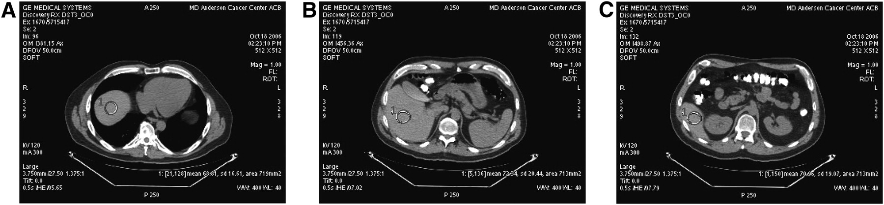
- FIGURE 1.
ROIs in patient's thorax for SD measurement with minimal area of 200 mm2: axial slice in aortic arch (A), descending aorta above liver in coronal view (B), and descending aorta at level of liver (C).
- FIGURE 2.
ROIs in patient's liver for SD measurement with minimal area of 200 mm2: dome of liver (A), middle level of liver, where no lung tissue can be seen (B), and tail of liver (C).
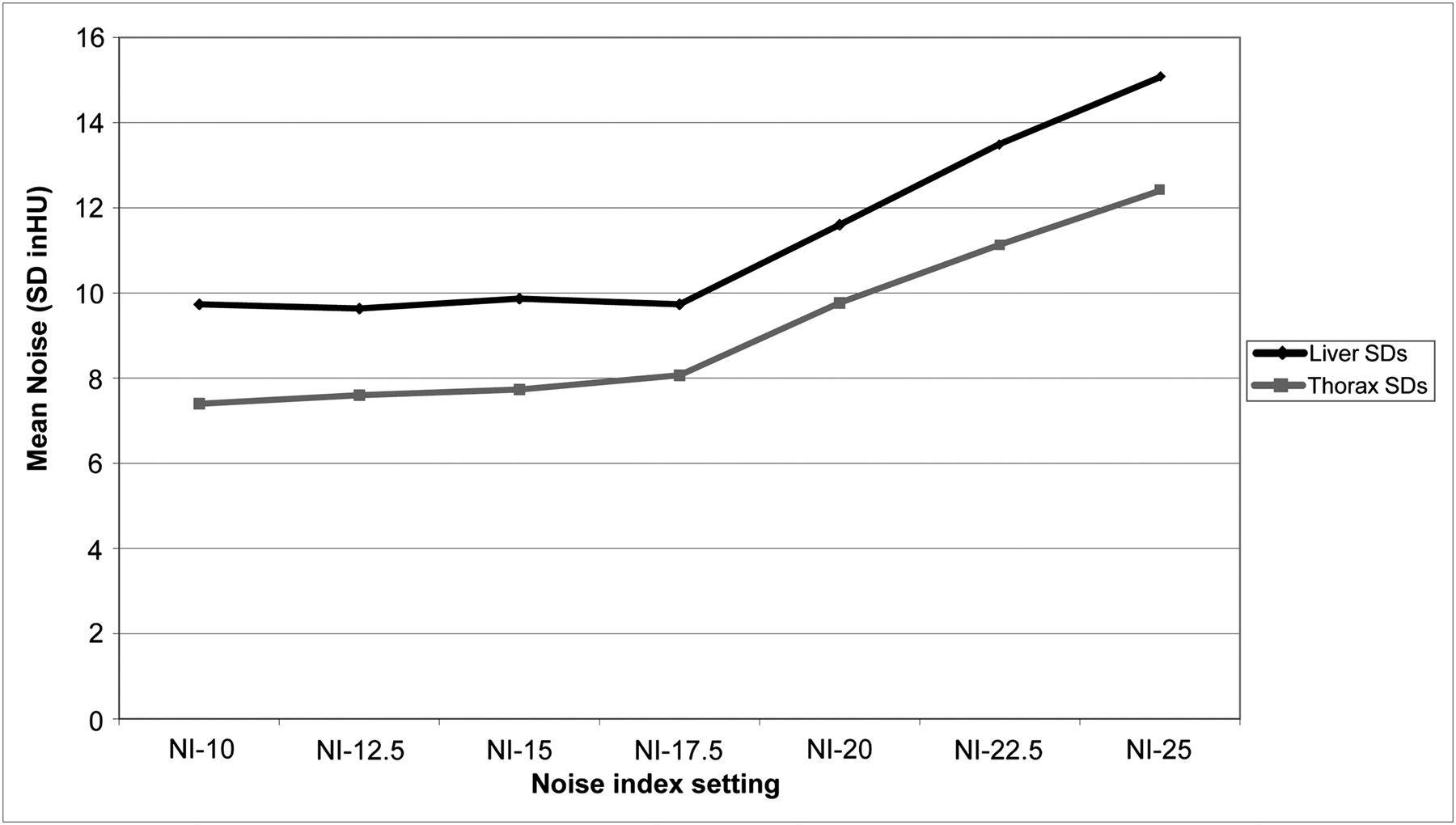
- FIGURE 3.
Average noise (mean of SDs of CT numbers in HU) in CT images at various NI settings for phantom acquisitions of 3 ROIs in thorax and 3 in liver.
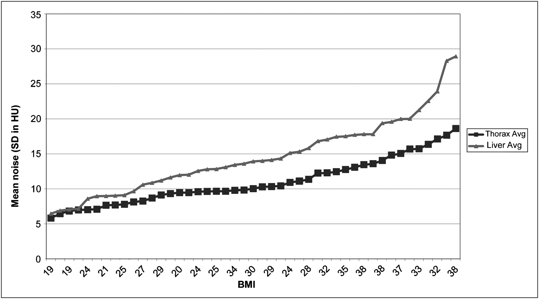
- FIGURE 4.
Average image noise (SD of CT number in HU) for CT images of 3 ROIs in thorax and 3 in liver for 45 patients analyzed retrospectively and grouped according to BMI. Avg = average.
- FIGURE 5.
Physician-determined CT image quality (on scale of 1–3) according to noise level in scans of thorax (A) and scans of liver (B). Black lines indicate best-fit linear functions for data.
- FIGURE 6.
Average decrease in tube current by patient BMI group in CT scans using ATCM with NI of 20 compared with fixed tube current of 300 mA.
- FIGURE 7.
Average image noise (SD of CT number in HU) in CT images of liver; n = 7 for each BMI range. Noise levels were not markedly different for patients with BMIs < 28 whose examinations had tube current modulation.


Tables
- TABLE 1
Noise Levels in Thorax CT Images Using ATCM and NI of 20 in Patients with BMI ≤ 28
Noise (SD of CT number in HU) Patient no. ROI 1 ROI 2 ROI 3 Mean 1 11.7 9.3 12.9 11.3 2 8.0 9.1 12.3 9.8 3 8.5 14 10.5 11.0 4 9.1 11.1 10.4 10.2 5 7.1 10.2 11.9 9.7 6 8.9 9.7 13.7 10.8 7 9.1 8.4 12.6 10.0 8 9.1 12 11.6 10.9 9 10.0 12.4 13.1 11.8 10 7.0 9.4 12.7 9.7 11 9.4 11.3 13.2 11.3 12 8.4 11 11.0 10.1 13 7.0 12.2 14.4 11.2 14 8.0 9.4 12.3 9.9 15 7.3 10.8 12.2 10.1 16 10.6 12.3 12.3 11.7 17 10.8 10.6 11.0 10.8 18 9.7 12.5 13.7 12.0 19 9.3 11.8 14.0 11.7 20 9.8 13.3 12.7 11.9 21 7.5 10.3 12.4 10.1 22 9.5 12.1 9.3 10.3 Mean noise in all thorax scans 10.7


Jump to section
Related Articles
Cited By...
- No citing articles found.