
Main menu

  • Home
  • Content
    • Current
      • JNMT Supplement
    • Ahead of print
    • Past Issues
    • Continuing Education
    • JNMT Podcast
    • SNMMI Annual Meeting Abstracts
  • Subscriptions
    • Subscribers
    • Rates
    • Journal Claims
    • Institutional and Non-member
  • Authors
    • Submit to JNMT
    • Information for Authors
    • Assignment of Copyright
    • AQARA Requirements
  • Info
    • Reviewers
    • Permissions
    • Advertisers
    • Corporate & Special Sales
  • About
    • About Us
    • Editorial Board
    • Contact Information
  • More
    • Alerts
    • Feedback
    • Help
    • SNMMI Journals
  • SNMMI
    • JNMT
    • JNM
    • SNMMI Journals
    • SNMMI

User menu

  • Subscribe
  • My alerts
  • Log in
  • My Cart

Search

  • Advanced search
Journal of Nuclear Medicine Technology
  • SNMMI
    • JNMT
    • JNM
    • SNMMI Journals
    • SNMMI
  • Subscribe
  • My alerts
  • Log in
  • My Cart
Journal of Nuclear Medicine Technology

Advanced Search

  • Home
  • Content
    • Current
    • Ahead of print
    • Past Issues
    • Continuing Education
    • JNMT Podcast
    • SNMMI Annual Meeting Abstracts
  • Subscriptions
    • Subscribers
    • Rates
    • Journal Claims
    • Institutional and Non-member
  • Authors
    • Submit to JNMT
    • Information for Authors
    • Assignment of Copyright
    • AQARA Requirements
  • Info
    • Reviewers
    • Permissions
    • Advertisers
    • Corporate & Special Sales
  • About
    • About Us
    • Editorial Board
    • Contact Information
  • More
    • Alerts
    • Feedback
    • Help
    • SNMMI Journals
  • Watch or Listen to JNMT Podcast
  • Visit SNMMI on Facebook
  • Join SNMMI on LinkedIn
  • Follow SNMMI on Twitter
  • Subscribe to JNMT RSS feeds
Research ArticleIMAGING

Patient Weight–Based Acquisition Protocols to Optimize18F-FDG PET/CT Image Quality

Akio Nagaki, Masahisa Onoguchi and Norikazu Matsutomo
Journal of Nuclear Medicine Technology June 2011, 39 (2) 72-76; DOI: https://doi.org/10.2967/jnmt.110.081661
Akio Nagaki
  • Find this author on Google Scholar
  • Find this author on PubMed
  • Search for this author on this site
Masahisa Onoguchi
  • Find this author on Google Scholar
  • Find this author on PubMed
  • Search for this author on this site
Norikazu Matsutomo
  • Find this author on Google Scholar
  • Find this author on PubMed
  • Search for this author on this site
  • Article
  • Figures & Data
  • Info & Metrics
  • PDF
Loading

Article Figures & Data

Figures

  • FIGURE 1.
    • Download figure
    • Open in new tab
    • Download powerpoint
    FIGURE 1.

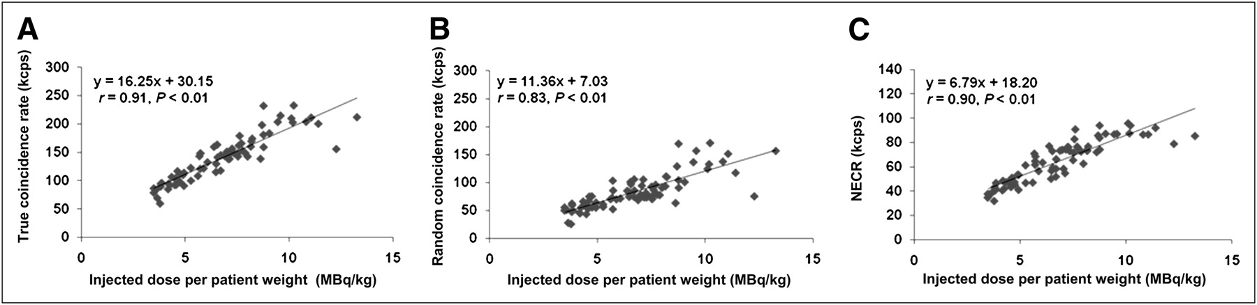
     Plots of true coincidence rate (A), random coincidence rate (B), and NECR (C) vs. injected dose per patient weight for 76 patient scans. True coincidence rate, random coincidence rate, and NECR significantly increased with increasing injected dose. NECR maximized at 10.11 MB/kg.

  • FIGURE 2.
    • Download figure
    • Open in new tab
    • Download powerpoint
    FIGURE 2.

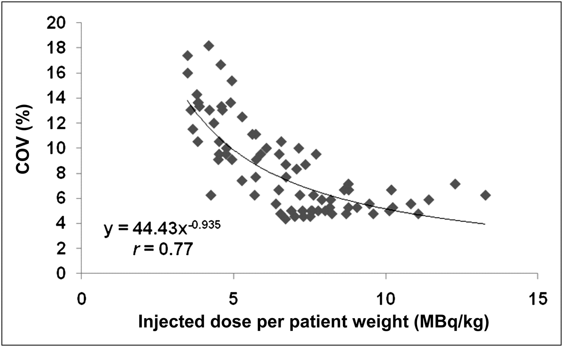
     Plots of COV vs. injected dose per patient weight. At lower injected dose, COV of PET images shows steep decline.
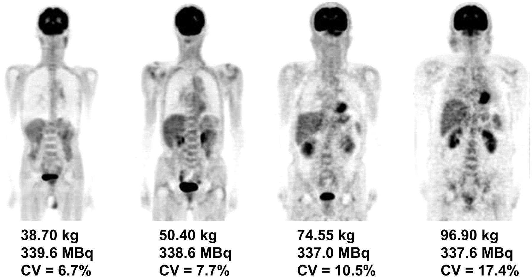
  • FIGURE 3.
    • Download figure
    • Open in new tab
    • Download powerpoint
    FIGURE 3.

     Image quality of approximately same injected dose of 18F-FDG in patients of different body weights. PET images of underweight patients had lower COV and excellent quality. In contrast, PET images of overweight patients were noisy and of poor quality.
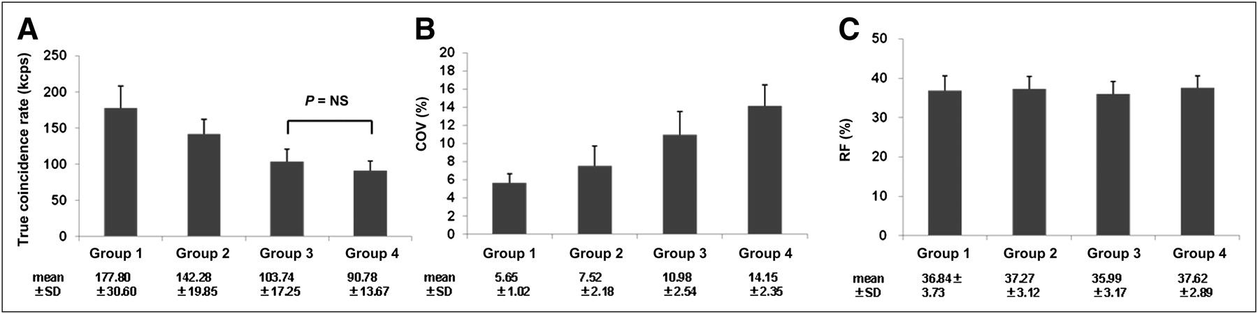
  • FIGURE 4.
    • Download figure
    • Open in new tab
    • Download powerpoint
    FIGURE 4.

     True coincidence rate (A), COV of PET images (B), and random fraction (C) for 4 patient-weight groups. Except for group 3 vs. group 4 (P < 0.01), true coincidence rate differed significantly among groups. COV of the 4 groups differed significantly for all pairs (P < 0.01). Average random fraction was approximately 35% in all groups (P = not statistically significant). RF = random fraction.

  • FIGURE 5.
    • Download figure
    • Open in new tab
    • Download powerpoint
    FIGURE 5.

     Follow-up images of 102-kg patient with lymphoma, injected with 388.7 MBq (A) and 254.1 MBq (B) of 18F-FDG. Because optimal protocols were regulated by acquisition time (A, 120 s/bed position; B, 190 s/bed position), COVs of PET images were nearly identical (A, 18.5%; B, 18.8%).

PreviousNext
Back to top

In this issue

Journal of Nuclear Medicine Technology: 39 (2)
Journal of Nuclear Medicine Technology
Vol. 39, Issue 2
June 1, 2011
  • Table of Contents
  • About the Cover
  • Index by author
Print
Download PDF
Article Alerts
Sign In to Email Alerts with your Email Address
Email Article

Thank you for your interest in spreading the word on Journal of Nuclear Medicine Technology.

NOTE: We only request your email address so that the person you are recommending the page to knows that you wanted them to see it, and that it is not junk mail. We do not capture any email address.

Enter multiple addresses on separate lines or separate them with commas.
Patient Weight–Based Acquisition Protocols to Optimize18F-FDG PET/CT Image Quality
(Your Name) has sent you a message from Journal of Nuclear Medicine Technology
(Your Name) thought you would like to see the Journal of Nuclear Medicine Technology web site.
Citation Tools
Patient Weight–Based Acquisition Protocols to Optimize18F-FDG PET/CT Image Quality
Akio Nagaki, Masahisa Onoguchi, Norikazu Matsutomo
Journal of Nuclear Medicine Technology Jun 2011, 39 (2) 72-76; DOI: 10.2967/jnmt.110.081661

Citation Manager Formats

  • BibTeX
  • Bookends
  • EasyBib
  • EndNote (tagged)
  • EndNote 8 (xml)
  • Medlars
  • Mendeley
  • Papers
  • RefWorks Tagged
  • Ref Manager
  • RIS
  • Zotero
Share
Patient Weight–Based Acquisition Protocols to Optimize18F-FDG PET/CT Image Quality
Akio Nagaki, Masahisa Onoguchi, Norikazu Matsutomo
Journal of Nuclear Medicine Technology Jun 2011, 39 (2) 72-76; DOI: 10.2967/jnmt.110.081661
Twitter logo Facebook logo LinkedIn logo Mendeley logo
  • Tweet Widget
  • Facebook Like
  • Google Plus One
Bookmark this article

Jump to section

  • Article
    • Abstract
    • MATERIALS AND METHODS
    • RESULTS
    • DISCUSSION
    • CONCLUSION
    • REFERENCES
  • Figures & Data
  • Info & Metrics
  • PDF

Related Articles

  • No related articles found.
  • PubMed
  • Google Scholar

Cited By...

  • Weight-Based Protocols Offer Significant Reduction in Radiation Dose Without Affecting PET-CT Image Quality
  • Added Value of Digital over Analog PET/CT: More Significant as Image Field of View and Body Mass Index Increase
  • Influence of Statistical Fluctuation on Reproducibility and Accuracy of SUVmax and SUVpeak: A Phantom Study
  • Improvement in PET/CT Image Quality with a Combination of Point-Spread Function and Time-of-Flight in Relation to Reconstruction Parameters
  • Google Scholar

More in this TOC Section

  • Early 10-Minute Postinjection [18F]F-FAPI-42 uEXPLORER Total-Body PET/CT Scanning Protocol for Staging Lung Cancer Using HYPER Iterative Reconstruction
  • Single- Versus Dual-Time-Point Imaging for Transthyretin Cardiac Amyloid Using 99mTc-Pyrophosphate
  • Does Arthrography Improve Accuracy of SPECT/CT for Diagnosis of Aseptic Loosening in Patients with Painful Knee Arthroplasty: A Systematic Review and Metaanalysis
Show more Imaging

Similar Articles

SNMMI

© 2025 SNMMI

Powered by HighWire