


Article Figures & Data
Figures
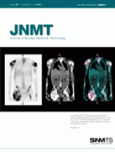
- FIGURE 1.
This figure shows 3 γ-rays being emitted from a point source at top of figure. γ-ray labeled “Good Detection” passes right through collimator hole and will be properly localized directly below point source. γ-ray labeled “Stopped by septa” hits septa and interacts, presumably by photoelectric effect, in septum and thus does not reach scintillation crystal.However, γ-ray labeled “Septal Penetration Bad Detection” passes through septum, reaching scintillation crystal at wrong location, not directly below point source. Probability for septal penetration depends on energy of incident γ-ray and thickness of septum. d = hole diameter; L = hole length; t = thickness of interhole septa.
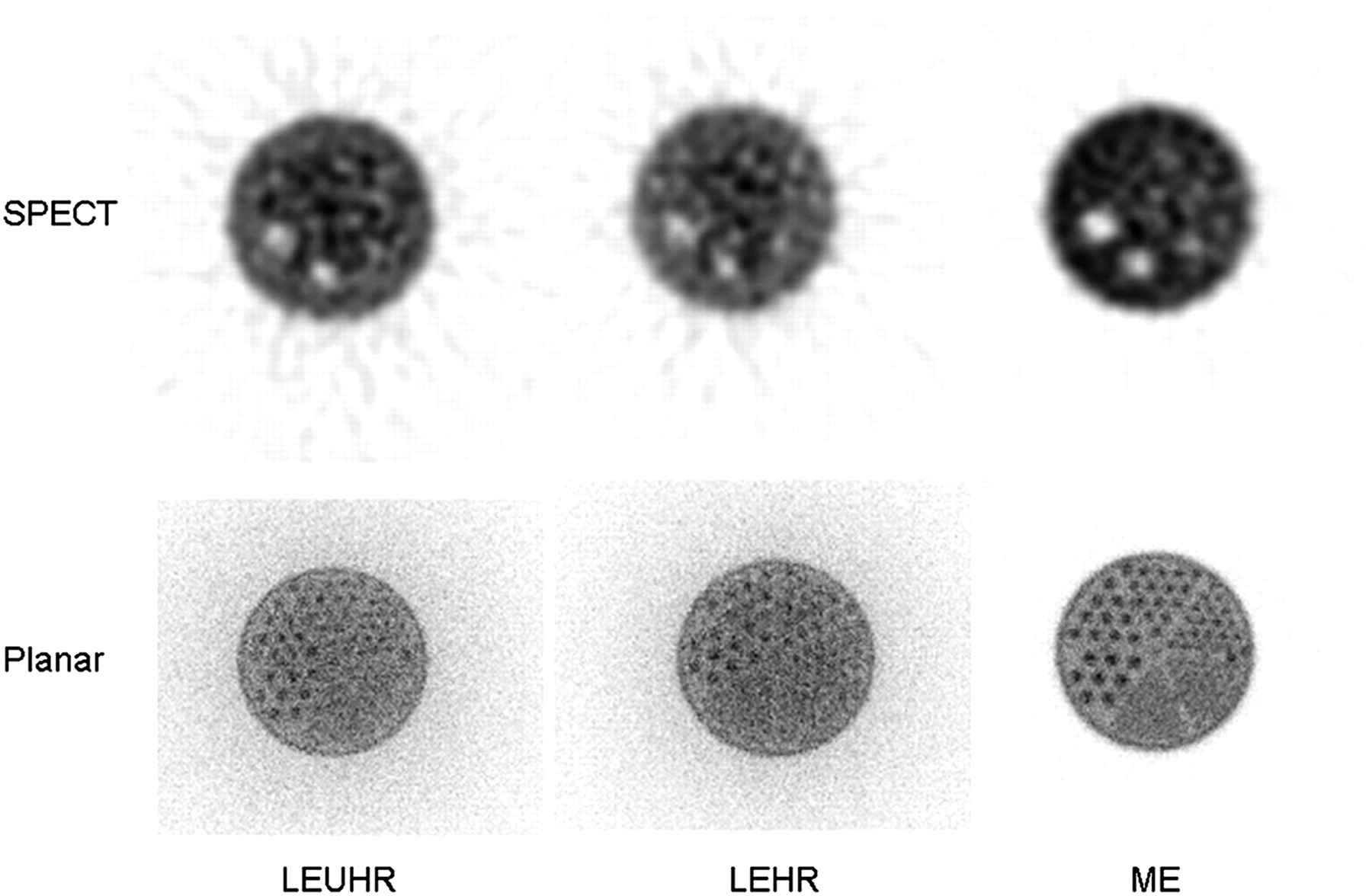
- FIGURE 2.
This figure shows SPECT and planar images acquired with a tomographic phantom filled with 123I. The phantom was imaged with LEUHR, LEHR, and medium-energy (ME) collimators. On the images using low-energy collimators, many events are localized outside the boundary of the phantom because of septal penetration. Image quality is better for the medium-energy-collimator images than for low-energy-collimator images because of higher contrast and lower noise.
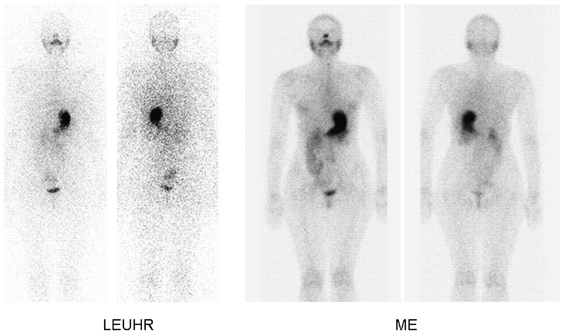
- FIGURE 3.
Whole-body planar (A) and SPECT (B) images of 123I-MIBG study in a 7-y-old patient with stage 4 neuroblastoma. Images on left were acquired during initial visit when we were using LEUHR collimators, and those on right were acquired several months later when we had modified our acquisition protocol to use medium-energy (ME) collimators. SPECT images are anterior maximum-intensity-projection images. Results are similar to the phantom results. For both whole-body planar imaging and SPECT, images obtained using medium-energy collimators demonstrated less septal penetration (fewer counts outside patient), higher contrast, and less noise.
- FIGURE 4.
Whole-body planar images of 123I-NaI study in a 17-y-old patient being evaluated for thyroid cancer. Images on left were acquired during initial visit when we were using LEUHR collimator, and those on right were acquired several months later when we had modified our acquisition protocol to use medium-energy (ME) collimator. Results are similar to the phantom results. Images obtained using medium-energy collimator demonstrated less septal penetration (fewer counts outside patient), higher contrast, and less noise.


Tables
Energy (keV) γ-rays per decay 159 0.828 248 0.0007 281 0.0008 346 0.0013 440 0.0043 505 0.0031 529 0.0138 539 0.0038 625 0.0008 688 0.0003 736 0.0006 784 0.0006 Parameter LEUHR LEHR Medium-energy Septal thickness (mm) 0.13 0.16 1.14 Hole diameter (mm) 1.16 1.11 2.94 Hole length (mm) 35.8 24.05 40.64 Extrinsic sensitivity (cpm/kBq)* 2.70 5.46 8.38 Spatial resolution (mm) 4.6 6.4 10.8 Septal penetration at 159 keV 2.0% 3.5% 0.0% Septal penetration at 300 keV 42% 48% 4.90% Septal penetration at 500 keV 71% 75% 30% ↵* At 140 keV.
- TABLE 3
System Sensitivity (in Counts per Minute per Kilobecquerel) of 123I Versus Distance for LEUHR, LEHR, and Medium-Energy Collimators
Distance (cm) LEUHR LEHR Medium-energy 0 6.59 14.32 8.00 10 5.51 12.30 7.97


