
Main menu

  • Home
  • Content
    • Current
      • JNMT Supplement
    • Ahead of print
    • Past Issues
    • SNMMI Annual Meeting Abstracts
  • Subscriptions
    • Subscribers
    • Rates
    • Journal Claims
    • Institutional and Non-member
  • Authors
    • Submit to JNMT
    • Information for Authors
    • Assignment of Copyright
    • AQARA Requirements
  • Info
    • Permissions
    • Continuing Education
    • Advertisers
    • Corporate & Special Sales
  • About
    • About Us
    • Editorial Board
    • Editorial Contact
  • More
    • Alerts
    • Feedback
    • Help
    • SNMMI Journals
  • SNMMI
    • JNMT
    • JNM
    • SNMMI Journals
    • SNMMI

User menu

  • Subscribe
  • My alerts
  • Log in
  • Log out
  • My Cart

Search

  • Advanced search
Journal of Nuclear Medicine Technology
  • SNMMI
    • JNMT
    • JNM
    • SNMMI Journals
    • SNMMI
  • Subscribe
  • My alerts
  • Log in
  • Log out
  • My Cart
Journal of Nuclear Medicine Technology

Advanced Search

  • Home
  • Content
    • Current
    • Ahead of print
    • Past Issues
    • SNMMI Annual Meeting Abstracts
  • Subscriptions
    • Subscribers
    • Rates
    • Journal Claims
    • Institutional and Non-member
  • Authors
    • Submit to JNMT
    • Information for Authors
    • Assignment of Copyright
    • AQARA Requirements
  • Info
    • Permissions
    • Continuing Education
    • Advertisers
    • Corporate & Special Sales
  • About
    • About Us
    • Editorial Board
    • Editorial Contact
  • More
    • Alerts
    • Feedback
    • Help
    • SNMMI Journals
  • Follow SNMMI on Twitter
  • Visit SNMMI on Facebook
  • Join SNMMI on LinkedIn
  • Subscribe to JNMT RSS feeds
OtherCONTINUING EDUCATION

Economic Evaluation of PET and PET/CT in Oncology: Evidence and Methodologic Approaches

Andreas K. Buck, Ken Herrmann, Tom Stargardt, Tobias Dechow, Bernd Joachim Krause and Jonas Schreyögg
Journal of Nuclear Medicine Technology March 2010, 38 (1) 6-17; DOI: https://doi.org/10.2967/jnmt.108.059584
Andreas K. Buck
  • Find this author on Google Scholar
  • Find this author on PubMed
  • Search for this author on this site
Ken Herrmann
  • Find this author on Google Scholar
  • Find this author on PubMed
  • Search for this author on this site
Tom Stargardt
  • Find this author on Google Scholar
  • Find this author on PubMed
  • Search for this author on this site
Tobias Dechow
  • Find this author on Google Scholar
  • Find this author on PubMed
  • Search for this author on this site
Bernd Joachim Krause
  • Find this author on Google Scholar
  • Find this author on PubMed
  • Search for this author on this site
Jonas Schreyögg
  • Find this author on Google Scholar
  • Find this author on PubMed
  • Search for this author on this site
  • Article
  • Figures & Data
  • Info & Metrics
  • PDF
Loading

Article Figures & Data

Figures

  • Tables
  • FIGURE 1. 
    • Download figure
    • Open in new tab
    • Download powerpoint
    FIGURE 1. 

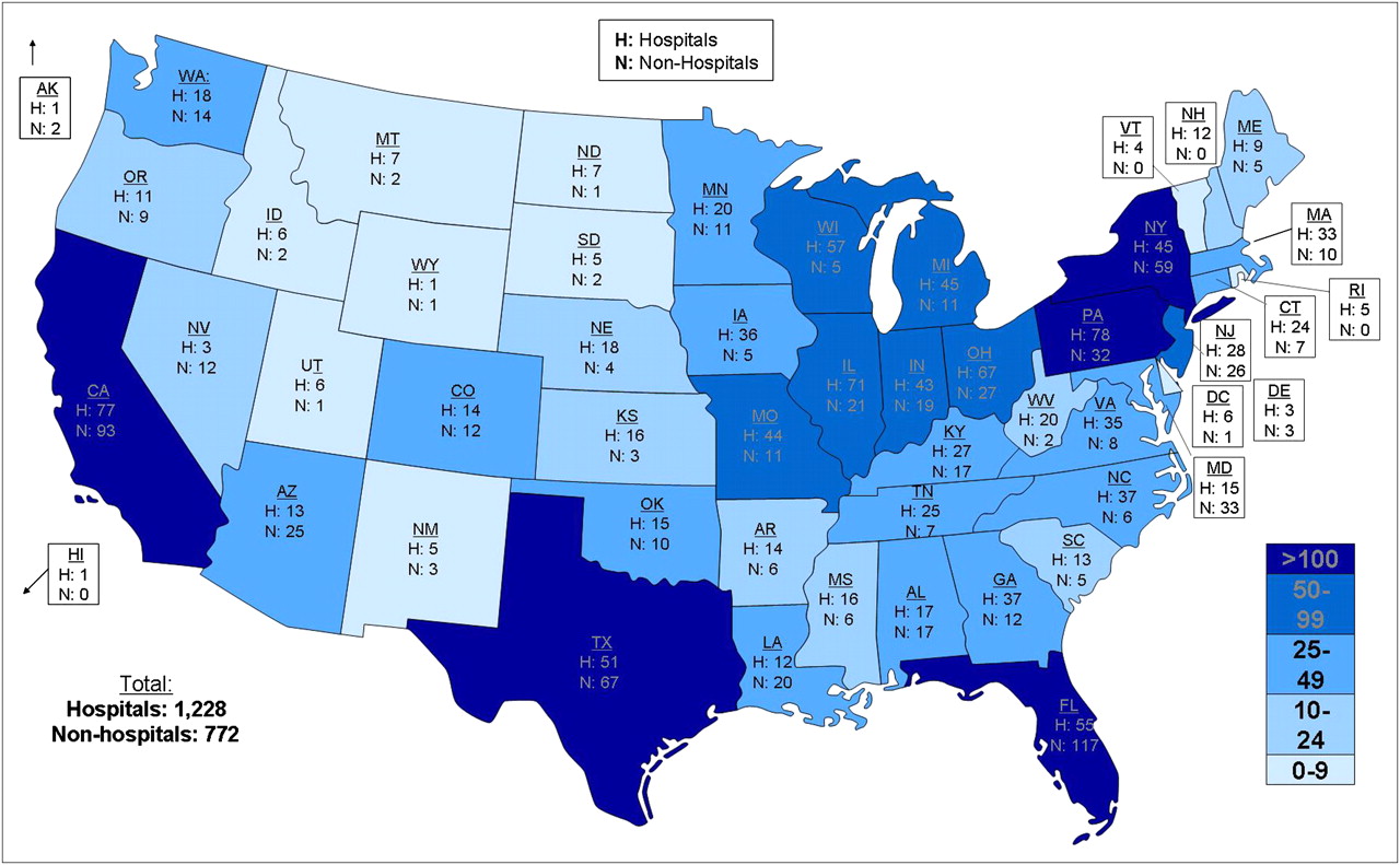
    PET/CT represents one of the medical imaging modalities with the largest growth worldwide. In 2009, approximately 2,000 PET/CT scanners were installed in the United States and approximately 350 were installed in Europe. Considering a population of about 307 million in the United States and 830 million in Europe, the United States has installed about 6 times as many scanners as all of Europe but has only one third the population. (Courtesy of Siemens/CTI.)

  • FIGURE 2. 
    • Download figure
    • Open in new tab
    • Download powerpoint
    FIGURE 2. 

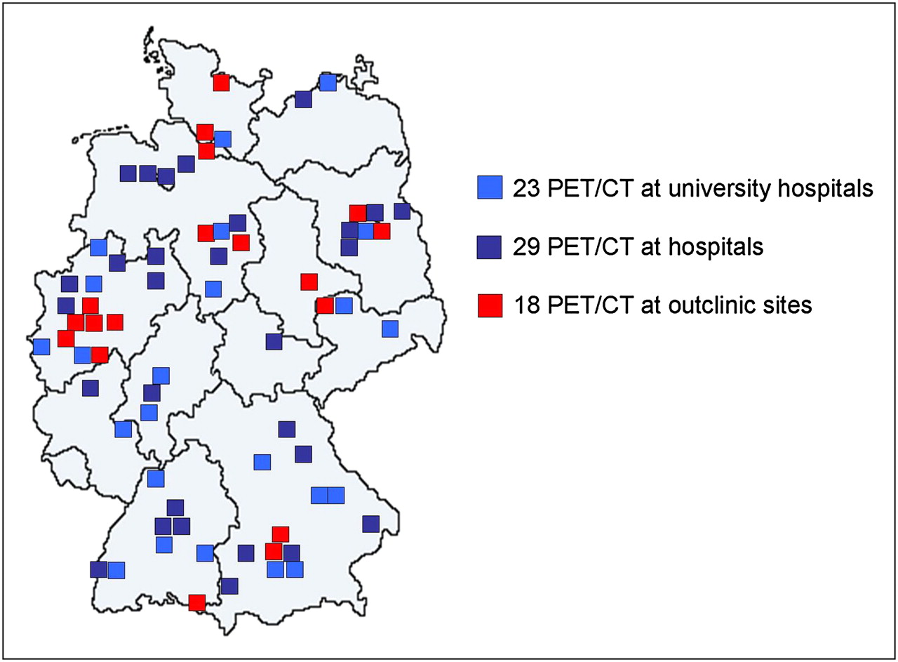
    In Europe, introduction of PET/CT hybrid scanners has also led to an increase in their installations. Compared with the United States, however, a less accelerated growth has been observed. In 2009, 70 scanners were installed in Germany and 350 in all of Europe. (Courtesy of Siemens AG.)

  • FIGURE 3. 
    • Download figure
    • Open in new tab
    • Download powerpoint
    FIGURE 3. 

    PET/CT has greater diagnostic accuracy than separately performed imaging modalities. In this patient at initial diagnosis of colorectal cancer, coronal (A) and sagittal (B) PET/CT images indicate increased metabolic activity of malignant primary (arrows); transaxial CT (C) and PET/CT (D) images indicate synchronous bone and liver metastases (arrows), leading to change from curative resection to systemic chemotherapy; and transaxial CT (E) and PET/CT (F) images at another level indicate primary tumor.

Tables

  • Figures
    • View popup
    TABLE 1

    Reimbursement of PET and PET/CT in United States (59) and Germany (60)

    United StatesGermany
    IndicationInitial treatment strategySubsequent treatment strategyInitial treatment strategySubsequent treatment strategy
    Head and neck cancerCC——
    Esophagus cancerCC——
    Gastric cancerCNOPR——
    Small intestinal cancerCNOPR——
    Colon and rectal cancerCC——
    Anal cancerCNOPR*——
    Hepatocellular carcinomaCNOPR——
    Gallbladder and cholangiocellular carcinomaCNOPR——
    Pancreatic cancerCNOPR——
    Cancers of retroperitoneum and peritoneumCNOPR——
    Non–small cell lung cancerCCCC
    Small cell lung cancerCNOPR——
    MesotheliomaCNOPR——
    Cancers of mediastinum; thymus carcinomaCNOPR——
    Sarcoma of boneCNOPR——
    Soft-tissue sarcomaCNOPR——
    MelanomaC/—†C——
    Skin cancers (nonmelanoma)CNOPR——
    Breast cancerC/—†‡C——
    Uterine cancerCNOPR——
    Cervix carcinomaC/NOPR§C——
    Ovarian cancerCC——
    Prostate cancer—NOPR——
    Bladder cancerCNOPR——
    Kidney and other urinary tract cancersCNOPR——
    Primary brain tumorsCNOPR——
    Thyroid cancerCC/NOPR‖——
    Other endocrine tumorsCNOPR——
    Cancer of unknown primaryCNOPR——
    LymphomaCC——
    MyelomaCC——
    LeukemiaNOPRNOPR——
    Neuroendocrine tumorsCNOPR——
    Other cancersCNOPR——
    • ↵* Some Medicare contractors include anal cancer in their local coverage of “colorectal cancer”; for PET facilities served by those carriers, PET for subsequent treatment evaluation of anal cancer would be a covered indication.

    • ↵† PET is not covered for initial staging of axillary lymph nodes in patients with breast cancer and of regional lymph nodes in patients with melanoma but is covered for detection of distant metastatic disease in high-risk patients with breast cancer or melanoma.

    • ↵‡ PET is not covered for “diagnosis” of breast cancer to evaluate suggestive breast mass. However, PET is covered for initial treatment-strategy evaluation of patient with axillary nodal metastasis of unknown primary origin or patient with paraneoplastic syndrome potentially caused by occult breast cancer.

    • ↵§ Patient must have prior CT or MRI negative for extrapelvic metastatic disease for PET to qualify as covered indication for initial treatment-strategy evaluation. Patients who do not qualify for this covered indication (e.g., because CT or MRI was not done or because either CT or MRI showed extrapelvic metastatic disease) can be entered on NOPR.

    • ↵‖ To qualify as covered indication for subsequent treatment-strategy evaluation, thyroid cancer must be of follicular cell origin and have been previously treated by thyroidectomy and radioiodine ablation and patient must have serum thyroglobulin level > 10 ng/mL and negative whole-body 131I findings. Patients who do not qualify for this covered indication (e.g., because tumor is not of follicular cell origin, thyroglobulin is not elevated, or 131I whole-body imaging was not performed or is positive) can be entered on NOPR.

    • C = covered (not eligible for entry in National Oncologic PET Registry [NOPR]); NOPR = covered only with entry in NOPR; — = not covered nationally (not eligible for entry in NOPR).

    • Modified from http://www.cancerpetregistry.org/indications_facilities.htm.

PreviousNext
Back to top

In this issue

Journal of Nuclear Medicine Technology: 38 (1)
Journal of Nuclear Medicine Technology
Vol. 38, Issue 1
March 2010
  • Table of Contents
  • About the Cover
  • Index by author
Print
Download PDF
Article Alerts
Sign In to Email Alerts with your Email Address
Email Article

Thank you for your interest in spreading the word on Journal of Nuclear Medicine Technology.

NOTE: We only request your email address so that the person you are recommending the page to knows that you wanted them to see it, and that it is not junk mail. We do not capture any email address.

Enter multiple addresses on separate lines or separate them with commas.
Economic Evaluation of PET and PET/CT in Oncology: Evidence and Methodologic Approaches
(Your Name) has sent you a message from Journal of Nuclear Medicine Technology
(Your Name) thought you would like to see the Journal of Nuclear Medicine Technology web site.
Citation Tools
Economic Evaluation of PET and PET/CT in Oncology: Evidence and Methodologic Approaches
Andreas K. Buck, Ken Herrmann, Tom Stargardt, Tobias Dechow, Bernd Joachim Krause, Jonas Schreyögg
Journal of Nuclear Medicine Technology Mar 2010, 38 (1) 6-17; DOI: 10.2967/jnmt.108.059584

Citation Manager Formats

  • BibTeX
  • Bookends
  • EasyBib
  • EndNote (tagged)
  • EndNote 8 (xml)
  • Medlars
  • Mendeley
  • Papers
  • RefWorks Tagged
  • Ref Manager
  • RIS
  • Zotero
Share
Economic Evaluation of PET and PET/CT in Oncology: Evidence and Methodologic Approaches
Andreas K. Buck, Ken Herrmann, Tom Stargardt, Tobias Dechow, Bernd Joachim Krause, Jonas Schreyögg
Journal of Nuclear Medicine Technology Mar 2010, 38 (1) 6-17; DOI: 10.2967/jnmt.108.059584
Twitter logo Facebook logo LinkedIn logo Mendeley logo
  • Tweet Widget
  • Facebook Like
  • Google Plus One
Bookmark this article

Jump to section

  • Article
    • Abstract
    • DIAGNOSTIC EFFECTIVENESS OF PET AND PET/CT IN ONCOLOGY
    • COSTS FOR PET AND PET/CT
    • METHODS FOR ECONOMIC EVALUATION
    • COST-EFFECTIVENESS OF PET AND PET/CT IN SELECTED CANCERS
    • DIFFERENTIAL DIAGNOSIS OF SOLITARY PULMONARY NODULES
    • SUGGESTED SETUP FOR ECONOMIC EVALUATION OF PET/CT
    • SUMMARY
    • Footnotes
    • References
  • Figures & Data
  • Info & Metrics
  • PDF

Related Articles

  • No related articles found.
  • PubMed
  • Google Scholar

Cited By...

  • Solitary pulmonary nodule imaging approaches and the role of optical fibre-based technologies
  • Abnormal brain metabolism on FDG-PET/CT is a common early finding in autoimmune encephalitis
  • Translation of New Molecular Imaging Approaches to the Clinical Setting: Bridging the Gap to Implementation
  • Cost-Utility of a Prognostic Test Guiding Adjuvant Chemotherapy Decisions in Early-Stage Non-Small Cell Lung Cancer
  • Review on Production of 89Zr in a Medical Cyclotron for PET Radiopharmaceuticals
  • Cost-Effectiveness Analysis of Amino Acid PET-Guided Surgery for Supratentorial High-Grade Gliomas
  • Global Contrast in Nuclear Medicine
  • Google Scholar

More in this TOC Section

  • SNMMI Clinical Trial Network Research Series for Technologists: Application of Good Clinical Practice to Clinical Research in Medical Imaging
  • Clinical Trials of Prostate-Specific Membrane Antigen Radiopharmaceutical Therapy
  • V/Q SPECT and SPECT/CT in Pulmonary Embolism
Show more Continuing Education

Similar Articles

SNMMI

© 2023 Journal of Nuclear Medicine Technology

Powered by HighWire