


Article Figures & Data
Figures
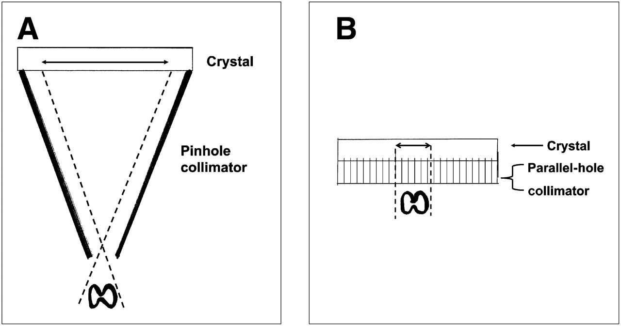
- FIGURE 1.
A larger area of the detector “sees” object when object is close to pinhole collimator (A) than when object is close to parallel-hole collimator (B).
- FIGURE 2.
Performance characteristics of various collimators (5).

Tables
Radiopharmaceutical Dose 131I 50–200 μCi 123I 200–600 μCi 99mTc-pertechnetate 2–10 mCi Parameter Number Percentage Surveys sent 350 Surveys returned 165 47.1 1. Radionuclide routinely used for thyroid imaging 123I 117 70.9 99mTc (pertechnetate) 41 24.8 Both 123I and 99mTc 6 3.6 131I 1 0.6 2. Routine dose administered 123I 100–199 μCi 11 200–299 μCi 85 300–399 μCi 9 400–499 μCi 1 500–599 μCi 2 99mTc (pertechnetate) 5 mCi 3 7 mCi 1 8 mCi 1 10 mCi 28 10–15 mCi 2 20 mCi 1 0.4–0.5 mCi 2 Did not answer 20 3. Neck positioning Neck hyperextended 142 86.1 Patient supine with neck extended but not hyperextended 13 7.9 Did not answer 10 6.1 4. Fasting required Yes 67 40.6 1–3 h 10 15 4–6 h 30 45 7–9 h 2 3 Overnight 22 33 No 98 59.4 5. History taken Yes 163 98.8 No 2 1.2 6. Views routinely taken Anterior view 162 1 anterior view without markers 37 22.8 1 anterior view with markers 13 8 2 anterior views: 1 with and 1 without markers 112 69.1 Oblique views in addition to the anterior view 151 93 Right and left anterior oblique without markers 149 Right and left anterior oblique with markers 2 Did not answer 3 7. Method used to acquire anterior view image* Clinics using 123I Counts reported 20,000–49,000 11 50,000–74,000 22 75,000–99,000 1 100,000–149,000 18 150,000–199,000 2 200,000–300,000 2 300,000+ 0 Time reported 5 min 22 7 min 1 8 min 6 10 min 50 15 min 5 20 min 3 Other 5 Answered only counts or time 84 Answered both counts and time 33 Clinics using 99mTc Counts reported 20,000–49,000 0 50,000–74,000 1 75,000–99,000 0 100,000–149,000 3 150,000–199,000 6 200,000–300,000 10 300,000+ 8 Time reported 5 min 12 10 min 5 15 min 0 20 min 0 Other 3 Did not answer 2 Answered only counts or time 30 Answered both counts and time 11 8. Method used to size gland Yes 72 43.6 No 93 56.4 9. Collimator used for imaging Pinhole 107 64.8 Parallel-hole 42 25.5 Both 16 9.7 10. Neck palpated for correlation with scan Yes 67 40.6 No 98 59.4 ↵* Many responses included count and time—whichever came first.
Radionuclide Advantages Disadvantages 99mTc-pertechnetate Less expensive Trapped but not organified More readily available Potentially misleading when activity is in esophagus or vessels Shorter examination time Poor image quality when uptake is low 123I-iodide Better for visualization of retrosternal thyroid tissue Higher cost Yields better images when uptake is low Potentially less convenient for delayed imaging at 24 h Less readily available Generally longer imaging times


Jump to section
Related Articles
Cited By...
- No citing articles found.