


Article Figures & Data
Figures
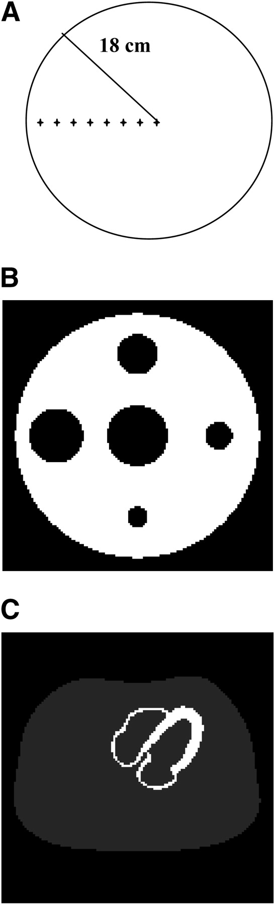
- FIGURE 1.
Computer simulations. (A) Uniform object used for 2D PSF estimations. Eight point sources with different locations are marked by plus signs. (B) Cold-lesion simulation. This object consists of 5 cylindric cold spots in uniformly attenuating cylinder of uniform activity. (C) Uniform cardiac simulation.
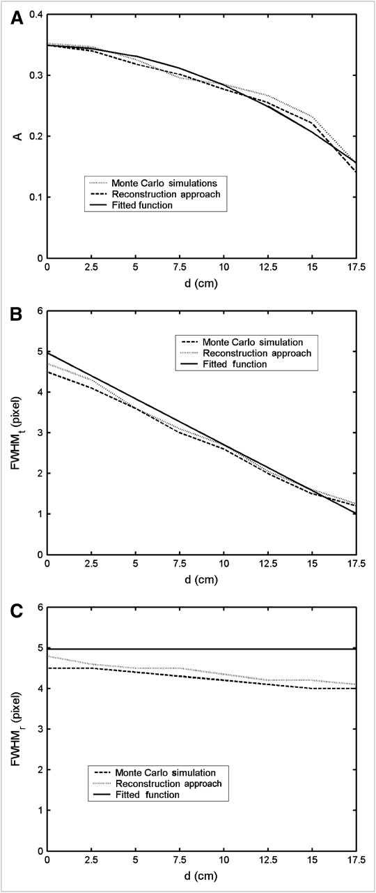
- FIGURE 2.
Variations of gaussian parameters for 8 point sources as function of radial distance d: relative volume A (A), FWHM in tangential direction of fitted gaussian function (B), and FWHM in radial direction of fitted gaussian function (C).
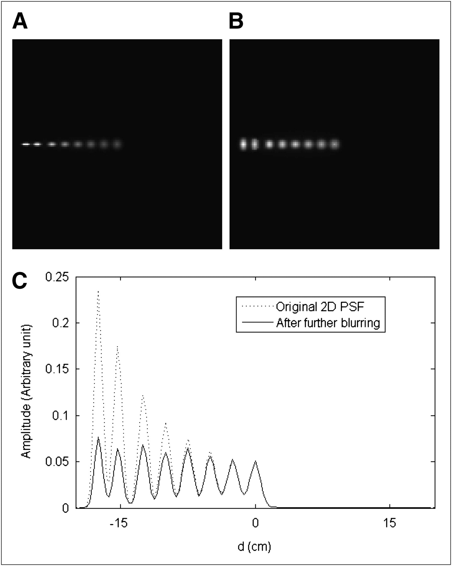
- FIGURE 3.
(A) Spatially variant 2D PSF along radial direction of uniform water filled cylinder; (B) further blurred image using rotational convolution; and (C) profile comparison of 2D PSF before and after rotational convolution.
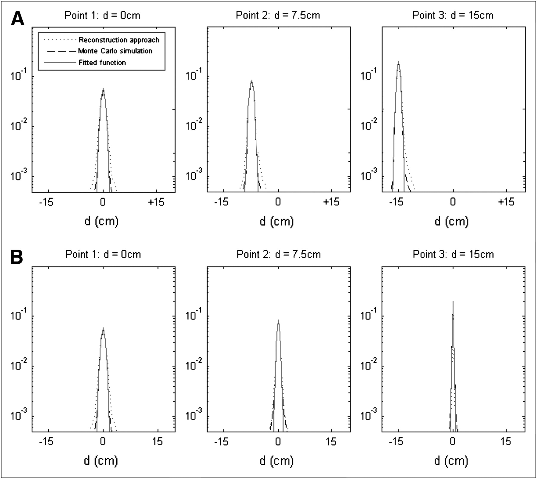
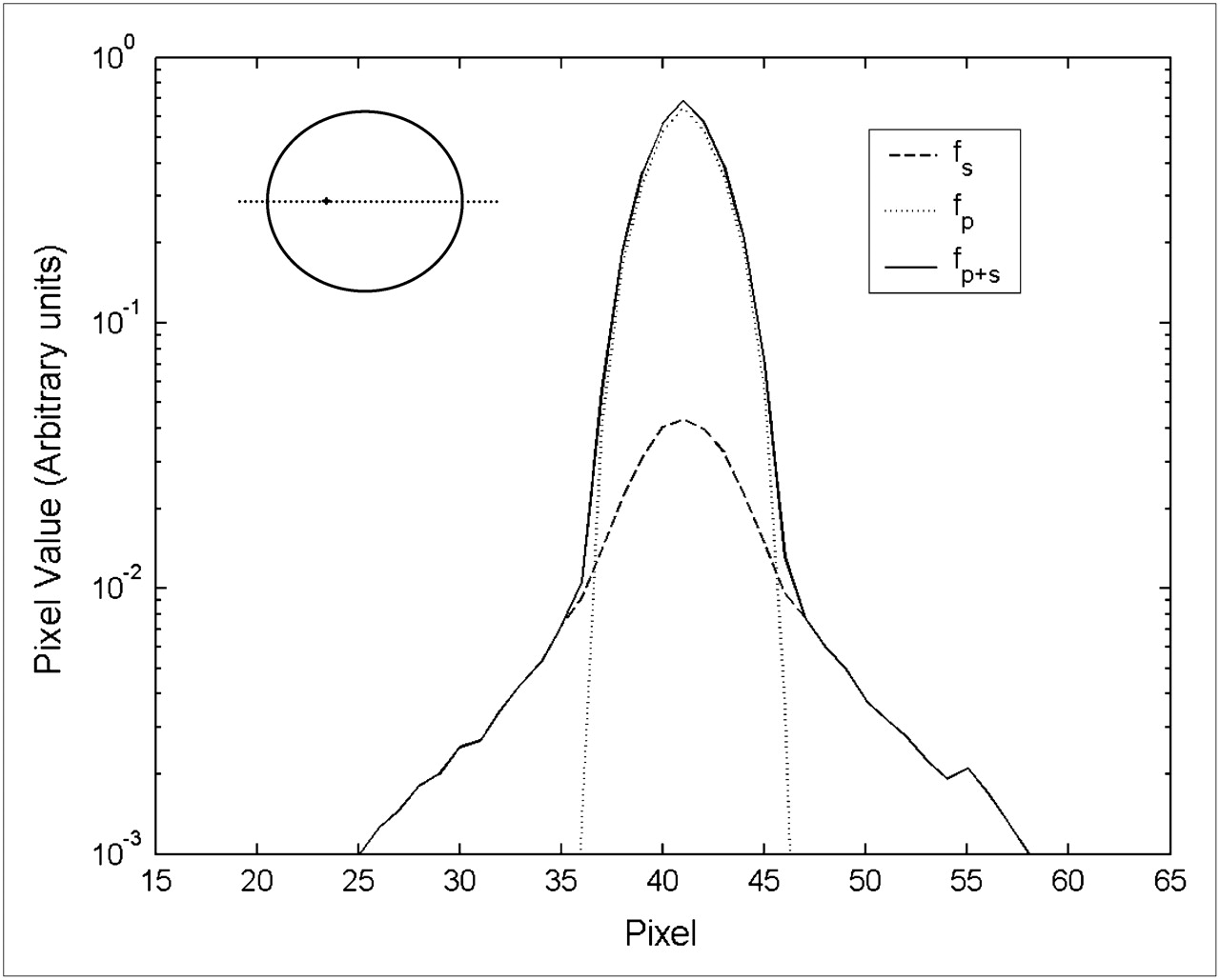
- FIGURE 4.
Comparison of 2D PSF calculated from Monte Carlo simulations, reconstruction approach, and fitted gaussian: radial direction (A); tangential direction (B). Profiles are plotted on logarithmic scales.
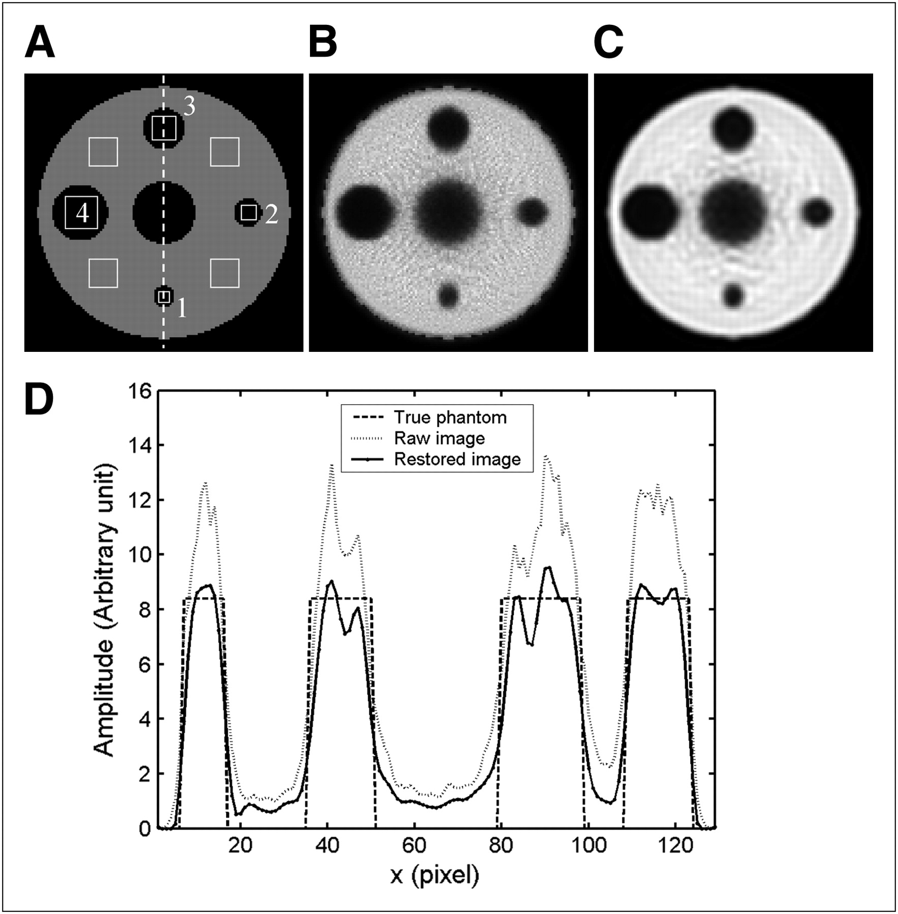
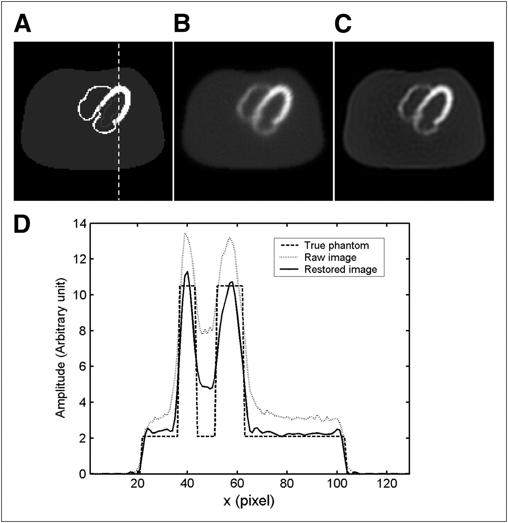
- FIGURE 5.
(A) Cold-lesion simulation with locations of region of interest used for contrast analysis, (B) raw reconstructed image with attenuation compensation but without scatter correction, (C) restored image with scatter compensation using our proposed method, and (D) vertical profiles through center of images.
- FIGURE 6.
(A) Uniform cardiac simulation with location of region of interest used for contrast analysis, (B) raw reconstructed image with attenuation compensation but without scatter correction, (C) restored image with scatter compensation using our proposed method, and (D) vertical profiles through center of images.
- FIGURE 7.
(A) Jaszczak phantom, (B) raw reconstructed image, and (C) restored image using proposed method.
- FIGURE 1A.
Profile of raw images along major axis in logarithmic scale.


Tables
Simulation True image Raw image Restored image Cold lesion SSE 0 6.96 3.51 Contrast 1 1 0.64 0.75 Contrast 2 1 0.72 0.78 Contrast 3 1 0.82 0.84 Contrast 4 1 0.87 0.89 Noise 0 0.066 0.027 Uniform cardiac SSE 0 8.33 4.48 Contrast 0.66 0.47 0.56 Noise 0 0.038 0.045


Jump to section
Related Articles
Cited By...
- No citing articles found.