


Article Figures & Data
Figures
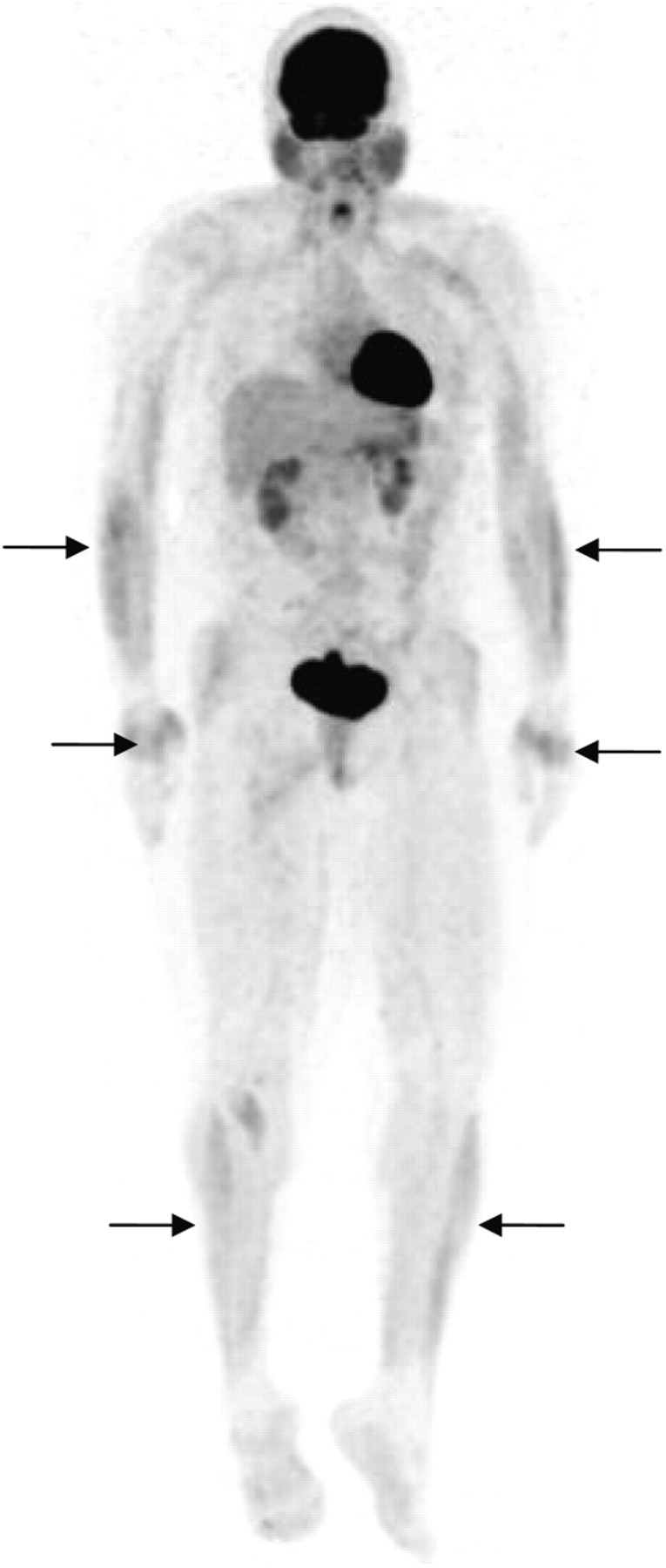
- FIGURE 1.
Representative 8F-FDG PET scan in coronal view of symmetric muscle uptake in forearms (arrows), hands (arrows), and anterior crural compartments (arrows) in control subject.
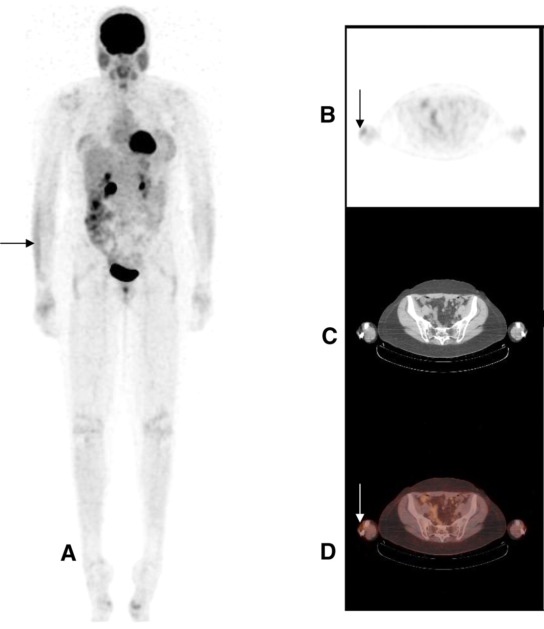
- FIGURE 2.
Representative 18F-FDG PET scan in coronal view (A) and transaxial view (B) of asymmetric muscle uptake in wrist or finger extensor muscles (arrow) in control subject. (C) Transaxial CT image used for muscle identification. (D) Fused PET/CT image showing asymmetric muscle uptake in wrist or finger extensor muscles (arrow).
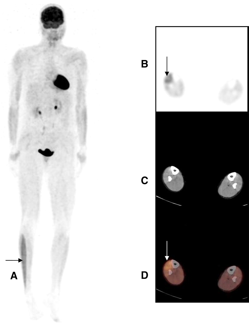
- FIGURE 3.
Representative 18F-FDG PET scan in coronal view (A) and transaxial view (B) of asymmetric muscle uptake in anterior crural compartment (arrow) in subject in activity group. (C) Transaxial CT image used for muscle identification. (D) Fused PET/CT image showing asymmetric muscle uptake in anterior crural compartment (arrow).

Tables
Value for the following subjects: Characteristic All Control group Exercise group Age (y) 47.8 ± 5.8 47.4 ± 6.4 48.1 ± 5.6 No. of women:men 16:4 8:2 8:2 Body mass (kg) 72.9 ± 8.1 70.2 ± 7.9 75.6 ± 7.7 Height (m) 1.69 ± 07 1.67 ± 09 1.71 ± 05 Body mass index (kg/m2) 25.6 ± 3.2 25.3 ± 3.7 25.9 ± 2.8 Values are reported as mean ± SD unless otherwise indicated.
18F-FDG uptake in: Subject Brain Heart Kidneys Bladder Liver Spleen Colon Parotid glands Testicles Vagina Breasts Other A Int Mod Mod B Int Mild Mild Mild Mild Mild esophagus B Int Int Int B Int Mild Mild Mod B Mild stomach C Int Mild Int B Int Mild Mild Mild Mild Mild B Mild B inguinal nodes D Int Int Mod B Int Mild Mild Mild B Mild B E Int Int Mod B Int Mild Mild Mild Mild B Mild stomach F Int Int Int B Int Mild Mild Mild G Int Int Mod B Int Mild Mild Mild B H Int Int Mod B Int Mild Mild Mod Mod B Mild B Mod B tonsils; mild stomach I Int Mild Int B Int Mild Mild J Int Mod Mild B Int Mild Mild Mild Mild B Mild B Int sublingual K Int Int Mod B Int Mild Mild Mild L Int Int Int B Int Mild Mild Mild Mild B M Int Int Int B Int Mild Mild Mild Mod focal R Mild stomach N Int Int Mod B Int Mild Mild Mild Mild O Int Int Mod B Int Mild Mild Mild Mild B Mild Mild B ovaries P Int Int Mild B Int Mild Mild Mild B Q Int Int Mod B Int Mild Mild R Int Mild Mod B Int Mild Mild Mild Mild B Mild Mild B S Int Mild Mod B Int Mild Mild Mild Mild B T Int Mild Int B Int Mild Mild Mild Mild Throat; lymph Int = intense; Mod = moderate; B = bilateral.
Uptake in the following muscle group: Subject Group Dominant side WE RC VC ACC LCC SPCC SCM PM A C R None None Mod None None None None None B C L Mild B None Int Mild B None None Mild L Mild B C C R Mild L > R None Mod Mild B None None None None D C R None None Mod Mod R; mild L None Mild B None None F C R Mild B None None Mild B None None None None G C R Mild B None Mild Mild B None None None None H C R Mild R > L Mild R Mod None None None None None J C R Mild R > L None None None None None None None L C R Mild R > L None None None None None None None O C R Mild R None None None None None None None I EL R Mild R > L None None None None None None None N EL R Mild R None None Mod R; mild L None None None None Q EL R None Mild L None Mod R; mild L None None None None R EL R None None None Mild R None None None None T EL R None None None Mild R > L None None None None E EU R Mild R > L None None None None None None None K EU R Mild R > L None Mod Mod R; mild L None None None None M EU R Mild R > L None Mild None Mod R None None None P EU R Mild R > L None Mild None None None None None S EU R Mild B None Mild None None None None None Group: C = control; EL = lower-extremity exercise; EU = upper-extremity exercise. Muscle groups: WE = wrist extensor; RC = rotator cuff; VC = vocal cords; ACC = anterior crural compartment (ankle dorsiflexor muscles); LCC = lateral crural compartment (ankle evertor muscles); SPCC = superficial posterior crural compartment (gastrocnemius); SCM = sternocleidomastoid; PM = pectoralis major. Uptake: Mod = moderate; B = bilateral; Int = intense.


