
Main menu

  • Home
  • Content
    • Current
      • JNMT Supplement
    • Ahead of print
    • Past Issues
    • Continuing Education
    • JNMT Podcast
    • SNMMI Annual Meeting Abstracts
  • Subscriptions
    • Subscribers
    • Rates
    • Journal Claims
    • Institutional and Non-member
  • Authors
    • Submit to JNMT
    • Information for Authors
    • Assignment of Copyright
    • AQARA Requirements
  • Info
    • Reviewers
    • Permissions
    • Advertisers
    • Corporate & Special Sales
  • About
    • About Us
    • Editorial Board
    • Contact Information
  • More
    • Alerts
    • Feedback
    • Help
    • SNMMI Journals
  • SNMMI
    • JNMT
    • JNM
    • SNMMI Journals
    • SNMMI

User menu

  • Subscribe
  • My alerts
  • Log in
  • My Cart

Search

  • Advanced search
Journal of Nuclear Medicine Technology
  • SNMMI
    • JNMT
    • JNM
    • SNMMI Journals
    • SNMMI
  • Subscribe
  • My alerts
  • Log in
  • My Cart
Journal of Nuclear Medicine Technology

Advanced Search

  • Home
  • Content
    • Current
    • Ahead of print
    • Past Issues
    • Continuing Education
    • JNMT Podcast
    • SNMMI Annual Meeting Abstracts
  • Subscriptions
    • Subscribers
    • Rates
    • Journal Claims
    • Institutional and Non-member
  • Authors
    • Submit to JNMT
    • Information for Authors
    • Assignment of Copyright
    • AQARA Requirements
  • Info
    • Reviewers
    • Permissions
    • Advertisers
    • Corporate & Special Sales
  • About
    • About Us
    • Editorial Board
    • Contact Information
  • More
    • Alerts
    • Feedback
    • Help
    • SNMMI Journals
  • Watch or Listen to JNMT Podcast
  • Visit SNMMI on Facebook
  • Join SNMMI on LinkedIn
  • Follow SNMMI on Twitter
  • Subscribe to JNMT RSS feeds
OtherIMAGING

Improving Evaluation of Primary Gastric Malignancies by Distending the Stomach with Milk Immediately Before 18F-FDG PET Scanning

Zhaohui Zhu, Fang Li, Yilei Mao, Wuying Cheng, Xin Cheng and Yonghong Dang
Journal of Nuclear Medicine Technology March 2008, 36 (1) 25-29; DOI: https://doi.org/10.2967/jnmt.107.044081
Zhaohui Zhu
  • Find this author on Google Scholar
  • Find this author on PubMed
  • Search for this author on this site
Fang Li
  • Find this author on Google Scholar
  • Find this author on PubMed
  • Search for this author on this site
Yilei Mao
  • Find this author on Google Scholar
  • Find this author on PubMed
  • Search for this author on this site
Wuying Cheng
  • Find this author on Google Scholar
  • Find this author on PubMed
  • Search for this author on this site
Xin Cheng
  • Find this author on Google Scholar
  • Find this author on PubMed
  • Search for this author on this site
Yonghong Dang
  • Find this author on Google Scholar
  • Find this author on PubMed
  • Search for this author on this site
  • Article
  • Figures & Data
  • Info & Metrics
  • PDF
Loading

Article Figures & Data

Figures

  • Tables
  • FIGURE 1. 
    • Download figure
    • Open in new tab
    • Download powerpoint
    FIGURE 1. 

    Comparison of empty-stomach and distended-stomach PET studies from same patient. Patient (F, 57 y) underwent serial follow-up of cervical carcinoma of uterus after curative surgical removal and radiotherapy. Interval between 2 studies was 7 mo. Stomach (arrow) showing moderate to intense 18F-FDG uptake in empty-stomach study (A, coronal images) changed to “hollow” region in distended-stomach study (B, coronal images). Note that heart, with intense uptake under empty-stomach condition, did not show prominent uptake after gastric distention with milk.
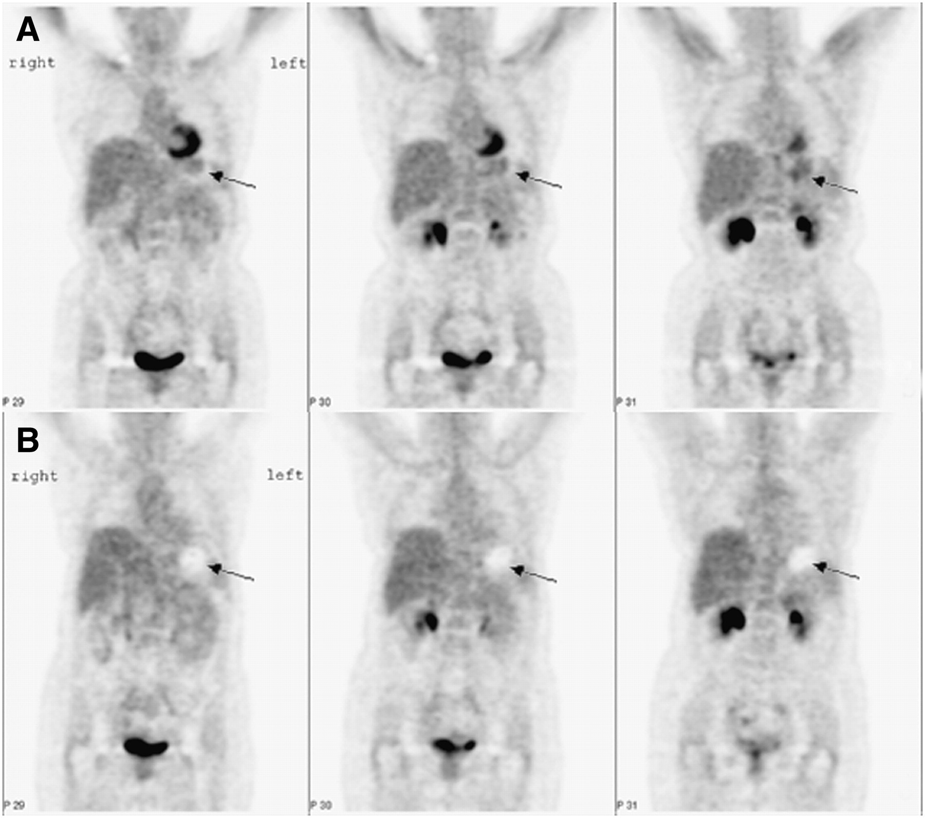
  • FIGURE 2. 
    • Download figure
    • Open in new tab
    • Download powerpoint
    FIGURE 2. 

    Comparison of empty-stomach and distended-stomach PET scans in 2 patients with moderately to poorly differentiated adenocarcinomas at antrum of stomach. First patient (M, 90 y) underwent empty-stomach PET scan. Coronal (a) and transaxial (b) images showed intense uptake in whole stomach (arrow), which obscured lesion located at antrum. Second patient (M, 62 y) underwent distended-stomach PET scan. On coronal (c) and transaxial (d) images, lesion (arrow) was clearly outlined, and normal gastric wall was distended.

Tables

  • Figures
    • View popup
    TABLE 1

    Groups and Diagnoses of Patients

    Groups and diagnosesNo. of patients
    Group 1: serial studies under both conditions (distended stomach [n = 72] and empty stomach [n = 79])43
     Lung cancer18
     Non-Hodgkin's lymphoma6
     Ovarian cancer6
     Breast cancer2
     Cervical cancer of uterus2
     Other9
    Group 2: gastric malignancies
     Underwent distended-stomach PET study24
      Adenocarcinoma, not mucinous15
      Adenocarcinoma, mucinous1
      Signet-ring cell carcinoma5
      Non-Hodgkin's lymphoma3
     Underwent empty-stomach PET study17
      Adenocarcinoma, not mucinous11
      Adenocarcinoma, mucinous2
      Signet-ring cell carcinoma3
      Non-Hodgkin's lymphoma1
    • View popup
    TABLE 2

    SUVs in Gastric Wall, Heart, Mediastinum, and Liver in Same Patient According to Scan Condition

    Mean ± SD SUV in:
    Organ or tissueEmpty stomach (n = 43)Stomach distended with milk (n = 43)FP
    Gastric wall2.21 ± 0.580.93 ± 0.32184.470.00
    Heart3.13 ± 1.852.71 ± 1.542.000.16
    Mediastinum1.38 ± 0.231.36 ± 0.220.470.50
    Liver1.85 ± 0.301.82 ± 0.290.480.49
PreviousNext
Back to top

In this issue

Journal of Nuclear Medicine Technology: 36 (1)
Journal of Nuclear Medicine Technology
Vol. 36, Issue 1
March 2008
  • Table of Contents
  • About the Cover
  • Index by author
Print
Download PDF
Article Alerts
Sign In to Email Alerts with your Email Address
Email Article

Thank you for your interest in spreading the word on Journal of Nuclear Medicine Technology.

NOTE: We only request your email address so that the person you are recommending the page to knows that you wanted them to see it, and that it is not junk mail. We do not capture any email address.

Enter multiple addresses on separate lines or separate them with commas.
Improving Evaluation of Primary Gastric Malignancies by Distending the Stomach with Milk Immediately Before 18F-FDG PET Scanning
(Your Name) has sent you a message from Journal of Nuclear Medicine Technology
(Your Name) thought you would like to see the Journal of Nuclear Medicine Technology web site.
Citation Tools
Improving Evaluation of Primary Gastric Malignancies by Distending the Stomach with Milk Immediately Before 18F-FDG PET Scanning
Zhaohui Zhu, Fang Li, Yilei Mao, Wuying Cheng, Xin Cheng, Yonghong Dang
Journal of Nuclear Medicine Technology Mar 2008, 36 (1) 25-29; DOI: 10.2967/jnmt.107.044081

Citation Manager Formats

  • BibTeX
  • Bookends
  • EasyBib
  • EndNote (tagged)
  • EndNote 8 (xml)
  • Medlars
  • Mendeley
  • Papers
  • RefWorks Tagged
  • Ref Manager
  • RIS
  • Zotero
Share
Improving Evaluation of Primary Gastric Malignancies by Distending the Stomach with Milk Immediately Before 18F-FDG PET Scanning
Zhaohui Zhu, Fang Li, Yilei Mao, Wuying Cheng, Xin Cheng, Yonghong Dang
Journal of Nuclear Medicine Technology Mar 2008, 36 (1) 25-29; DOI: 10.2967/jnmt.107.044081
Twitter logo Facebook logo LinkedIn logo Mendeley logo
  • Tweet Widget
  • Facebook Like
  • Google Plus One
Bookmark this article

Jump to section

  • Article
    • Abstract
    • MATERIALS AND METHODS
    • RESULTS
    • DISCUSSION
    • CONCLUSION
    • Footnotes
    • References
  • Figures & Data
  • Info & Metrics
  • PDF

Related Articles

  • No related articles found.
  • PubMed
  • Google Scholar

Cited By...

  • No citing articles found.
  • Google Scholar

More in this TOC Section

  • Early 10-Minute Postinjection [18F]F-FAPI-42 uEXPLORER Total-Body PET/CT Scanning Protocol for Staging Lung Cancer Using HYPER Iterative Reconstruction
  • Single- Versus Dual-Time-Point Imaging for Transthyretin Cardiac Amyloid Using 99mTc-Pyrophosphate
  • Software Discrepancies in Radionuclide-Derived Left Ventricular Ejection Fraction
Show more Imaging

Similar Articles

SNMMI

© 2025 SNMMI

Powered by HighWire