


Article Figures & Data
Figures
- FIGURE 1.
Projection of 31 parathyroid adenomas on planar pinhole images. Half of P4-derived adenomas were located in midportion of thyroid lobe, at medial margin, close to isthmus. In contrast, 95% of P3-derived adenomas were located at tip of inferior lobe or along thyrothymic tract. Most frequent sites are represented by shading. Note that there are potential areas of overlap between P3- and P4-derived adenomas.
- FIGURE 2.
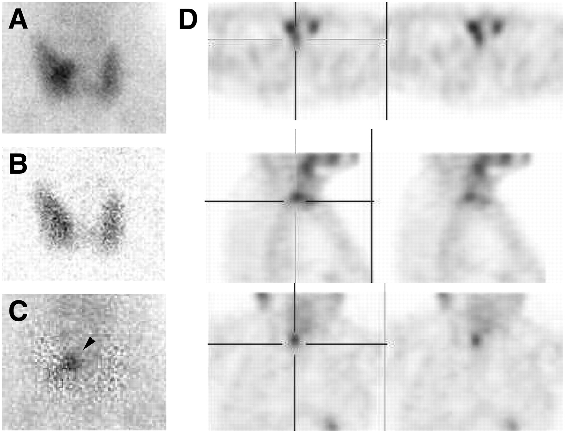
Planar pinhole images (A: 99mTc-sestamibi; B: 123I; C: subtraction) and SPECT images (D) of parathyroid adenomas. Posterior extension of adenoma on SPECT images is highly suggestive of P4 origin, despite its apparently right inferior origin on planar images (arrowhead).
- FIGURE 3.
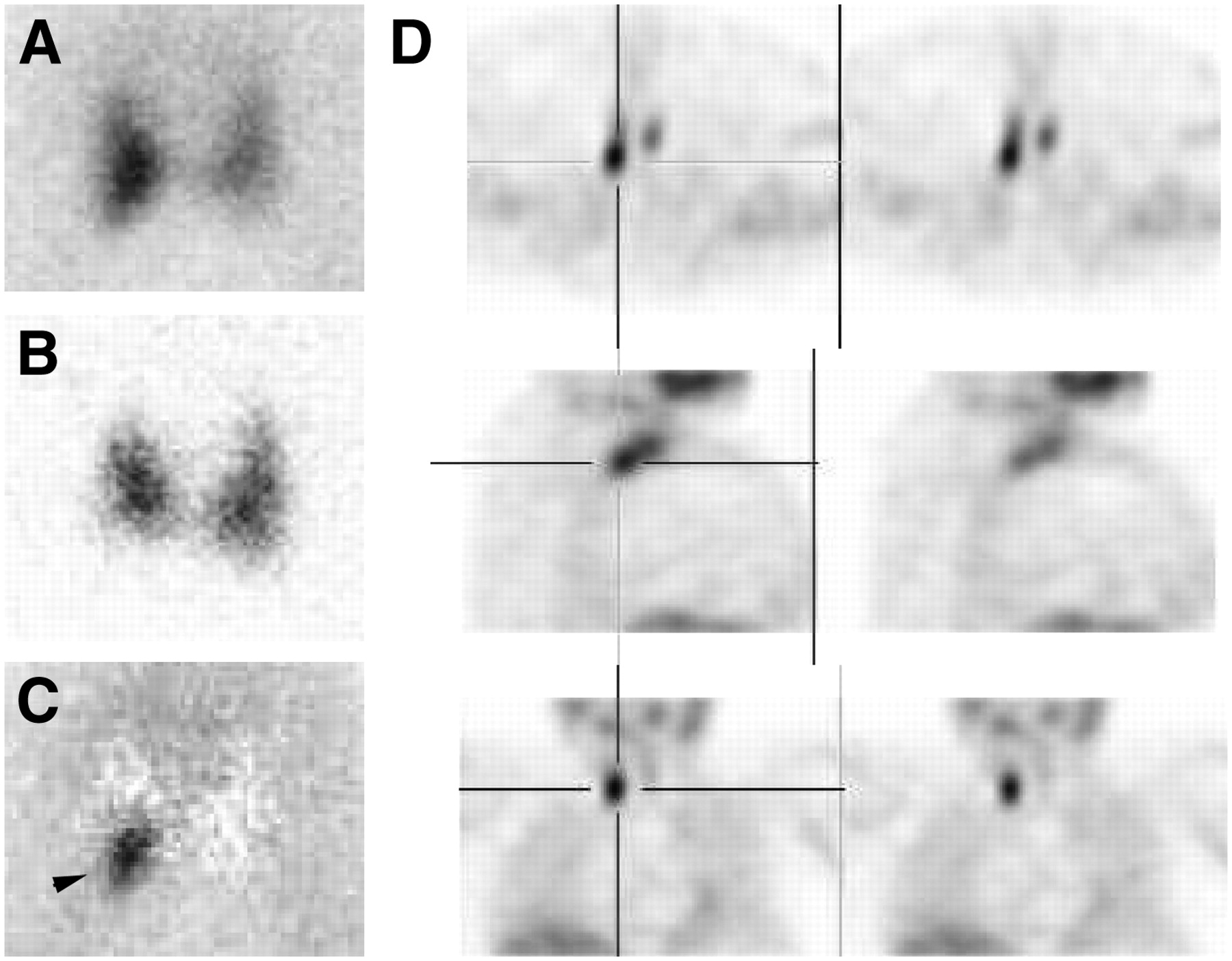
Planar pinhole images (A: 99mTc-sestamibi; B: 123I; C: subtraction) and SPECT images (D) of parathyroid adenomas. Adenoma is located at posteromedial part of right thyroid lobe (arrowhead), above isthmus; this location is characteristic of P4 origin.
- FIGURE 4.
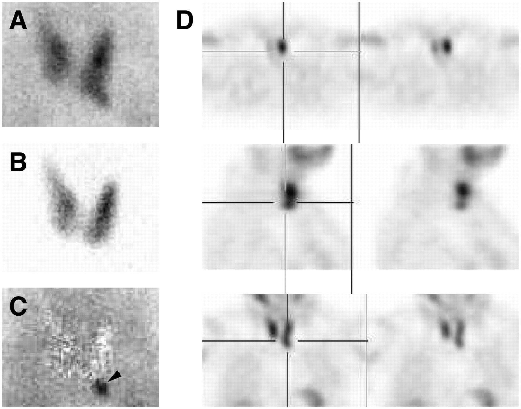
Planar pinhole images (A: 99mTc-sestamibi; B: 123I; C: subtraction) and SPECT images (D) of parathyroid adenomas. Typical P3 adenoma is located at tip of left inferior lobe on planar images (arrowhead) and in anterior position on SPECT images.


