
Main menu

  • Home
  • Content
    • Current
      • JNMT Supplement
    • Ahead of print
    • Past Issues
    • Continuing Education
    • JNMT Podcast
    • SNMMI Annual Meeting Abstracts
  • Subscriptions
    • Subscribers
    • Rates
    • Journal Claims
    • Institutional and Non-member
  • Authors
    • Submit to JNMT
    • Information for Authors
    • Assignment of Copyright
    • AQARA Requirements
  • Info
    • Reviewers
    • Permissions
    • Advertisers
    • Corporate & Special Sales
  • About
    • About Us
    • Editorial Board
    • Contact Information
  • More
    • Alerts
    • Feedback
    • Help
    • SNMMI Journals
  • SNMMI
    • JNMT
    • JNM
    • SNMMI Journals
    • SNMMI

User menu

  • Subscribe
  • My alerts
  • Log in
  • My Cart

Search

  • Advanced search
Journal of Nuclear Medicine Technology
  • SNMMI
    • JNMT
    • JNM
    • SNMMI Journals
    • SNMMI
  • Subscribe
  • My alerts
  • Log in
  • My Cart
Journal of Nuclear Medicine Technology

Advanced Search

  • Home
  • Content
    • Current
    • Ahead of print
    • Past Issues
    • Continuing Education
    • JNMT Podcast
    • SNMMI Annual Meeting Abstracts
  • Subscriptions
    • Subscribers
    • Rates
    • Journal Claims
    • Institutional and Non-member
  • Authors
    • Submit to JNMT
    • Information for Authors
    • Assignment of Copyright
    • AQARA Requirements
  • Info
    • Reviewers
    • Permissions
    • Advertisers
    • Corporate & Special Sales
  • About
    • About Us
    • Editorial Board
    • Contact Information
  • More
    • Alerts
    • Feedback
    • Help
    • SNMMI Journals
  • Watch or Listen to JNMT Podcast
  • Visit SNMMI on Facebook
  • Join SNMMI on LinkedIn
  • Follow SNMMI on Twitter
  • Subscribe to JNMT RSS feeds
OtherIMAGING

Easy Method of Patient Positioning for Convergent-Beam Cardiac SPECT

Gengsheng L. Zeng and Paul E. Christian
Journal of Nuclear Medicine Technology September 2007, 35 (3) 131-134; DOI: https://doi.org/10.2967/jnmt.107.041160
Gengsheng L. Zeng
  • Find this author on Google Scholar
  • Find this author on PubMed
  • Search for this author on this site
Paul E. Christian
  • Find this author on Google Scholar
  • Find this author on PubMed
  • Search for this author on this site
  • Article
  • Figures & Data
  • Info & Metrics
  • PDF
Loading

Article Figures & Data

Figures

  • FIGURE 1. 
    • Download figure
    • Open in new tab
    • Download powerpoint
    FIGURE 1. 

    When parallel-hole collimators are used to image heart, detector area is not efficiently used, especially when detector area is large.

  • FIGURE 2. 
    • Download figure
    • Open in new tab
    • Download powerpoint
    FIGURE 2. 

    Convergent-beam collimators can magnify image size on detector or generate multiple images of object on detector, so that large detector area is more efficiently used for imaging of small organs. (Left) Fanbeam collimator. (Middle) Combined parallel-hole and slant-hole collimator. (Right) Collimator with varying focal length.
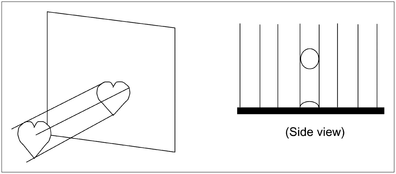
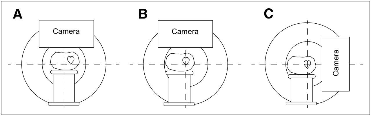
  • FIGURE 3. 
    • Download figure
    • Open in new tab
    • Download powerpoint
    FIGURE 3. 

    Patient-positioning procedure. (A) Default patient table position, in which table is centered. (B) Suggested left locking position of patient table. Top-view camera is used to monitor whether heart is centered. (C) Adjustment of table height so that heart is at center of side-view camera.
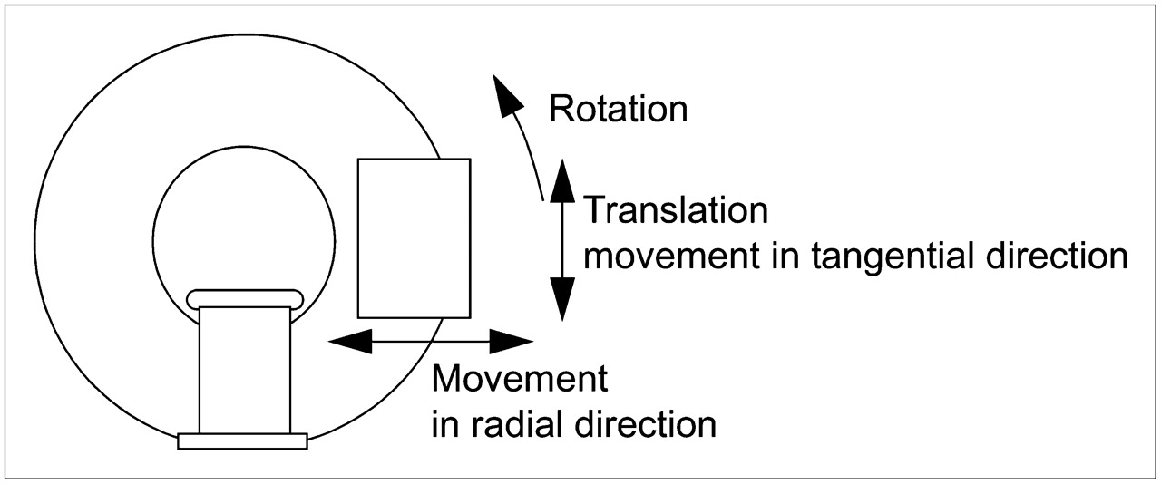
  • FIGURE 4. 
    • Download figure
    • Open in new tab
    • Download powerpoint
    FIGURE 4. 

    In modern SPECT systems, detector head can rotate and move radially and tangentially.
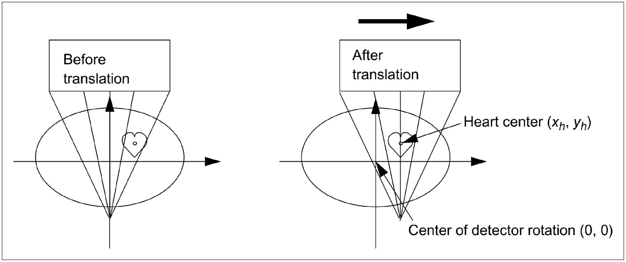
  • FIGURE 5. 
    • Download figure
    • Open in new tab
    • Download powerpoint
    FIGURE 5. 

    In modern SPECT systems, detector head can translate at each view, so that central ray passes through center of heart.
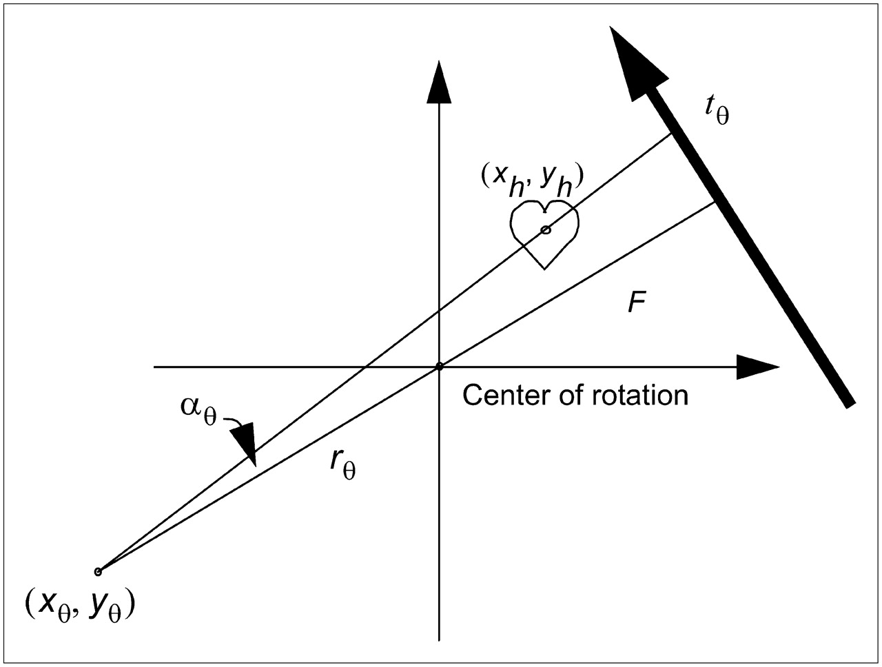
  • FIGURE 6. 
    • Download figure
    • Open in new tab
    • Download powerpoint
    FIGURE 6. 

    Coordinate system for fanbeam imaging geometry. During patient setup, center of heart on detector is marked as tθ and stored in computer. Two angles of θ are required to determine center of heart.
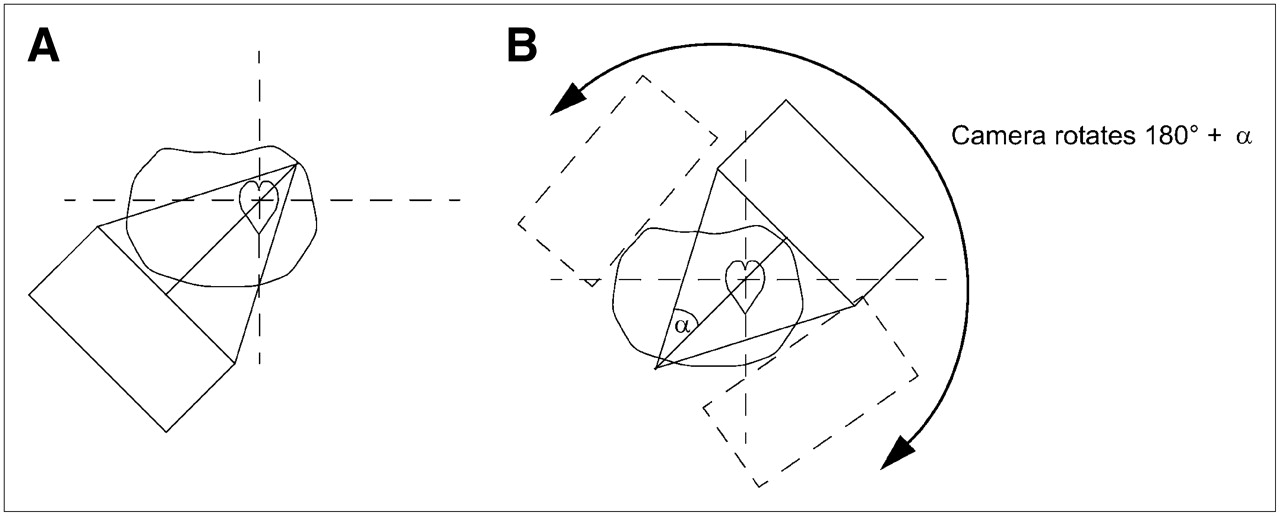
  • FIGURE 7. 
    • Download figure
    • Open in new tab
    • Download powerpoint
    FIGURE 7. 

    (A) Worst-case situation, at which truncation of heart is most likely to happen. (B) For patients weighing more than 135 kg, short scan can be used to avoid worst-case situation.

PreviousNext
Back to top

In this issue

Journal of Nuclear Medicine Technology: 35 (3)
Journal of Nuclear Medicine Technology
Vol. 35, Issue 3
September 2007
  • Table of Contents
  • About the Cover
  • Index by author
Print
Download PDF
Article Alerts
Sign In to Email Alerts with your Email Address
Email Article

Thank you for your interest in spreading the word on Journal of Nuclear Medicine Technology.

NOTE: We only request your email address so that the person you are recommending the page to knows that you wanted them to see it, and that it is not junk mail. We do not capture any email address.

Enter multiple addresses on separate lines or separate them with commas.
Easy Method of Patient Positioning for Convergent-Beam Cardiac SPECT
(Your Name) has sent you a message from Journal of Nuclear Medicine Technology
(Your Name) thought you would like to see the Journal of Nuclear Medicine Technology web site.
Citation Tools
Easy Method of Patient Positioning for Convergent-Beam Cardiac SPECT
Gengsheng L. Zeng, Paul E. Christian
Journal of Nuclear Medicine Technology Sep 2007, 35 (3) 131-134; DOI: 10.2967/jnmt.107.041160

Citation Manager Formats

  • BibTeX
  • Bookends
  • EasyBib
  • EndNote (tagged)
  • EndNote 8 (xml)
  • Medlars
  • Mendeley
  • Papers
  • RefWorks Tagged
  • Ref Manager
  • RIS
  • Zotero
Share
Easy Method of Patient Positioning for Convergent-Beam Cardiac SPECT
Gengsheng L. Zeng, Paul E. Christian
Journal of Nuclear Medicine Technology Sep 2007, 35 (3) 131-134; DOI: 10.2967/jnmt.107.041160
Twitter logo Facebook logo LinkedIn logo Mendeley logo
  • Tweet Widget
  • Facebook Like
  • Google Plus One
Bookmark this article

Jump to section

  • Article
    • Abstract
    • MATERIALS AND METHODS
    • RESULTS AND DISCUSSION
    • CONCLUSION
    • Acknowledgments
    • Footnotes
    • References
  • Figures & Data
  • Info & Metrics
  • PDF

Related Articles

  • No related articles found.
  • PubMed
  • Google Scholar

Cited By...

  • No citing articles found.
  • Google Scholar

More in this TOC Section

  • Early 10-Minute Postinjection [18F]F-FAPI-42 uEXPLORER Total-Body PET/CT Scanning Protocol for Staging Lung Cancer Using HYPER Iterative Reconstruction
  • Single- Versus Dual-Time-Point Imaging for Transthyretin Cardiac Amyloid Using 99mTc-Pyrophosphate
  • Robustness of 18F-FDG PET Radiomic Features in Lung Cancer: Impact of Advanced Reconstruction Algorithm
Show more Imaging

Similar Articles

SNMMI

© 2025 SNMMI

Powered by HighWire