
Main menu

  • Home
  • Content
    • Current
      • JNMT Supplement
    • Ahead of print
    • Past Issues
    • Continuing Education
    • JNMT Podcast
    • SNMMI Annual Meeting Abstracts
  • Subscriptions
    • Subscribers
    • Rates
    • Journal Claims
    • Institutional and Non-member
  • Authors
    • Submit to JNMT
    • Information for Authors
    • Assignment of Copyright
    • AQARA Requirements
  • Info
    • Reviewers
    • Permissions
    • Advertisers
    • Corporate & Special Sales
  • About
    • About Us
    • Editorial Board
    • Contact Information
  • More
    • Alerts
    • Feedback
    • Help
    • SNMMI Journals
  • SNMMI
    • JNMT
    • JNM
    • SNMMI Journals
    • SNMMI

User menu

  • Subscribe
  • My alerts
  • Log in
  • My Cart

Search

  • Advanced search
Journal of Nuclear Medicine Technology
  • SNMMI
    • JNMT
    • JNM
    • SNMMI Journals
    • SNMMI
  • Subscribe
  • My alerts
  • Log in
  • My Cart
Journal of Nuclear Medicine Technology

Advanced Search

  • Home
  • Content
    • Current
    • Ahead of print
    • Past Issues
    • Continuing Education
    • JNMT Podcast
    • SNMMI Annual Meeting Abstracts
  • Subscriptions
    • Subscribers
    • Rates
    • Journal Claims
    • Institutional and Non-member
  • Authors
    • Submit to JNMT
    • Information for Authors
    • Assignment of Copyright
    • AQARA Requirements
  • Info
    • Reviewers
    • Permissions
    • Advertisers
    • Corporate & Special Sales
  • About
    • About Us
    • Editorial Board
    • Contact Information
  • More
    • Alerts
    • Feedback
    • Help
    • SNMMI Journals
  • Watch or Listen to JNMT Podcast
  • Visit SNMMI on Facebook
  • Join SNMMI on LinkedIn
  • Follow SNMMI on Twitter
  • Subscribe to JNMT RSS feeds
OtherIMAGING

Subtraction SPECT for Parathyroid Scintigraphy Based on Maximization of Mutual Information

Narihiro Hara, Teruhiko Takayama, Masahisa Onoguchi, Norikazu Obane, Toshiaki Miyati, Toshiaki Yoshioka, Katsuhiko Sakaguchi and Minoru Honda
Journal of Nuclear Medicine Technology June 2007, 35 (2) 84-90; DOI: https://doi.org/10.2967/jnmt.106.033480
Narihiro Hara
  • Find this author on Google Scholar
  • Find this author on PubMed
  • Search for this author on this site
Teruhiko Takayama
  • Find this author on Google Scholar
  • Find this author on PubMed
  • Search for this author on this site
Masahisa Onoguchi
  • Find this author on Google Scholar
  • Find this author on PubMed
  • Search for this author on this site
Norikazu Obane
  • Find this author on Google Scholar
  • Find this author on PubMed
  • Search for this author on this site
Toshiaki Miyati
  • Find this author on Google Scholar
  • Find this author on PubMed
  • Search for this author on this site
Toshiaki Yoshioka
  • Find this author on Google Scholar
  • Find this author on PubMed
  • Search for this author on this site
Katsuhiko Sakaguchi
  • Find this author on Google Scholar
  • Find this author on PubMed
  • Search for this author on this site
Minoru Honda
  • Find this author on Google Scholar
  • Find this author on PubMed
  • Search for this author on this site
  • Article
  • Figures & Data
  • Info & Metrics
  • PDF
Loading

Article Figures & Data

Figures

  • Tables
  • FIGURE 1. 
    • Download figure
    • Open in new tab
    • Download powerpoint
    FIGURE 1. 

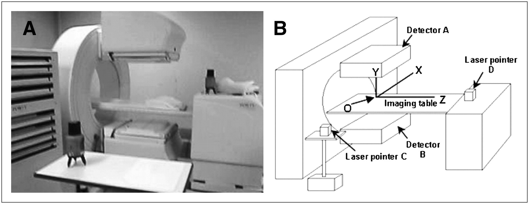
    Appearance (A) and illustration (B) of pair of laser pointers aligned at right angles. Point O corresponds to center of rotation of detectors.
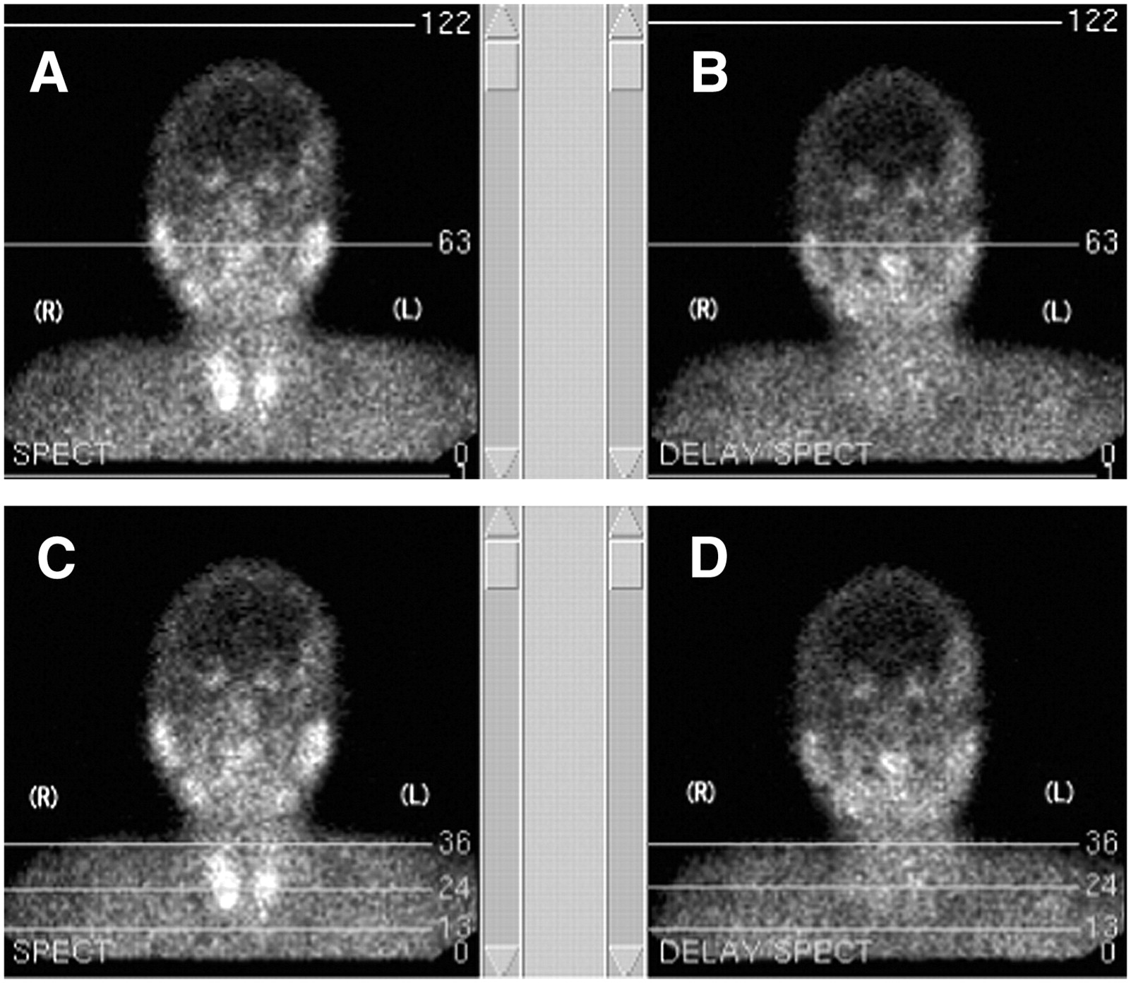
  • FIGURE 2. 
    • Download figure
    • Open in new tab
    • Download powerpoint
    FIGURE 2. 

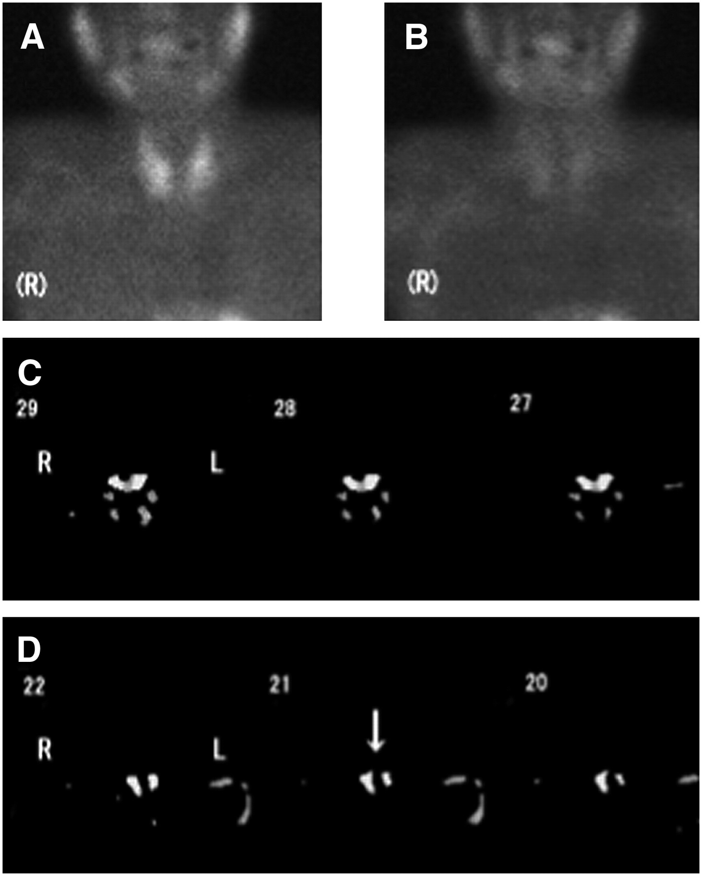
    Planar images used to select reconstructed regions for SPECT images. (A and C) Early planar images. Line marked 63 in A is centerline in 128 matrix. Centerline passes through right and left external auditory canals. Region between lines 13 and 36 is reconstructed region on SPECT images, excluding salivary glands. (B and D) Delayed planar images. Similarly, centerline passes through both external auditory canals. Reconstructed region for delayed SPECT image is similarly selected between lines 13 and 36.

  • FIGURE 3. 
    • Download figure
    • Open in new tab
    • Download powerpoint
    FIGURE 3. 

    SPECT images of a patient: early (A), delayed (B), fusion (C), and subtraction (D).

  • FIGURE 4. 
    • Download figure
    • Open in new tab
    • Download powerpoint
    FIGURE 4. 

    A 61-y-old man with 4 hyperplastic glands (patient 10). Glands weighed 300–700 mg. In this patient, 24 slices (1–24) were reconstructed. Slice numbers more and less than 24 were considered to represent superior and inferior parathyroid glands, respectively. (A–D) Early planar image (A), delayed planar image (B), and subtraction SPECT images showing upper (C) and lower (D) parathyroid hyperplasia. Arrow indicates hyperplastic lesion.

  • FIGURE 5. 
    • Download figure
    • Open in new tab
    • Download powerpoint
    FIGURE 5. 

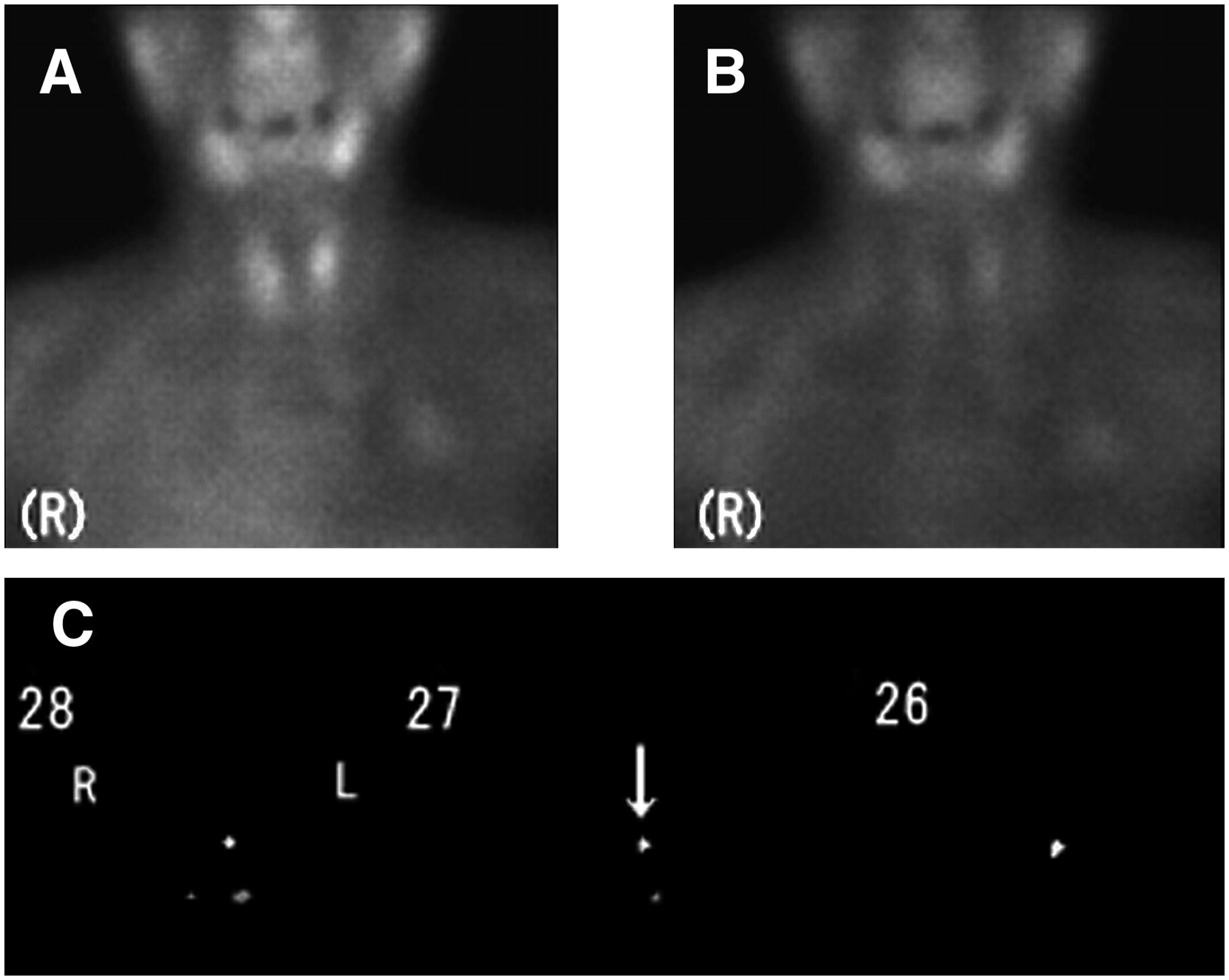
    A 74-y-old woman with adenoma (patient 12). Lesion weighed 300 mg. (A–C) Early planar image (A), delayed planar image (B), and subtraction SPECT images (C). Lesion (arrow) was seen in left upper region.

Tables

  • Figures
    • View popup
    TABLE 1

    Summary of Clinical Data

    Blood test
    Patient no.Age (y)Calcium (mg/dL) (normal, 8.2–10.2 mg/dL)Phosphorus (mg/dL) (normal, 2.7–4.4 mg/dL)ALP (IU/L) (normal, 115–359 IU/L)Internal secretion, PTH (pg/mL) (normal, 10–60 pg/mL)
    SexDialysisBeforeAfterBeforeAfterBeforeAfterBeforeAfter
    137M+8.87.07.53.9483889101510
    237M+7.16.44.64.335437127460
    354M+9.78.45.83.0403626762140
    460F+9.78.04.63.9298304933136
    562M+8.27.74.53.844383777812
    658M+9.99.87.33.93635844977
    753M+10.27.28.56.21752631,258126
    863M+9.87.87.94.15427487807
    952M+10.08.76.13.427331153312
    1061M+10.07.66.34.32452484905
    1159M+9.48.75.03.94434391,23610
    1274F−10.99.03.63.123323918074
    1364F−11.19.02.72.737032718040
    1456F−11.49.72.92.52872596239
    • ALP = alkaline phosphatase; PTH = parathyroid hormone; before = data within 2 wk before surgery; after = data within 5 d after surgery.

    • View popup
    TABLE 2

    Results of Scintigraphy, Ultrasonography, and Surgery

    ScintigraphyUltrasonographySurgery/pathology
    Patient no.Location+/−Size (mm)+/−HistopathologyWeight (mg)
    1RU+−Parathyroid hyperplasia350
    RL+16 × 15 × 15+Parathyroid hyperplasia1,650
    LU+−Parathyroid hyperplasia800
    LL+18 × 13 × 11+Parathyroid hyperplasia1,100
    2RU+−Parathyroid hyperplasia1,600
    RL+23 × 10 × 9+Parathyroid hyperplasia200
    LU+−Parathyroid hyperplasia1,300
    LL+6 × 5 × 5+Parathyroid hyperplasia300
    3RU+21 × 14 × 9+Parathyroid hyperplasia1,600
    RL+13 × 11 × 10+Parathyroid hyperplasia500
    LU+−No excision
    LL−12 × 10 × 6+Parathyroid hyperplasia300
    4RU+−Parathyroid hyperplasia500
    RL+10 × 6 × 5+Parathyroid hyperplasia250
    LU+−Parathyroid hyperplasia450
    LL+8 × 8 × 7+Parathyroid hyperplasia400
    5RU−−No excision
    RL−10 × 6 × 5+No excision
    LU+−Parathyroid hyperplasia1,600
    LL+16 × 12 × 10+Parathyroid hyperplasia400
    6RU+−Parathyroid hyperplasia1,450
    RL+11 × 10 × 8+Parathyroid hyperplasia600
    LU+−Parathyroid hyperplasia2,250
    LL+10 × 8 × 7+Parathyroid hyperplasia700
    7RU+6 × 6 × 3+Parathyroid hyperplasia950
    RL+11 × 4 × 3+Parathyroid hyperplasia350
    LU+−Parathyroid hyperplasia2,050
    LL+12 × 11 × 3+Parathyroid hyperplasia350
    8RU+9 × 8 × 8+Parathyroid hyperplasia1,950
    RL+20 × 11 × 14+Parathyroid hyperplasia300
    LU+11 × 7 × 7+Parathyroid hyperplasia250
    LL+11 × 11 × 6+Parathyroid hyperplasia400
    9RU+−Parathyroid hyperplasia1,500
    RL−23 × 13 × 8+Parathyroid hyperplasia900
    LU−−Parathyroid hyperplasia600
    LL−−Parathyroid hyperplasia200
    10RU+10 × 8 × 5+Parathyroid hyperplasia400
    RL+12 × 9 × 7+Parathyroid hyperplasia700
    LU+6 × 6 × 8+Parathyroid hyperplasia300
    LL+10 × 10 × 4+Parathyroid hyperplasia500
    11RU+8 × 7 × 5+Parathyroid hyperplasia100
    RL+13 × 9 × 7+Parathyroid hyperplasia200
    LU+6 × 5 × 4+Parathyroid hyperplasia50
    LL+17 × 13 × 6+Parathyroid hyperplasia500
    12RU−−No excision
    RL−−No excision
    LU+15 × 5 × 3+Adenoma300
    LL+16 × 5 × 3+No excision
    13RU+20 × 7 × 4+Adenoma500
    RL−−No excision
    LU−−No excision
    LL−8 × 8 × 4+No excision
    14RU−−No excision
    RL+6 × 4 × 4+Adenoma300
    LU−−No excision
    LL−−No excision
    • RU = right upper; RL = right lower; LU = left upper; LL = left lower; + = positive; − = negative.

    • View popup
    TABLE 3

    Comparison of Lesion Detection Between Scintigraphy and Ultrasonography

    Ultrasonography(+) (n = 31)Ultrasonography(−) (n = 13)
    Scintigraphy(+/−)LocationRURLLULLRURLLULL
    + (n = 40)RU65
    RL10
    LU46
    LL9
    − (n = 4)RU
    RL1
    LU1
    LL11
    • RU = right upper; RL = right lower; LU = left upper; LL = left lower; + = positive; − = negative.

PreviousNext
Back to top

In this issue

Journal of Nuclear Medicine Technology: 35 (2)
Journal of Nuclear Medicine Technology
Vol. 35, Issue 2
June 2007
  • Table of Contents
  • About the Cover
  • Index by author
Print
Download PDF
Article Alerts
Sign In to Email Alerts with your Email Address
Email Article

Thank you for your interest in spreading the word on Journal of Nuclear Medicine Technology.

NOTE: We only request your email address so that the person you are recommending the page to knows that you wanted them to see it, and that it is not junk mail. We do not capture any email address.

Enter multiple addresses on separate lines or separate them with commas.
Subtraction SPECT for Parathyroid Scintigraphy Based on Maximization of Mutual Information
(Your Name) has sent you a message from Journal of Nuclear Medicine Technology
(Your Name) thought you would like to see the Journal of Nuclear Medicine Technology web site.
Citation Tools
Subtraction SPECT for Parathyroid Scintigraphy Based on Maximization of Mutual Information
Narihiro Hara, Teruhiko Takayama, Masahisa Onoguchi, Norikazu Obane, Toshiaki Miyati, Toshiaki Yoshioka, Katsuhiko Sakaguchi, Minoru Honda
Journal of Nuclear Medicine Technology Jun 2007, 35 (2) 84-90; DOI: 10.2967/jnmt.106.033480

Citation Manager Formats

  • BibTeX
  • Bookends
  • EasyBib
  • EndNote (tagged)
  • EndNote 8 (xml)
  • Medlars
  • Mendeley
  • Papers
  • RefWorks Tagged
  • Ref Manager
  • RIS
  • Zotero
Share
Subtraction SPECT for Parathyroid Scintigraphy Based on Maximization of Mutual Information
Narihiro Hara, Teruhiko Takayama, Masahisa Onoguchi, Norikazu Obane, Toshiaki Miyati, Toshiaki Yoshioka, Katsuhiko Sakaguchi, Minoru Honda
Journal of Nuclear Medicine Technology Jun 2007, 35 (2) 84-90; DOI: 10.2967/jnmt.106.033480
Twitter logo Facebook logo LinkedIn logo Mendeley logo
  • Tweet Widget
  • Facebook Like
  • Google Plus One
Bookmark this article

Jump to section

  • Article
    • Abstract
    • MATERIALS AND METHODS
    • RESULTS
    • DISCUSSION
    • CONCLUSION
    • Acknowledgments
    • Footnotes
    • References
  • Figures & Data
  • Info & Metrics
  • PDF

Related Articles

  • No related articles found.
  • PubMed
  • Google Scholar

Cited By...

  • No citing articles found.
  • Google Scholar

More in this TOC Section

  • Early 10-Minute Postinjection [18F]F-FAPI-42 uEXPLORER Total-Body PET/CT Scanning Protocol for Staging Lung Cancer Using HYPER Iterative Reconstruction
  • Single- Versus Dual-Time-Point Imaging for Transthyretin Cardiac Amyloid Using 99mTc-Pyrophosphate
  • Robustness of 18F-FDG PET Radiomic Features in Lung Cancer: Impact of Advanced Reconstruction Algorithm
Show more Imaging

Similar Articles

SNMMI

© 2025 SNMMI

Powered by HighWire