


Article Figures & Data
Figures
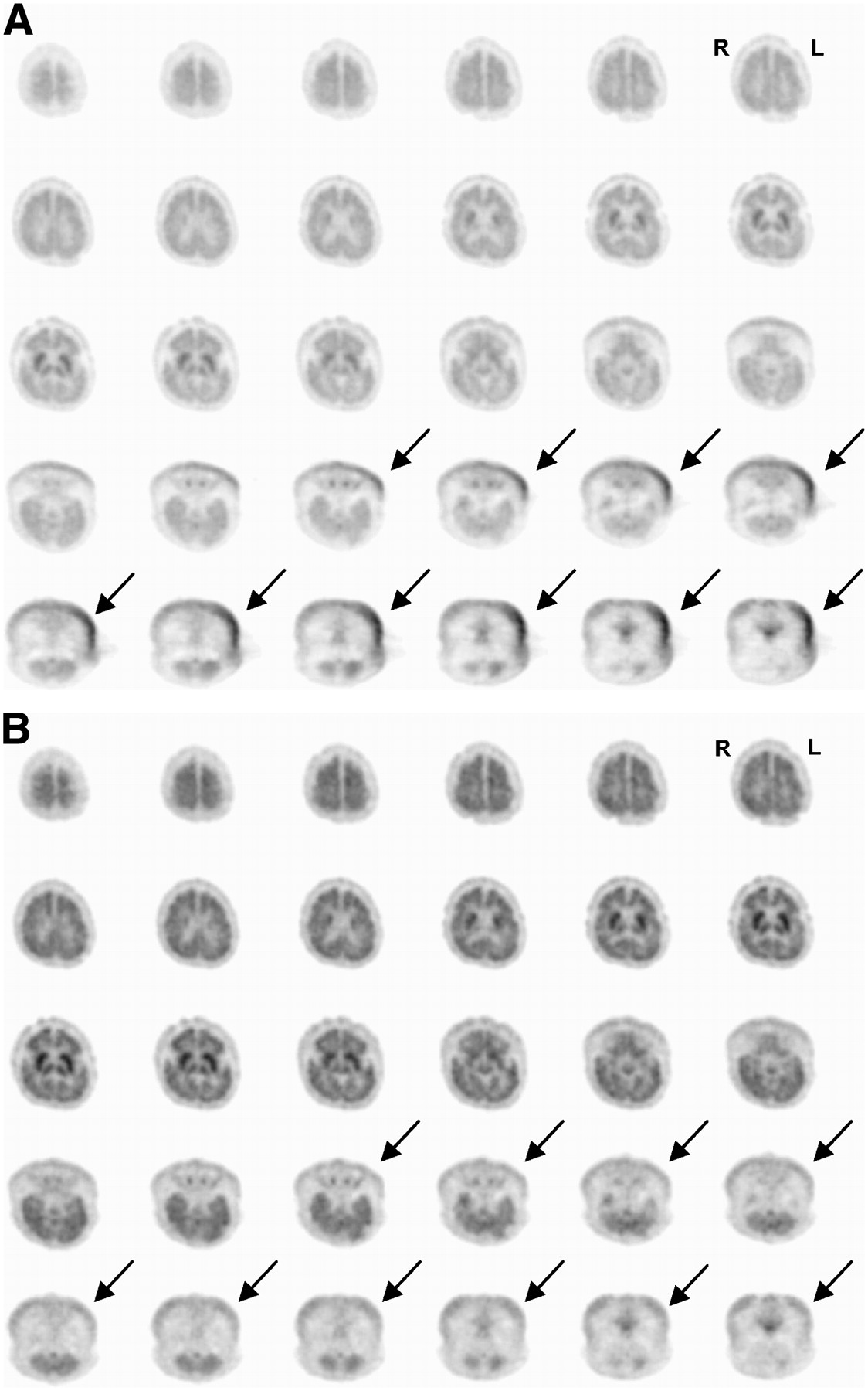
- FIGURE 1.
Transverse slices of brain 18F-FDG PET study of 11-mo-old girl with epileptic seizures. Attenuation correction was performed using method for calculated attenuation correction provided by scanner software (A) and using postinjection transmission scan (measured attenuation correction) (B). Calculated attenuation correction showed lesion of apparent hypermetabolism at level of skin on left side of neck (arrows). Comparison with measured attenuation correction, which did not show hypermetabolism at neck, suggested that this finding was artifact related to calculated attenuation correction.
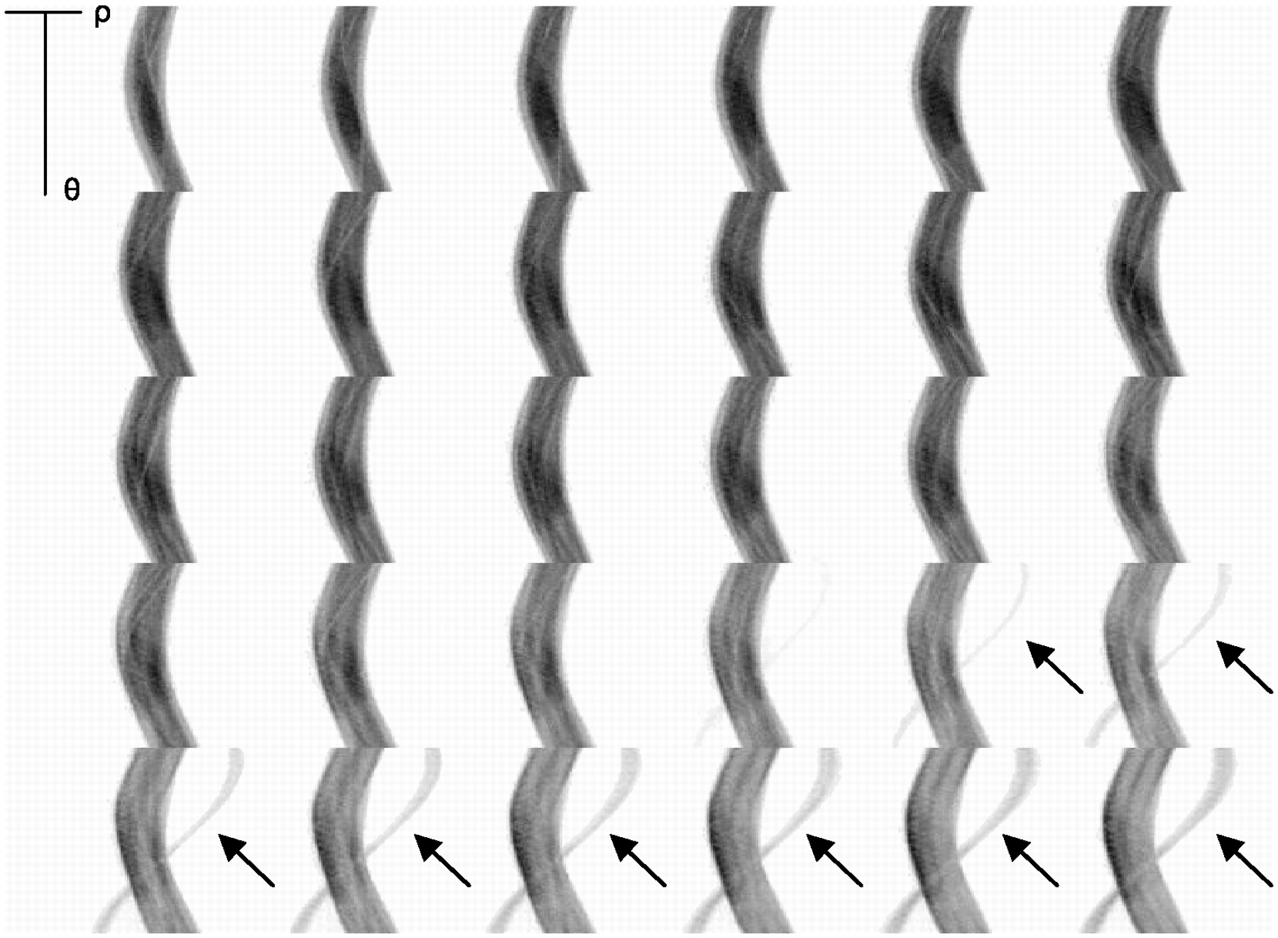
- FIGURE 2.
Sinograms corresponding to transverse slices in Figure 1. Sinograms are corrected for varying detector efficiency (normalized) but not for attenuation. Obvious extracranial hot object is seen within field of view at level of calculated attenuation correction lesion in neck (arrows). Object is assessed more easily on projection images (Fig. 3).
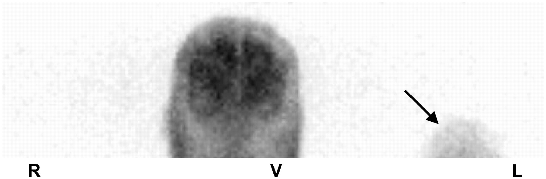
- FIGURE 3.
Ventral (V) projection of brain 18F-FDG PET shown in Figure 1. Hot object to left of head most likely is infant's left fist.
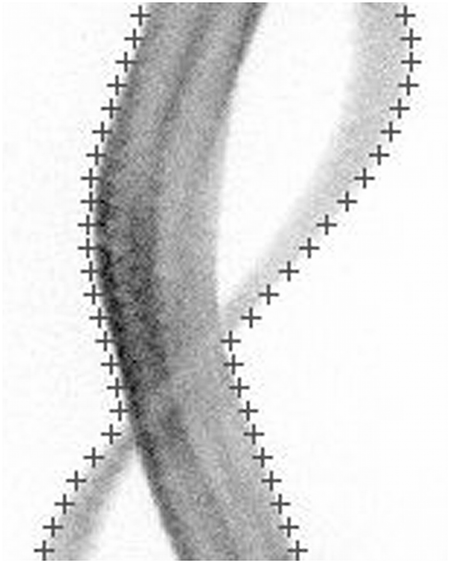
- FIGURE 4.
Sinogram corresponding to fourth slice from the end in Figure 1. Crosses indicate boundary detected by calculated attenuation correction method. Method assumes that detected boundary defines surface of skull, skull has constant thickness and homogeneous attenuation coefficient, and medium under skull has homogeneous attenuation coefficient characteristic of brain tissue. Hot object to left of head caused calculated attenuation correction to significantly overestimate size of head and, thus, to overestimate attenuation factors. This overestimation resulted in overcorrection of 18F-FDG uptake, predominantly in areas close to hot extracranial object.


Jump to section
Related Articles
Cited By...
- No citing articles found.