


Abstract
This case report involves a 45-y-old woman after cadaveric renal transplantation. The initial renal scan on the first postsurgical day showed normal findings. Subsequently, on the sixth postsurgical day, the patient had decreased urine output; 99mTc-diethylenetriaminepentaacetic acid renal scanning was performed again and did not show definite scintigraphic evidence of a urine leak. The patient’s condition was not improving; renal scanning was repeated on day 20 and demonstrated a large photopenic region surrounding the superior, medial, and inferior aspects of the kidney. Delayed imaging was performed 2.5 h after injection of radiotracer, and activity was observed in the previously cold region.
Complications after renal transplantation include vascular or ureteral obstruction or stenosis, hematoma, lymphocele, and urine leakage or urinoma (1). Urine leakage or urinoma is a rare and early complication after kidney transplantation. Renography can play an important role in the evaluation of these complications. For urine leaks, early scintigraphic images can show cold areas. Delayed images are the key to the diagnosis and often show a gradual fill-in of these cold regions (2). Hematomas usually present as nonspecific cold areas and are difficult to diagnose with scintigraphy. Ureteral obstruction can present in many ways and often requires a multimodality approach. Ultrasound also plays a crucial role in diagnosis of postoperative complications, often directly revealing fluid collections, dilated collecting systems, and vascular stenosis (3,4). However, ultrasound cannot differentiate the nature of the collection easily, and such differentiation is important for management and treatment planning. Our case stresses the need and importance of repeated follow-up studies and the value of delayed imaging in radionuclide renography.
CASE REPORT
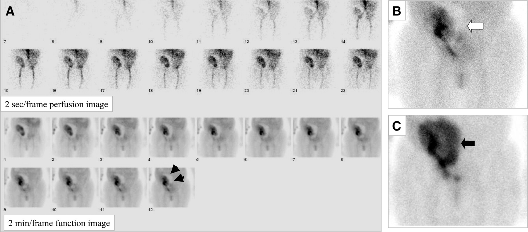
The patient was a 45-y-old woman who received a cadaveric renal transplant during an unremarkable procedure. The patient underwent 99mTc-diethylenetriaminepentaacetic acid (DTPA) renography immediately after transplantation to evaluate the perfusion, which showed good flow and reasonable function (Fig. 1A). However, during the next few days, she had less urine production than expected and her creatinine levels were persistently elevated. Renal ultrasound was performed on the 6th day after transplantation and showed a perinephric fluid collection (Figs. 1B and 1C). However, it was difficult to determine whether the perinephric fluid was from a urine leak or other postsurgical complications. Therefore, 99mTc-DTPA renography was performed on the same day as ultrasound, and no definite scintigraphic evidence of urine leakage was noted. Two weeks later, the urine output of the patient did not improve and creatinine levels remained high. Follow-up renal scanning was therefore performed, and the initial images demonstrated a large area of photopenia around the medial aspects of the kidney (Figs. 2A and 2B). Hematoma, lymphocele, abscess, and urinoma by urine leakage were on the differential list. A 2.5-h delayed image of the abdomen and pelvis was obtained, and significant activity was observed in the originally cold area (Fig. 2C)—compatible with a urine leak. The appropriate treatment was given and the patient recovered uneventfully.
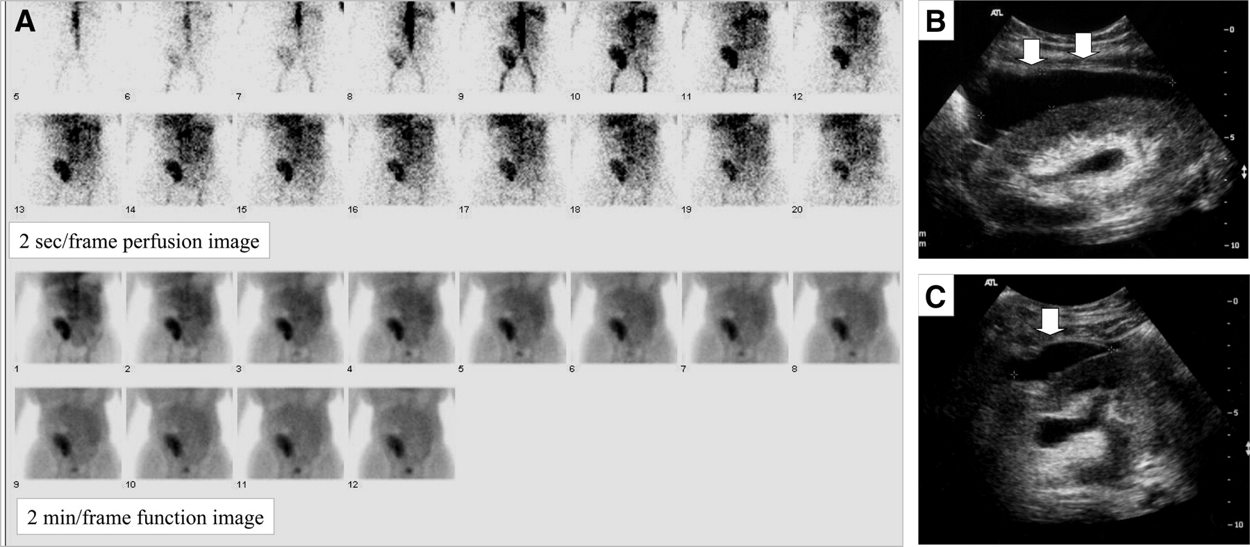
(A) Renal scintigraphy performed immediately after transplantation surgery. Top 2 rows are initial blood-flow images (2 s/frame). Bottom 2 rows are subsequent 24-min dynamic images (2 min/frame). (B and C) Sagittal (B) and transverse (C) ultrasound images demonstrate fluid collection (arrows), which appears to be simple fluid.

(A) Dynamic renal scan demonstrates large photopenic region surrounding kidney medially (arrows). Top 2 rows are initial blood-flow images (2 s/frame). Bottom 2 rows are subsequent 24-min dynamic images (2 min/frame). (B) Postvoiding image after dynamic renal scanning demonstrates large photopenic region (white arrow). (C) Delayed image obtained at 2.5 h demonstrates increased activity in previously photopenic region (black arrow).
DISCUSSION
Use of renal scintigraphy in the evaluation of a urine leak after renal transplantation has been described by many authors (2,5–8). Urine leakage usually requires invasive treatment by either interventional radiology or early surgery (9–13). Lymphocele is less common and has more complicated presentations (14,15). For all causes of fluid collection, early detection helps with clinical decision making and promotes patient recovery.
A perinephric fluid collection after renal transplantation can represent multiple etiologies, including urinoma, lymphocele, hematoma, and abscess. In general, a persistent photopenic defect likely represents hematoma, lymphocele, or abscess. On the other hand, an initially photopenic region that becomes hotter than background levels over time is likely due to urine extravasation. The problem is that when the urine leak is small, the change in intensity might not be noticeable on the initial images, as in our case. Therefore, delayed images can be valuable in the detection of a small urine leak.
Scintigraphic renography can play a vital role in the diagnosis of complications after renal transplantation. The importance of delayed images cannot be overstated when a small urine leak is suspected.
Footnotes
For correspondence or reprints contact: Hongming Zhuang, MD, PhD, Division of Nuclear Medicine; Department of Radiology; Hospital of the University of Pennsylvania; 110 Donner Bldg.; 3400 Spruce St.; Philadelphia, PA 19104.
E-mail: zhuang{at}oasis.rad.upenn.edu


Jump to section
Related Articles
Cited By...
- No citing articles found.