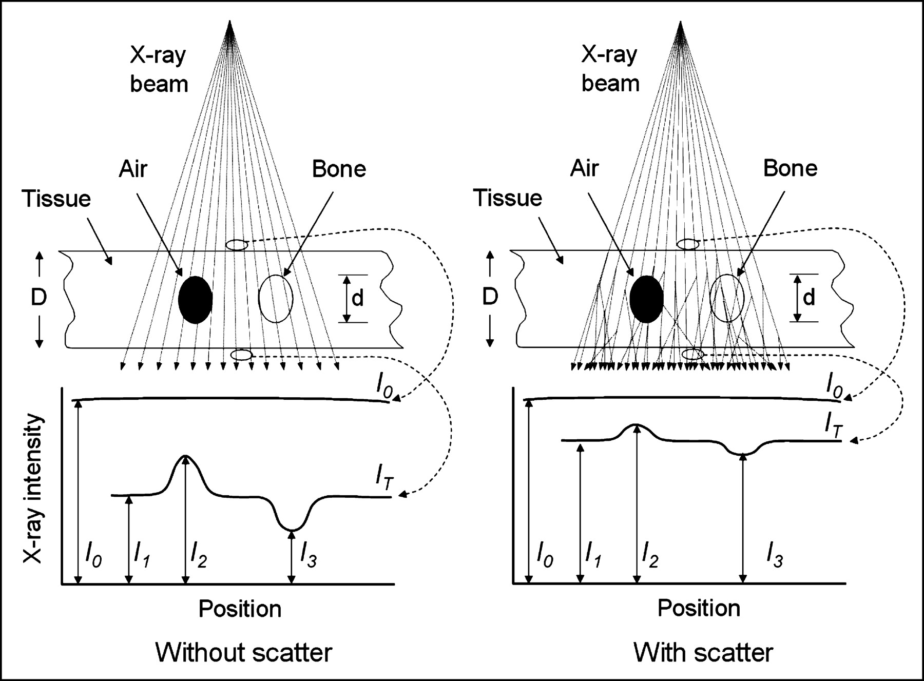
Main menu

  • Home
  • Content
    • Current
      • JNMT Supplement
    • Ahead of print
    • Past Issues
    • Continuing Education
    • JNMT Podcast
    • SNMMI Annual Meeting Abstracts
  • Subscriptions
    • Subscribers
    • Rates
    • Journal Claims
    • Institutional and Non-member
  • Authors
    • Submit to JNMT
    • Information for Authors
    • Assignment of Copyright
    • AQARA Requirements
  • Info
    • Reviewers
    • Permissions
    • Advertisers
    • Corporate & Special Sales
  • About
    • About Us
    • Editorial Board
    • Contact Information
  • More
    • Alerts
    • Feedback
    • Help
    • SNMMI Journals
  • SNMMI
    • JNMT
    • JNM
    • SNMMI Journals
    • SNMMI

User menu

  • Subscribe
  • My alerts
  • Log in
  • My Cart

Search

  • Advanced search
Journal of Nuclear Medicine Technology
  • SNMMI
    • JNMT
    • JNM
    • SNMMI Journals
    • SNMMI
  • Subscribe
  • My alerts
  • Log in
  • My Cart
Journal of Nuclear Medicine Technology

Advanced Search

  • Home
  • Content
    • Current
    • Ahead of print
    • Past Issues
    • Continuing Education
    • JNMT Podcast
    • SNMMI Annual Meeting Abstracts
  • Subscriptions
    • Subscribers
    • Rates
    • Journal Claims
    • Institutional and Non-member
  • Authors
    • Submit to JNMT
    • Information for Authors
    • Assignment of Copyright
    • AQARA Requirements
  • Info
    • Reviewers
    • Permissions
    • Advertisers
    • Corporate & Special Sales
  • About
    • About Us
    • Editorial Board
    • Contact Information
  • More
    • Alerts
    • Feedback
    • Help
    • SNMMI Journals
  • Watch or Listen to JNMT Podcast
  • Visit SNMMI on Facebook
  • Join SNMMI on LinkedIn
  • Follow SNMMI on Twitter
  • Subscribe to JNMT RSS feeds
OtherCONTINUING EDUCATION

X-Ray Imaging Physics for Nuclear Medicine Technologists. Part 2: X-Ray Interactions and Image Formation

J. Anthony Seibert and John M. Boone
Journal of Nuclear Medicine Technology March 2005, 33 (1) 3-18;
J. Anthony Seibert
  • Find this author on Google Scholar
  • Find this author on PubMed
  • Search for this author on this site
John M. Boone
  • Find this author on Google Scholar
  • Find this author on PubMed
  • Search for this author on this site

FIGURE 8.

FIGURE 8.
  • Download figure
  • Open in new tab
  • Download powerpoint
FIGURE 8.

Ideal projection radiograph is representation of transmitted primary x-ray fluence from point source through object and incident on detector, as depicted on left for a uniform incident fluence, I0, and transmitted I1, I2, and I3 fluences through tissue, air, and bone, respectively. Subject contrast is difference in signals of an object to background—for example, (I1 − I2)/I1 and (I1 − I3)/I1. On right is typical situation in presence of scatter, demonstrating loss of subject contrast and smaller difference between incident and transmitted radiation intensity.

  View article

In this issue

Journal of Nuclear Medicine Technology: 53 (1)
Journal of Nuclear Medicine Technology
Vol. 53, Issue 1
March 1, 2025
  • Table of Contents
  • About the Cover
  • Index by author
  • Complete Issue (PDF)
Print
Download PDF
Article Alerts
Sign In to Email Alerts with your Email Address
Email Article

Thank you for your interest in spreading the word on Journal of Nuclear Medicine Technology.

NOTE: We only request your email address so that the person you are recommending the page to knows that you wanted them to see it, and that it is not junk mail. We do not capture any email address.

Enter multiple addresses on separate lines or separate them with commas.
FIGURE 8.
(Your Name) has sent you a message from Journal of Nuclear Medicine Technology
(Your Name) thought you would like to see the Journal of Nuclear Medicine Technology web site.
Citation Tools
X-Ray Imaging Physics for Nuclear Medicine Technologists. Part 2: X-Ray Interactions and Image Formation
J. Anthony Seibert, John M. Boone
Journal of Nuclear Medicine Technology Mar 2005, 33 (1) 3-18;

Citation Manager Formats

  • BibTeX
  • Bookends
  • EasyBib
  • EndNote (tagged)
  • EndNote 8 (xml)
  • Medlars
  • Mendeley
  • Papers
  • RefWorks Tagged
  • Ref Manager
  • RIS
  • Zotero
Share
X-Ray Imaging Physics for Nuclear Medicine Technologists. Part 2: X-Ray Interactions and Image Formation
J. Anthony Seibert, John M. Boone
Journal of Nuclear Medicine Technology Mar 2005, 33 (1) 3-18;
Twitter logo Facebook logo LinkedIn logo Mendeley logo
  • Tweet Widget
  • Facebook Like
  • Google Plus One
Bookmark this article

Related Articles

  • No related articles found.
  • PubMed
  • Google Scholar

Cited By...

  • Tau deposition is associated with imaging patterns of tissue calcification in the P301L mouse model of human tauopathy
  • Pharmacology, Part 5: CT and MRI Contrast Media
  • Principles of CT: Radiation Dose and Image Quality
  • Principles of CT and CT Technology
  • Google Scholar

More in this TOC Section

  • Illuminating the Hidden: Standardizing Cardiac MIBG Imaging for Sympathetic Dysfunction
  • PET/CT Case Series: Unmasking the Mystery of Cardiac Sarcoidosis
  • Delivery Methods of Radiopharmaceuticals: Exploring Global Strategies to Minimize Occupational Radiation Exposure
Show more Continuing Education

Similar Articles

SNMMI

© 2025 SNMMI

Powered by HighWire