


Article Figures & Data
Figures
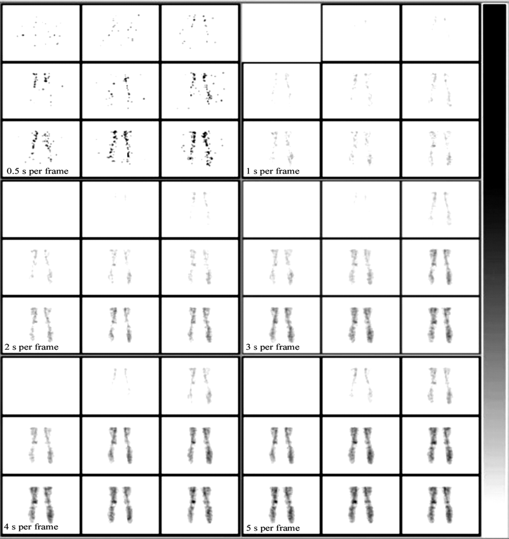
- FIGURE 1.
Effect of framing rate on a flow study. Framing rates of 2 or more seconds per frame display more anatomic structure than is seen at 0.5 s per frame, while preserving much of the sequential changes in perfusion.
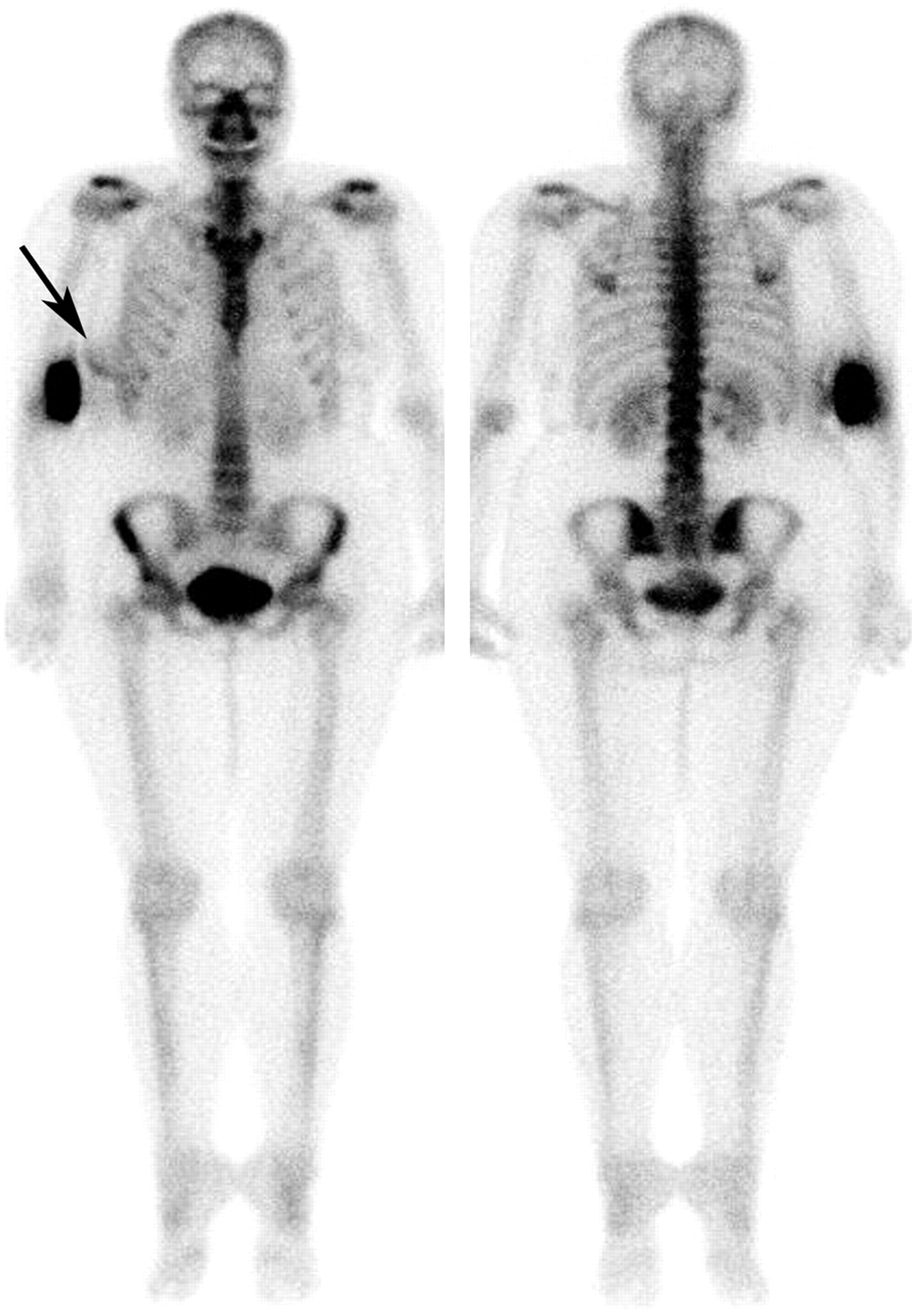
- FIGURE 2.
Compton scatter from a partially infiltrated dose (anterior view on left, posterior view on right). Because of this scatter from the site of infiltration in the right arm, there is apparent increased uptake in the adjacent right breast (arrow) that might erroneously be interpreted as a pathologic breast finding.
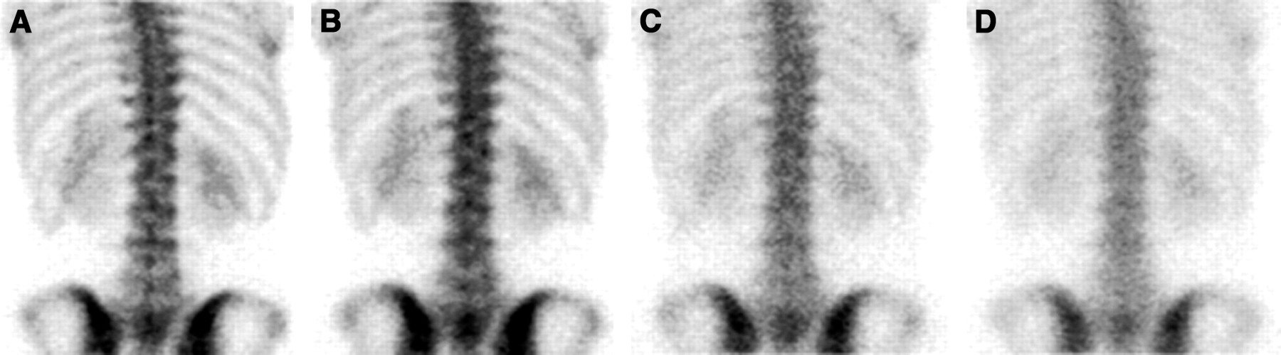
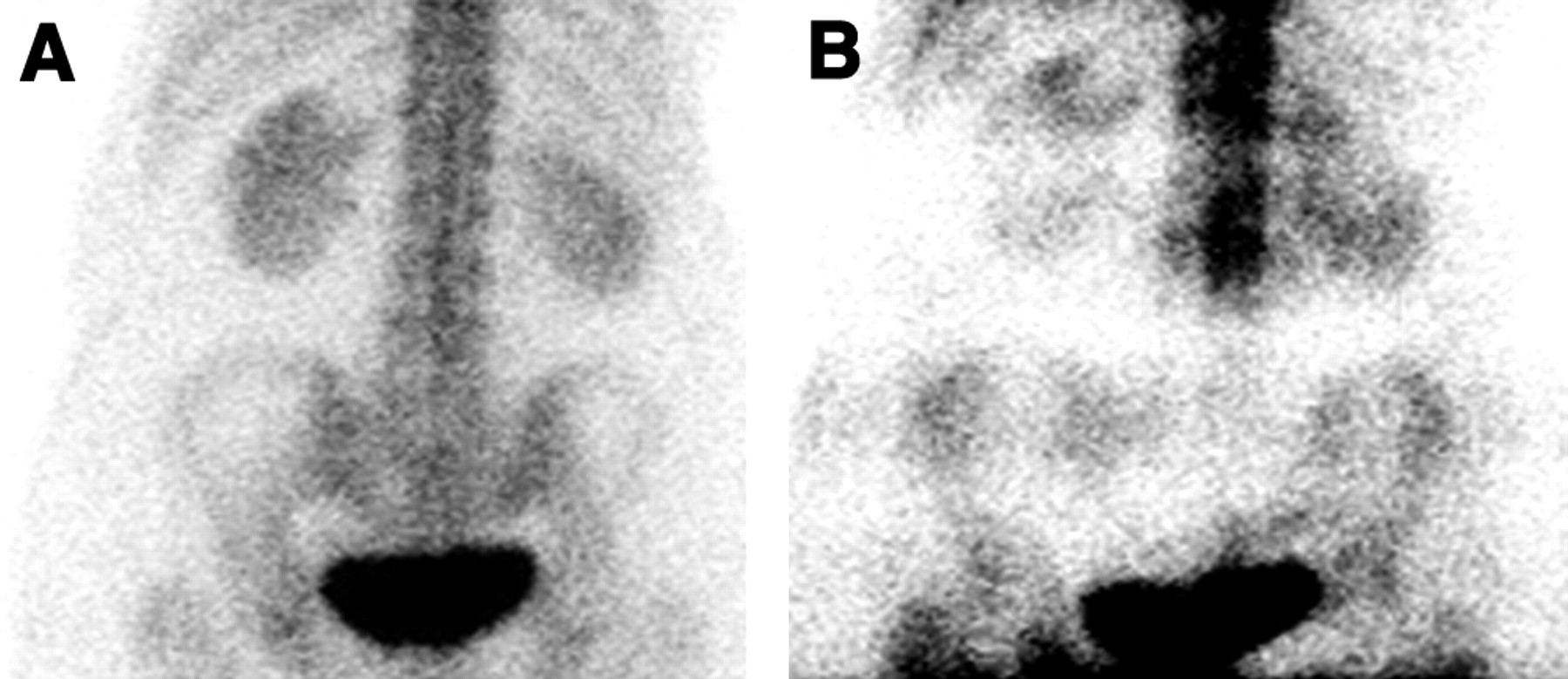
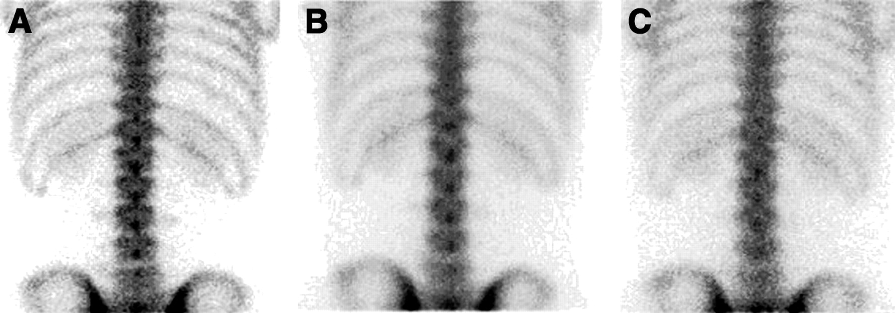
- FIGURE 3.
Effect of collimation on the resolution of planar bone scans: low-energy high-resolution collimator (A), low-energy general-purpose collimator (B), and medium-energy collimator (C).
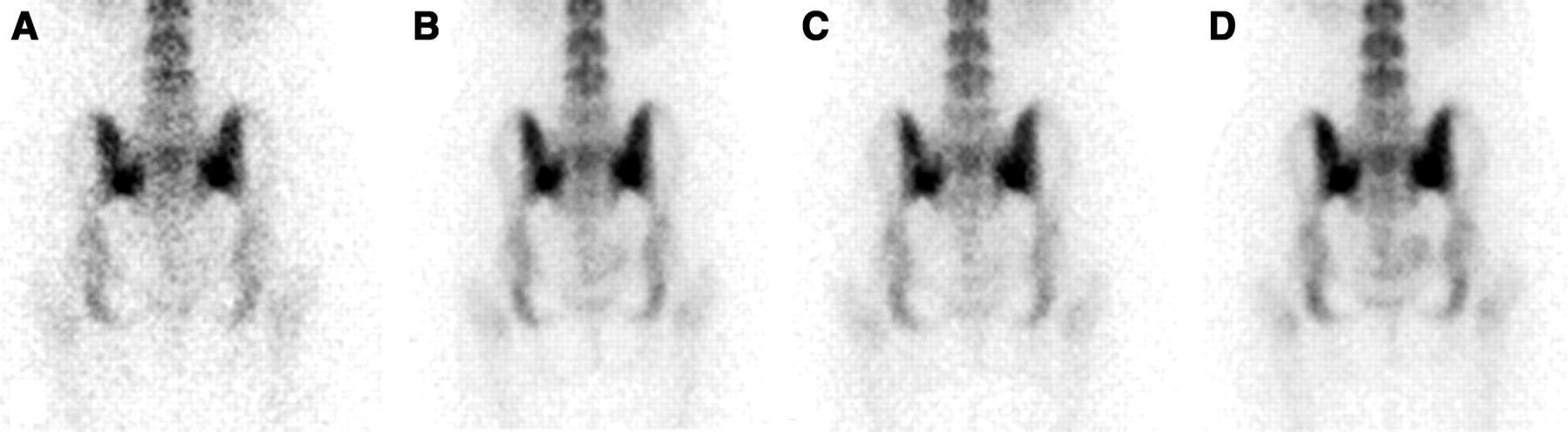
- FIGURE 4.
Effect of distance from the patient on the resolution of planar bone scans: just touching the back (A), 10 cm behind the back (B), 20 cm behind the back (C), and 30 cm behind the back (D). The anatomic detail of the lumbar spine progressively degrades as the distance between the patient and the collimator increases.
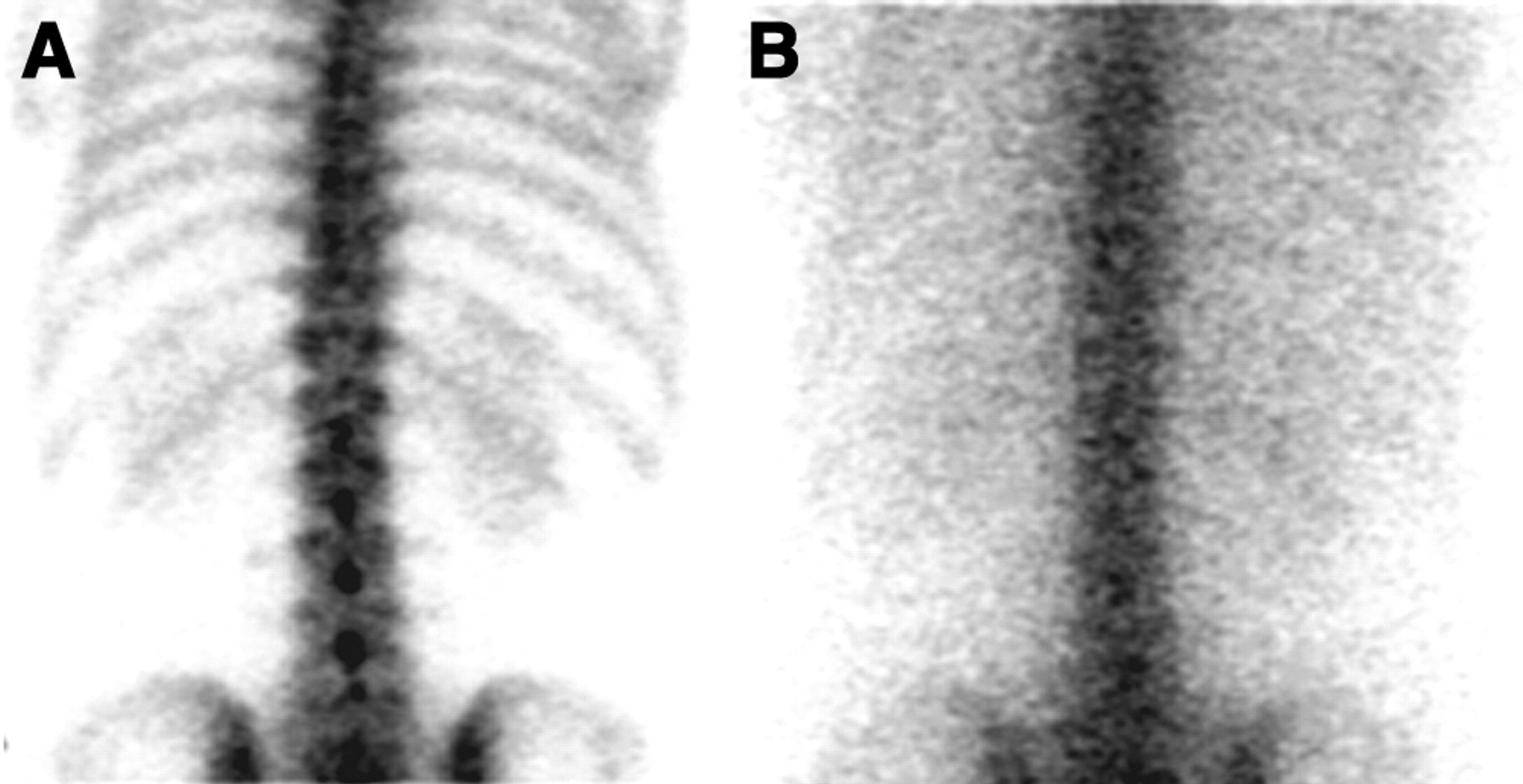
- FIGURE 5.
Effect of using a 57Co photopeak on planar bone scans: a 20% 140-keV photopeak (A) and a 20% 122-keV photopeak (B). Using a 57Co photopeak of 122 keV both increases soft-tissue scatter in the image and causes a loss of 99mTc photons. Image quality is dramatically reduced.
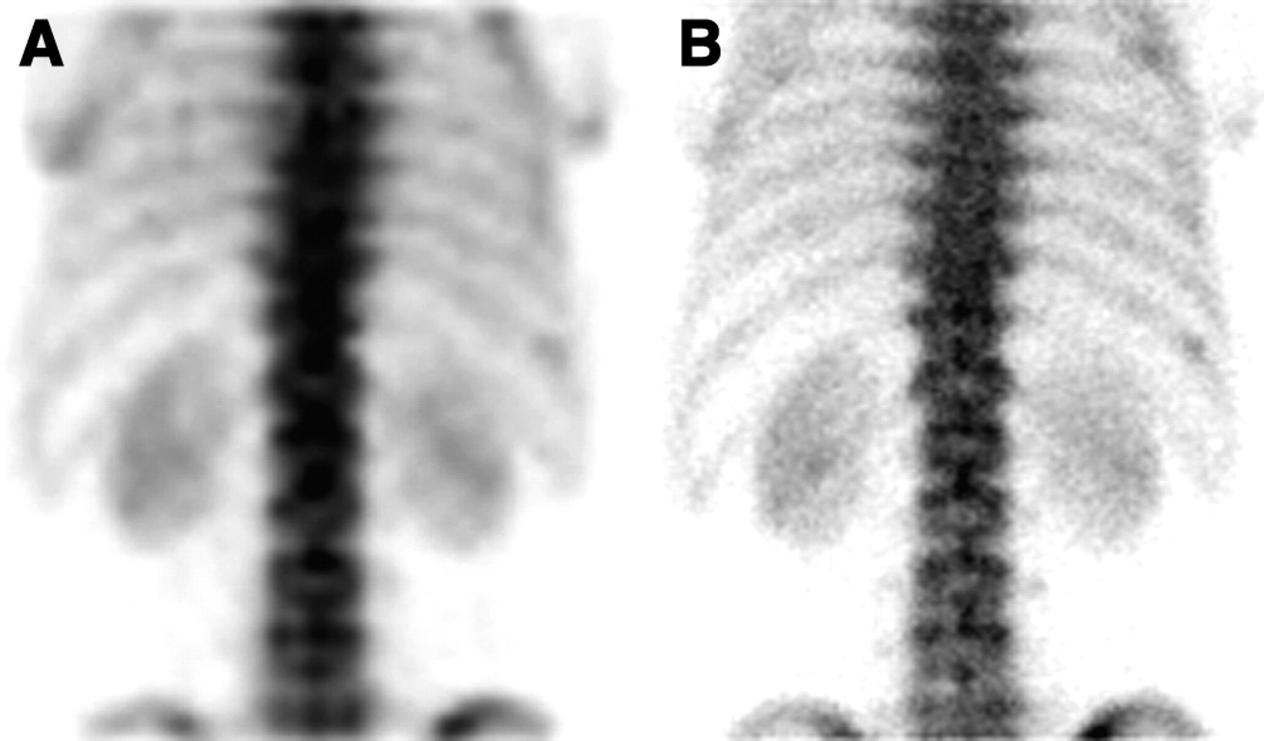
- FIGURE 6.
Effect of matrix size on planar bone scans. Posterior views of the lumbar spine obtained using a 64 × 64 (A) and a 256 × 256 (B) matrix demonstrate a loss of anatomic detail for the former.
- FIGURE 7.
Effect of count density on planar bone scans. Posterior views of the pelvis were acquired for 100,000 counts (A), 250,000 counts (B), 350,000 counts (C), and 500,000 counts (D). With increasing count density, image quality improves.
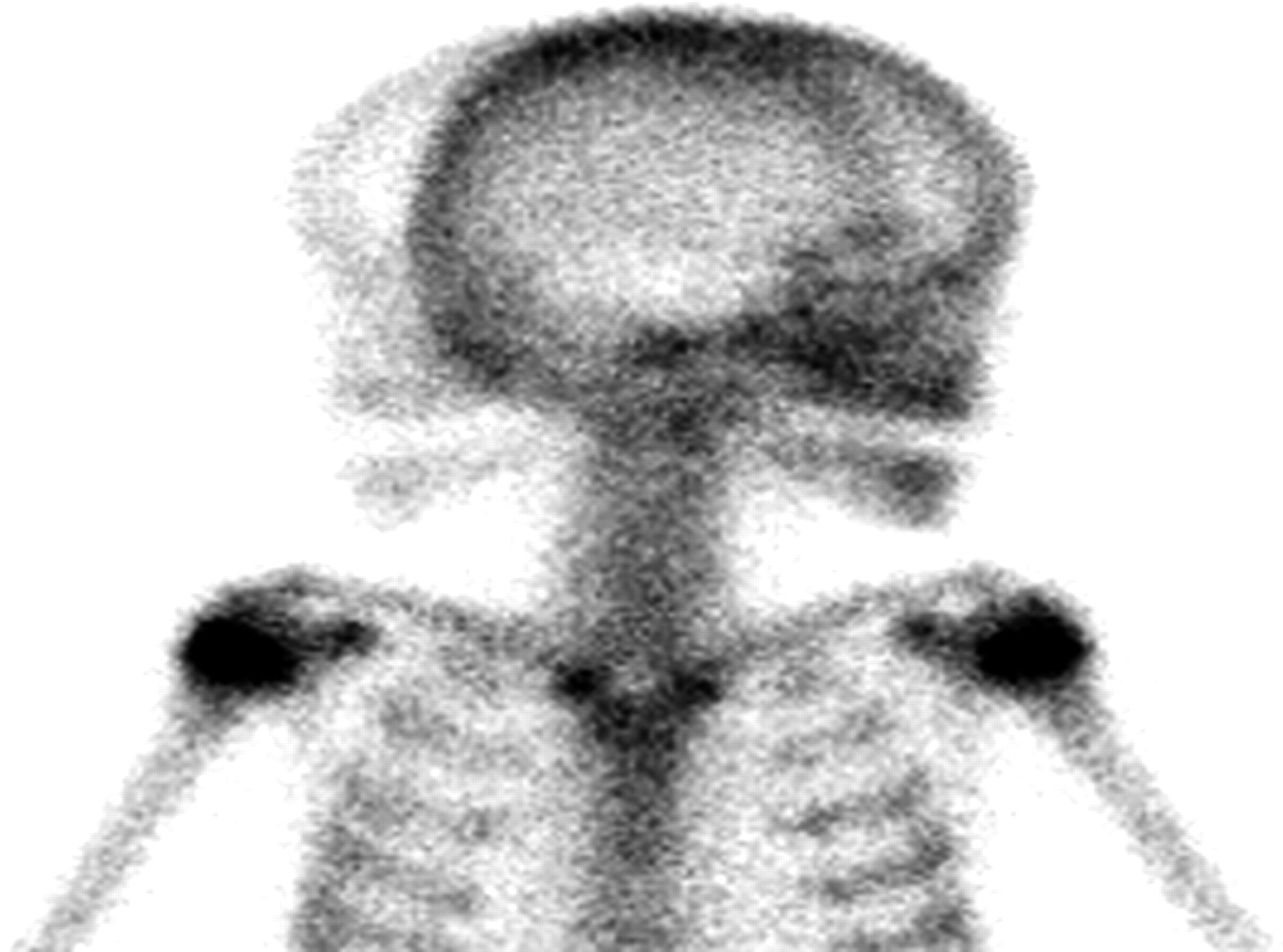
- FIGURE 8.
Movement of the patient’s head during planar bone scanning.


Tables
Intravenous dose of 99mTc-MDP or -HDP Acquisition phase Adult Child Flow Blood pool 2- to 4-h delayed* 740–1,110 MBq (20–30 mCi) 9.25 MBq/kg (250 μCi/kg) Low-energy all-purpose (or high-resolution) collimator Low-energy all-purpose (or high-resolution) collimator Low-energy high-resolution collimator 20% energy window centered at 140 keV 128 × 128 matrix 2–5 s/frame for 60 s 256 × 256 matrix 60-s images 256 × 256 matrix 150 to 1,000 kcts, depending on body part (Table 2) For limited studies on patients with localized pain, acquisition of at least 1 view to include major joints proximal and distal to the painful site ↵* Delay is greater for elderly, debilitated, or diabetic patients.
HDP = hydroxymethylene diphosphonate.
Projection Count Anterior chest, thoracic spine, lumbar spine, anterior abdomen, anterior and posterior pelvis 500,000–1,000,000 Skull (all 4 views) 400,000–500,000 Femur, humerus 500,000–600,000 Knee 350,000–450,000 Forearm, lower leg 250,000–300,000 Hand, foot 150,000–200,000


