


Abstract
Objective:
Scatter correction is an important factor in quantitative SPECT. In this study, we evaluated 2 methods of scatter correction for brain SPECT. The first is based on thresholding the energy spectrum (ES), and the second is based on a modification of the transmission-dependent convolution subtraction (TDCS) method.
Methods:
SPECT imaging of a skull striatal phantom was performed using a triple-head camera with and without scatter correction. The striatal compartments were filled with 123I, and the brain shell cavity (background) was filled with varying concentrations of 123I to obtain striatal-to-background ratios of 2, 5, 10, 15, 20, and 25 to 1, respectively, which were considered to be the expected ratios. SPECT-measured ratios of striatal-to-background counts were determined with scatter correction (both ES and TDCS methods) and without scatter correction and were then compared with the expected ratios.
Results:
Without scatter correction, measured striatal-to-background ratios were underestimated by an average of 41.7%, compared with the expected ratios. The ES method of scatter correction underestimated the striatal-to-background ratios by an average of 27.4%, a significant improvement (P < 0.04) over those without scatter correction. With the TDCS method of scatter correction, the ratios were underestimated by only 3.3% (P < 0.03). TDCS ratios were significantly (P < 0.04) higher than ES ratios and were nearly identical to the expected ratios.
Conclusion:
These results suggest that scatter correction significantly improves the striatal-to-background ratios. The TDCS method appears to correct scatter more effectively than does the ES method for the striatal phantom, thus providing more accurate quantification.
Brain SPECT imaging of dopamine, benzodiazepine, and cholinergic systems is becoming more widely used and has several potential clinical applications (1,2). Quantification of SPECT data is necessary to compare patients with age-matched healthy subjects (3,4) and to monitor disease progression or the effects of treatment (2,5,6).
To accurately perform quantitative SPECT, certain corrections are required (5,7). One of the most important is correction for scatter (6,8–10). Scatter is basically photons that have changed direction from their original trajectory but still have enough energy to be accepted into the energy window and detected as primary photons (5,11). The scatter fraction varies from subject to subject and can account for more than 30% of the total detected photons in 99mTc SPECT (5,11,12). Scatter adds low-frequency bias to the projection data, reducing contrast in the reconstructed images and therefore limiting accurate quantification (6,8,13). Scatter correction is necessary to restore the image contrast and improve the accuracy of quantitation. To evaluate the effects of scatter correction methods on SPECT, the true distribution of radioactivity in the object being imaged should be known (14). A striatal phantom was used in this study because true distribution is not known in human studies. The purpose of this study was to evaluate 2 methods of correction for scatter in quantitative brain SPECT. One method estimates the scatter based on a single threshold of the energy spectrum (ES). The second method is based on transmission-dependent convolution subtraction (TDCS), in which scatter is estimated from the transmission projections.
MATERIALS AND METHODS
Striatal Phantom
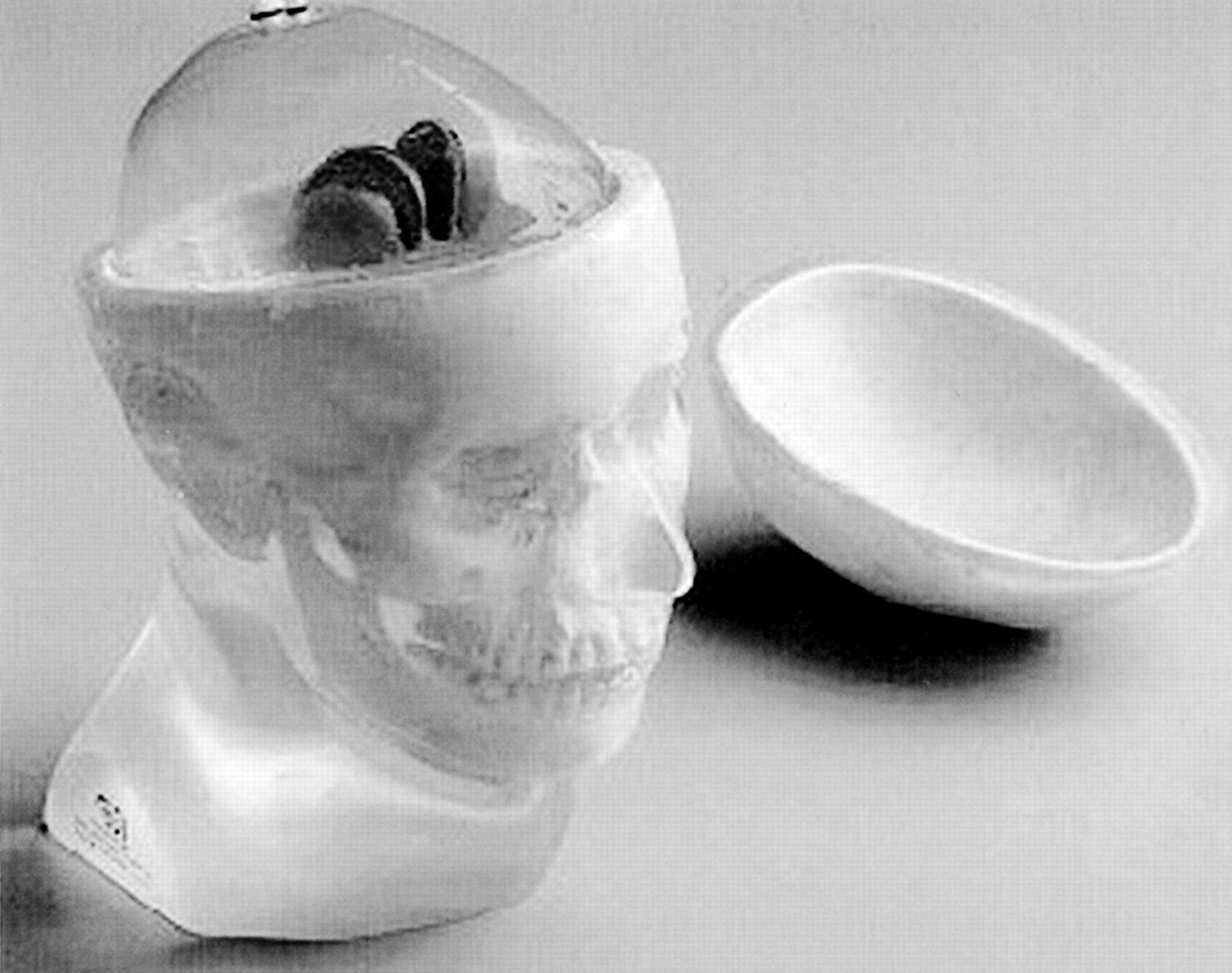
The striatal phantom (RS-901T; Radiology Support Devices Inc.; Fig. 1) imaged in this study is both a tissue-equivalent anthropomorphic phantom and an anatomically accurate model of the human striatum (12,15). The phantom can simulate nonuniform brain uptake, as in neurotransmission studies, if the caudate, putamen, and brain shell compartments are filled with differing concentrations of radioactivity. In this experiment, the caudate and putamen (striatal compartments) were filled with 134–187 KBq/mL (3.6–5.1 μCi/mL) of a solution of 123I, the most commonly used radionuclide in SPECT tracers for neurotransmission studies. The brain shell cavity (background) was filled with water initially, and then varying amounts of 123I solution were added to obtain striatal-to-background concentration ratios of 2, 5, 10, 15, 20, and 25 to 1. These were considered to be the expected ratios.

Photograph of RS-901T striatal phantom. (Courtesy of Radiology Support Devices Inc.)
SPECT Imaging
SPECT emission scans were obtained using a triple-head camera (Triad XLT-20; Trionix Research Laboratory Inc.) equipped with low-energy high-resolution parallel-hole collimators. The data were acquired using step-and-shoot motion of 3° intervals on a 128 × 128 matrix (pixel size, 4.48 mm) for 360° (each head rotating 120°), at a fixed radius of 15.0 cm for 15 min. All emission scans were reconstructed using a 360° ordered-subset expectation maximization algorithm with 5 iterations and 5 subsets. No attenuation correction was performed.
Transmission data were acquired separately (with the phantom still filled with radioactivity) by using a single scanning line source of 153Gd opposite to head 1 for 180°. All other parameters were the same as those used for emission scans.
Scatter Correction Methods
Two scatter correction methods, ES and TDCS, were applied. The ES method was provided by the camera manufacturer. The camera has the ability to acquire counts into an ES for each pixel. Scatter is corrected by selecting a threshold (cutoff) value below the photopeak and, for each pixel, subtracting counts that were lower than this threshold (11). The parameters used for ES were the defaults recommended by the manufacturer.
The TDCS method was a modification of the method of Iida et al. (8), which estimates scatter from the transmission projections based on a convolution model. The estimated scatter is then subtracted from the emission projections.
Data Analysis
An elliptic region of interest (ROI) was used as a master ROI and placed on both striata on 5 consecutive slices of the reconstructed images. Within this master ROI, a threshold was applied to obtain striatal counts from only pixels in the top 2%. This was done to minimize partial-volume effects (6,16). A background ROI was then drawn and placed on 2 consecutive slices. The background counts were calculated from all pixels within this ROI. An example of the striatal and background ROIs is illustrated in Figure 2. Ratios of striatal counts to background counts were determined from images with and without scatter correction and compared with the expected ratios. Paired t tests were performed to compare the ratios with and without the 2 scatter correction methods.

Transaxial images of striatal phantom with 10:1 striatal-to-background ratio: with no scatter correction (A), with ES scatter correction (B), and with TDCS scatter correction (C). Striatal ROI indicates only pixels in top 2% of striatum and may vary in location for different methods. Background (BKG) ROIs are the same for all methods.
RESULTS
The striatal and background counts and ratios are listed in Table 1. Without scatter correction, measured striatal-to-background ratios were underestimated by an average of 41.7% when compared with the expected ratios. With the ES method of scatter correction, the ratios were underestimated by 27.4%, a significant improvement (P < 0.04) over those without scatter correction. With TDCS, ratios were underestimated by only 3.3%, yielding a significant (P < 0.03) improvement over no scatter correction. TDCS ratios were also significantly (P < 0.04) higher than ES ratios and were nearly identical to the expected ratios (Fig. 3).
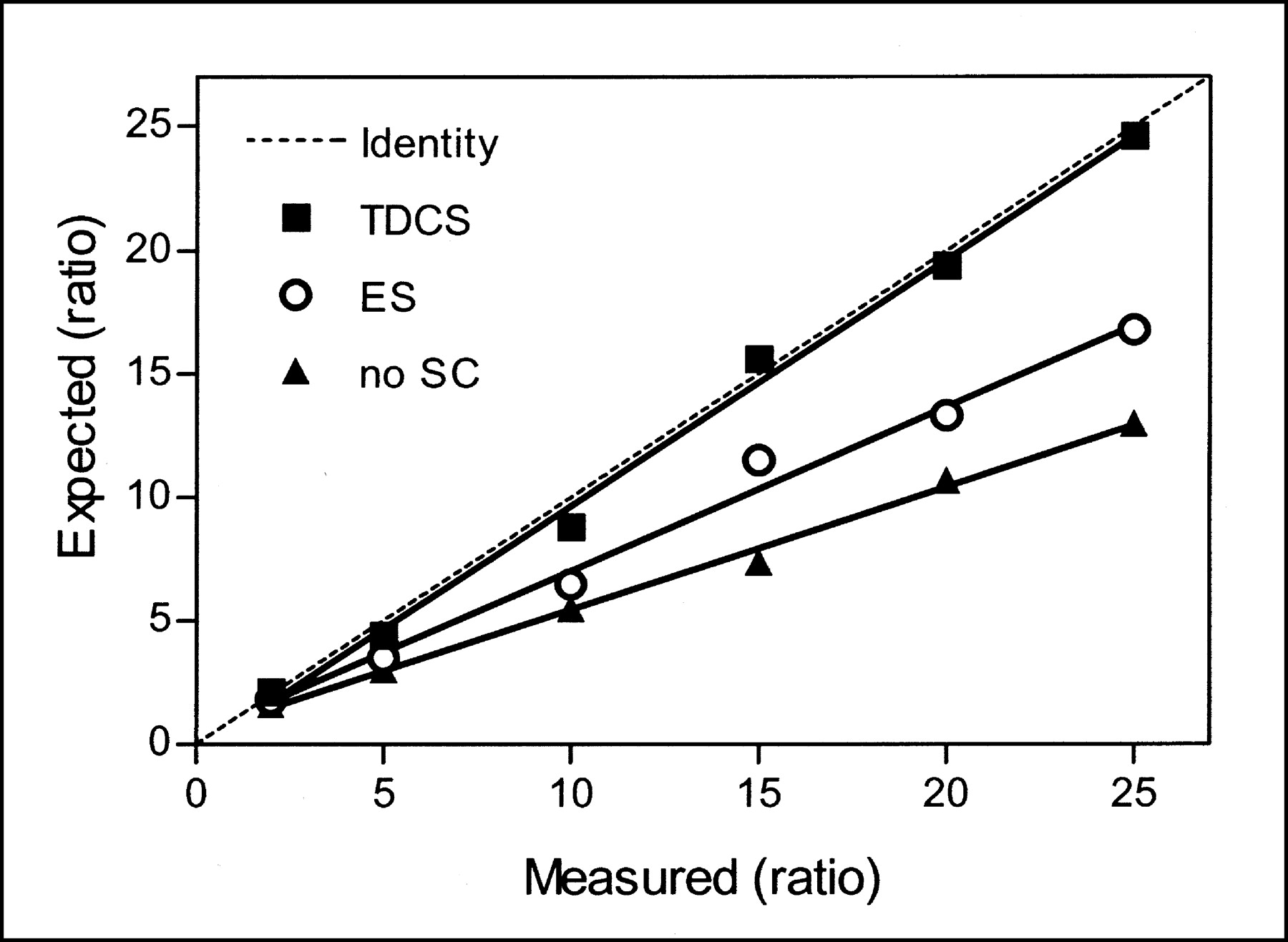
Relationship between expected and measured striatal-to-background ratios. Each series was fitted with linear regression line. SC = scatter correction.
DISCUSSION
Quantitative analysis of brain SPECT is important so that comparisons can be made within and between subjects (2,3,6). Scatter correction is one of the most important factors for contrast measurements (6,8–10). In this study, we have shown that compared with no scatter correction, both scatter correction methods improve the quantification of brain SPECT images (Fig. 3). The TDCS method appears to correct scatter more effectively than does ES.
These 2 scatter correction methods reduce the scatter in the images; thus, both the striatal and the background ROIs have fewer total counts with scatter correction than without, and the ratios are thus increased (Table 1). The superiority of TDCS over ES is due to the disproportionate subtraction of scatter from the striatum and background by the TDCS algorithm compared with the ES method. For example, in Table 1, the TDCS algorithm has subtracted less scatter in the striatum but more in the background than has the ES method, thus yielding higher total counts in the striatum and lower counts in the background for TDCS than for ES. Therefore, contrast is increased with TDCS as compared with ES.
With the ES method, we used the default parameters recommended by the manufacturer. However, changing these parameters would likely change the performance of the ES method and would need to be evaluated in a further study.
Several factors other than scatter correction also affect quantification, including attenuation correction and partial-volume correction. We attempted to eliminate these potentially confounding factors in evaluating scatter correction by not using them. However, to evaluate the effects of attenuation correction only, we determined striatal-to-background ratios after performing measured attenuation correction (using the transmission data) without scatter correction. Compared with ratios without either scatter or attenuation correction (41.7%), ratios with only attenuation correction were further underestimated (44.9%). Compared with ES (27.4%) and TDCS (3.3%), ratios with only attenuation correction were more markedly underestimated. This finding confirms, as reported by others (6,8–10), that scatter correction is perhaps the most important factor in contrast measurements.
To minimize partial-volume effects within the striatal ROIs, we used only pixels in the top 2% of the master ROI. To evaluate scatter correction with partial-volume effects, we also analyzed data using all pixels within the master ROI. With partial-volume effects included and no scatter correction, the striatal-to-background ratios were further underestimated, from 41.7% to 49.1%, as compared with the expected ratios. For ES and TDCS, ratio underestimation changed from 27.4% to 38.7% and from 3.3% to 19.5%, respectively. However, improvements in quantitation similar to those for the top 2% data were observed as a result of scatter correction, with TDCS again appearing more effective than ES.
In this study, we used the striatal phantom as a gold standard because it allowed expected activity ratios to be measured directly and the effects of scatter correction methods to be monitored with varying ratios. However, scatter correction is dependent on the various structures in the human brain, and the phantom may not represent the true scatter distribution in the human brain. This limitation is, however, unavoidable, as the true distribution cannot be measured in human studies.
CONCLUSION
The TDCS method appears to correct scatter more effectively than does the pixel-wise ES thresholding method for the striatal phantom using the default parameter values. However, both scatter correction methods significantly improved the measured striatal-to-background ratios, thus providing a more accurate quantitative estimation of the radioactivity in the striatal phantom.
Acknowledgments
This work was presented in part at the 49th annual meeting of the Society of Nuclear Medicine, Los Angeles, CA, June 15–19, 2002, and was awarded second place for a technologist article on brain imaging by the Brain Imaging Council. We thank Roberto Maass-Moreno, PhD (Nuclear Medicine Department, National Institutes of Health), for his helpful suggestions and participation in the data acquisition.
Footnotes
For correspondence or reprints contact: Douglass C. Vines, BSc, CNMT, Molecular Imaging Branch, NIMH, NIH, Building 1, Room B3-10, 1 Center Dr., Bethesda, MD 20892-0135.
E-mail: douglass.vines{at}nih.gov
REFERENCES


Jump to section
Related Articles
Cited By...
- No citing articles found.