


Article Figures & Data
Figures
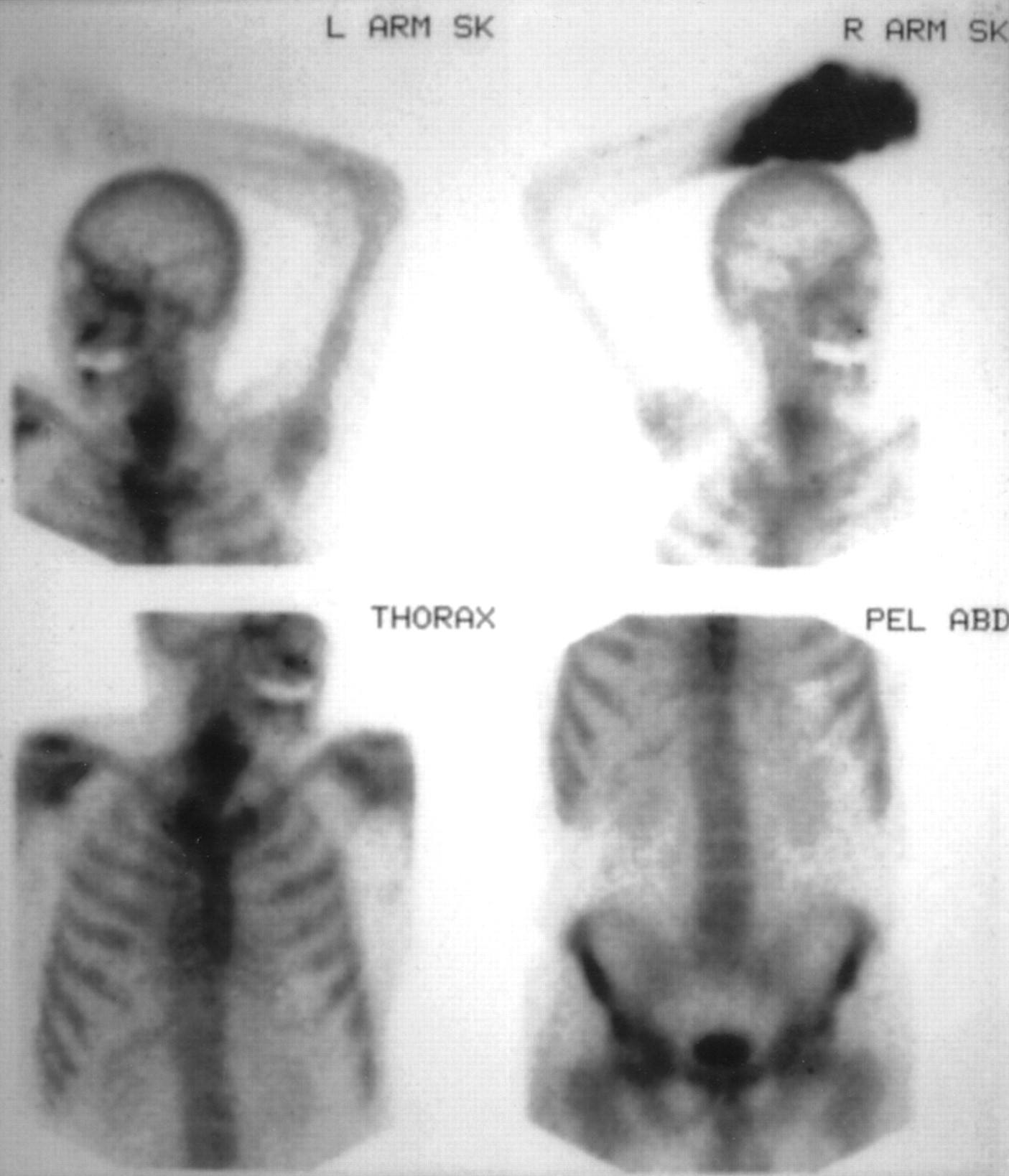
- FIGURE 1.
Injection of 99mTc-MDP into radial artery of right hand produces dramatic soft-tissue uptake in arterial distribution along lateral side of hand and wrist (anterior view). SK = skull; PEL ABD = pelvis and abdomen.
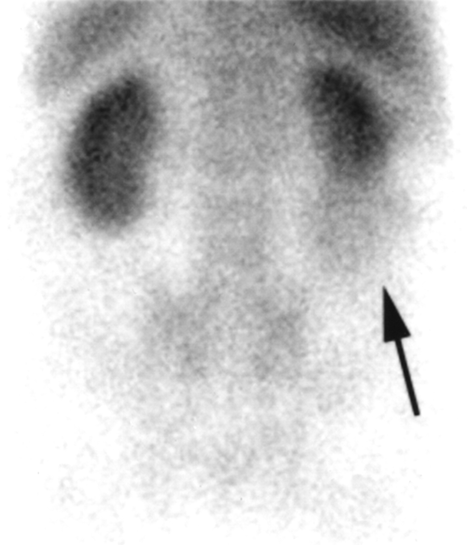
- FIGURE 2.
Blood-pool image of posterior lower back shows blunting of lower pole of right kidney and uptake in soft-tissue mass (arrow), which proved to be renal cell carcinoma.
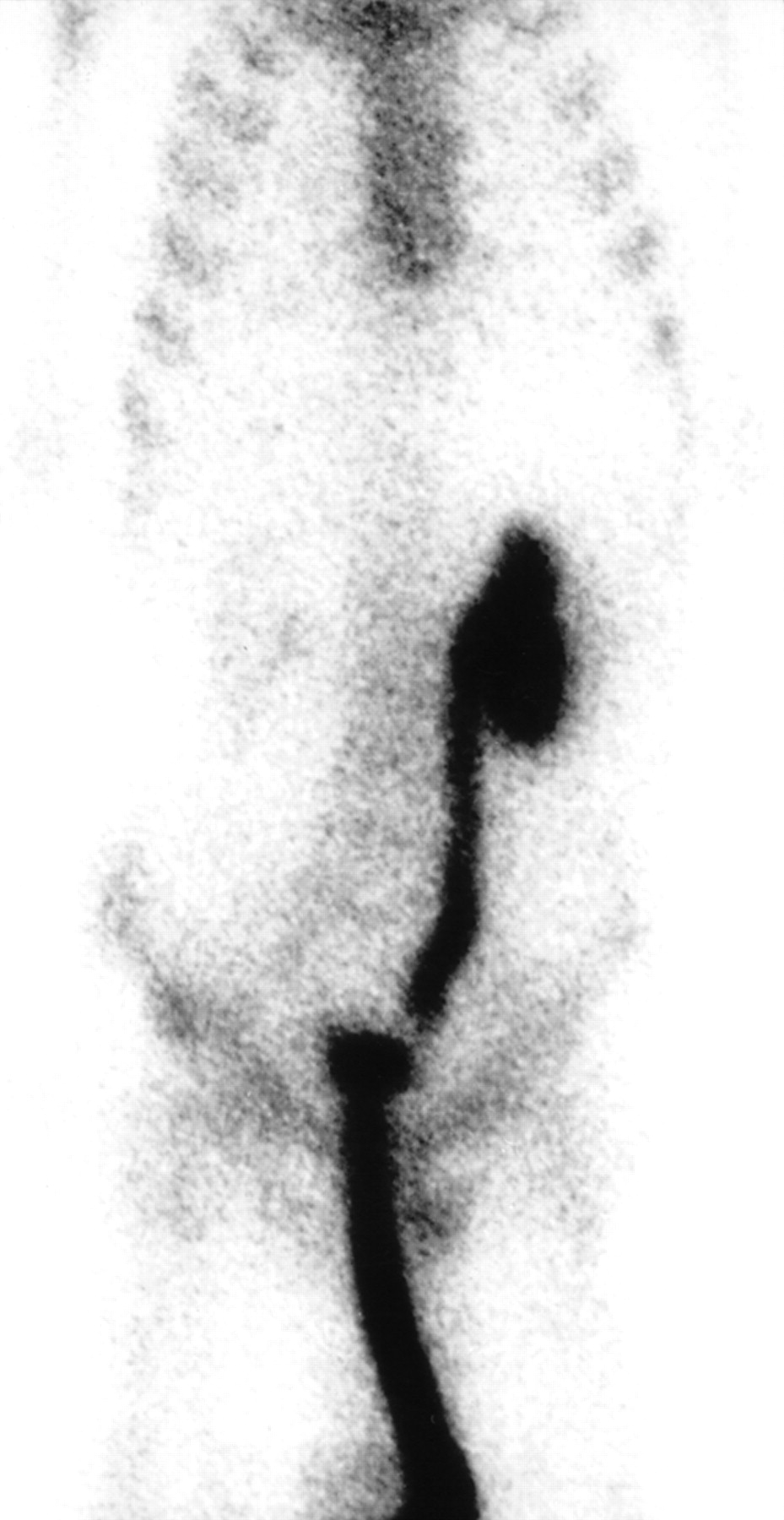
- FIGURE 3.
Grossly dilated left ureter and renal collecting system seen on anterior whole-body 99mTc-MDP bone scan of patient with prostate cancer. Indwelling Foley catheter was in place at time of scanning.
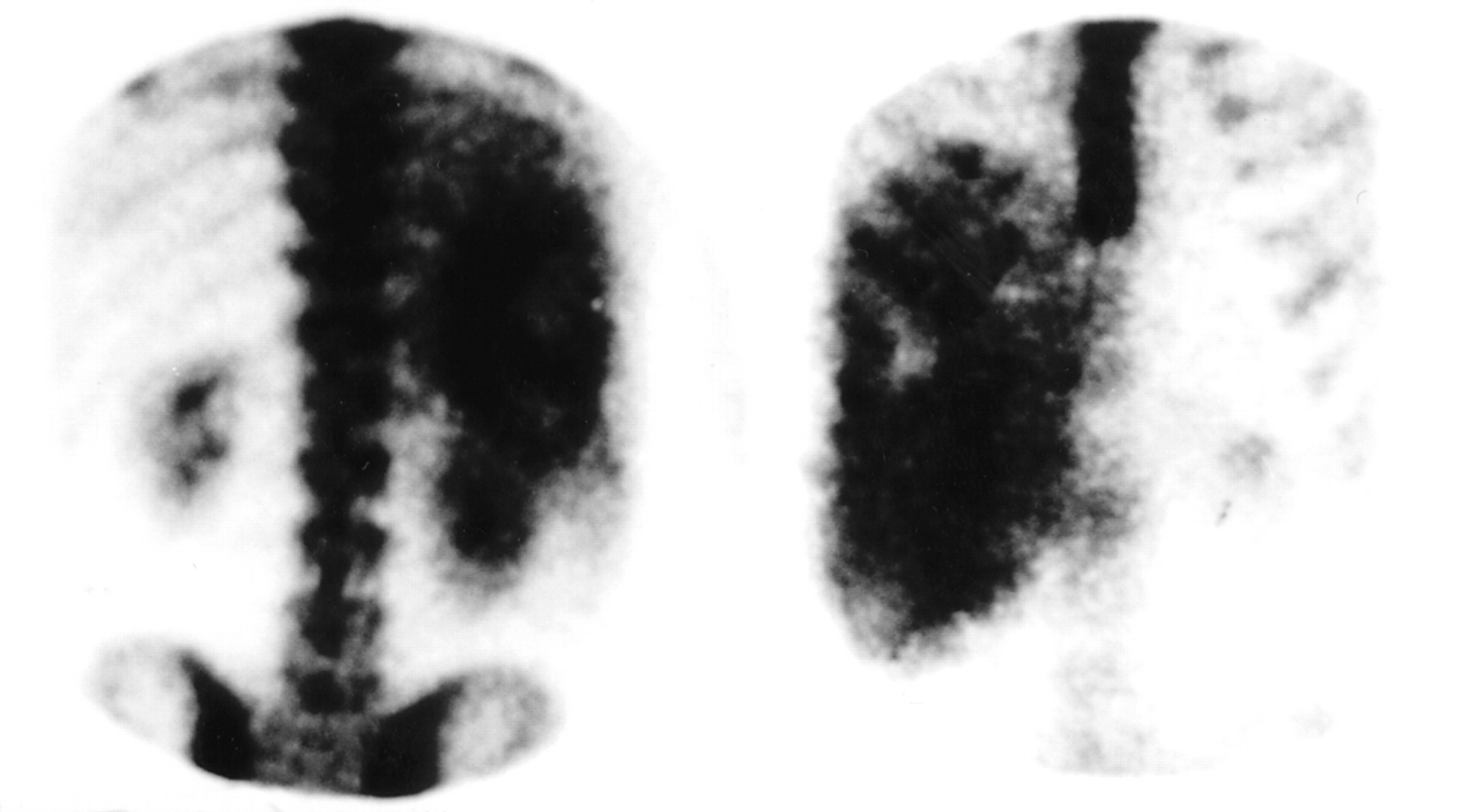
- FIGURE 4.
Anterior (right) and posterior (left) abdominal views of 99mTc-MDP bone scan of patient with hepatic metastasis from colon carcinoma show intense uptake of radioactivity in liver, which is heavily involved with tumor.
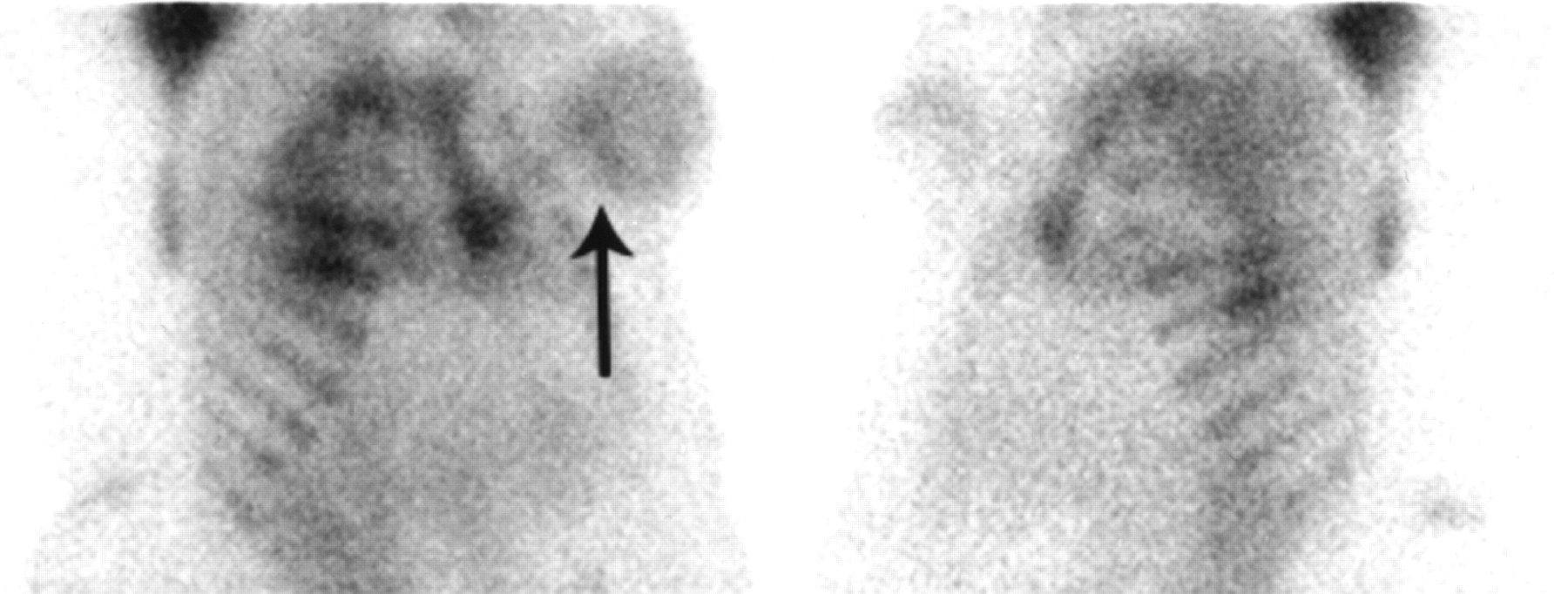
- FIGURE 5.
Left anterior oblique (right) and right anterior oblique (left) views of chest from 99mTc-MDP bone scan of patient with left-breast cancer show soft-tissue uptake in both breasts. Uptake in left breast is more extensive (arrow) and corresponds to tumor mass on that side.
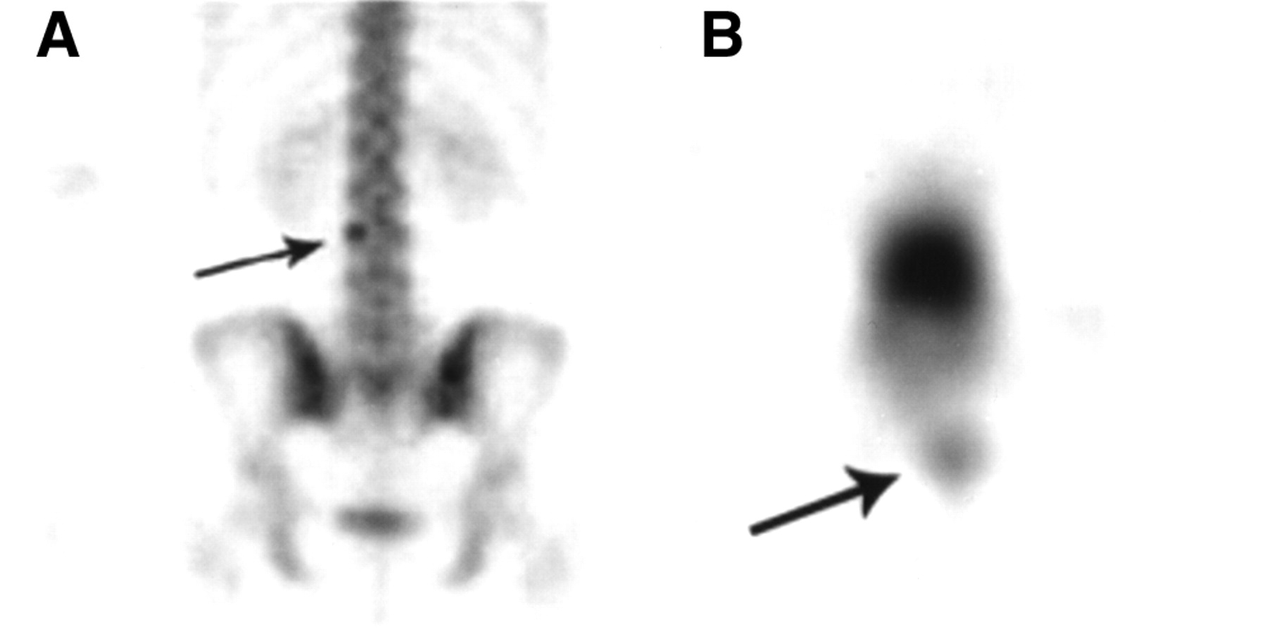
- FIGURE 6.
(A) Planar posterior image of lower back from 99mTc-MDP bone scan of patient with chronic back pain. Focus of increased uptake is seen in region of left pedicle of 3rd lumbar vertebra (arrow). (B) SPECT image shows this uptake to be more superficial in overlying soft tissue (arrow). Patient had received injections of antiinflammatory drugs at this site a few weeks before scan.


Tables
Fault Effect on bone scan Free pertechnetate due to presence of air in container, a long-standing preparation, an inappropriate amount of stannous ion, or altered preparation Thyroid uptake on early images (blood pool) and stomach, gastrointestinal tract, and salivary gland uptake Colloid formation due to aluminum Diffuse liver uptake and reduced bone uptake High pH in the preparation Liver, gallbladder, and gastrointestinal tract uptake Drug interaction: Diphosphonates, etidronate Decreased bone uptake Iron Increased soft-tissue uptake; renal cortex uptake Chemotherapy Renal cortex uptake and diffuse skull uptake Organ or tissue Pathologic condition Breast Breast cancer Pleural space Malignant effusion Liver Calcified or necrotic metastases Heart Amyloidosis or infarction Spleen Sickle cell disease Brain Infarction Muscle Heterotopic ossification or myositis Stomach Hypercalcemia, metastatic calcification Lung Hypercalcemia, metastatic calcification Renal cortex Hypercalcemia Joints Osteochondromatosis Pericardium Pericarditis or malignant effusion Soft-tissue tumor Osteogenic sarcoma or malignancy


