
Main menu

  • Home
  • Content
    • Current
      • JNMT Supplement
    • Ahead of print
    • Past Issues
    • Continuing Education
    • JNMT Podcast
    • SNMMI Annual Meeting Abstracts
  • Subscriptions
    • Subscribers
    • Rates
    • Journal Claims
    • Institutional and Non-member
  • Authors
    • Submit to JNMT
    • Information for Authors
    • Assignment of Copyright
    • AQARA Requirements
  • Info
    • Reviewers
    • Permissions
    • Advertisers
    • Corporate & Special Sales
  • About
    • About Us
    • Editorial Board
    • Contact Information
  • More
    • Alerts
    • Feedback
    • Help
    • SNMMI Journals
  • SNMMI
    • JNMT
    • JNM
    • SNMMI Journals
    • SNMMI

User menu

  • Subscribe
  • My alerts
  • Log in
  • Log out
  • My Cart

Search

  • Advanced search
Journal of Nuclear Medicine Technology
  • SNMMI
    • JNMT
    • JNM
    • SNMMI Journals
    • SNMMI
  • Subscribe
  • My alerts
  • Log in
  • Log out
  • My Cart
Journal of Nuclear Medicine Technology

Advanced Search

  • Home
  • Content
    • Current
    • Ahead of print
    • Past Issues
    • Continuing Education
    • JNMT Podcast
    • SNMMI Annual Meeting Abstracts
  • Subscriptions
    • Subscribers
    • Rates
    • Journal Claims
    • Institutional and Non-member
  • Authors
    • Submit to JNMT
    • Information for Authors
    • Assignment of Copyright
    • AQARA Requirements
  • Info
    • Reviewers
    • Permissions
    • Advertisers
    • Corporate & Special Sales
  • About
    • About Us
    • Editorial Board
    • Contact Information
  • More
    • Alerts
    • Feedback
    • Help
    • SNMMI Journals
  • Watch or Listen to JNMT Podcast
  • Visit SNMMI on Facebook
  • Join SNMMI on LinkedIn
  • Follow SNMMI on Twitter
  • Subscribe to JNMT RSS feeds
Review ArticleTeaching Case Studies

Liver Uptake on Bone Scanning: A Diagnostic Algorithm

Sayf Al-katib, Zaid Al-faham and Helena Balon
Journal of Nuclear Medicine Technology June 2015, 43 (2) 135-136; DOI: https://doi.org/10.2967/jnmt.114.146449
Sayf Al-katib
Beaumont Health System, Royal Oak, Michigan
  • Find this author on Google Scholar
  • Find this author on PubMed
  • Search for this author on this site
Zaid Al-faham
Beaumont Health System, Royal Oak, Michigan
  • Find this author on Google Scholar
  • Find this author on PubMed
  • Search for this author on this site
Helena Balon
Beaumont Health System, Royal Oak, Michigan
  • Find this author on Google Scholar
  • Find this author on PubMed
  • Search for this author on this site
  • Article
  • Figures & Data
  • Info & Metrics
  • PDF
Loading

Abstract

In this report, we present a case of liver uptake seen on a bone scan that was due to diffuse metastatic disease from breast carcinoma. We discuss possible etiologies for the uptake and offer an algorithm to narrow the differential diagnosis.

  • bone scan
  • liver metastasis
  • breast carcinoma

Hepatic uptake of 99mTc-methylene diphosphonate is an unexpected finding and should trigger a set of questions by the interpreting physician, as the distinction between technical and pathologic causes is critical to direct further evaluation of the patient.

CASE REPORT

A 54-y-old woman had a past medical history of right breast cancer and had undergone mastectomy with breast reconstruction and chemotherapy 2 y before presentation. She developed a fever and was admitted to the hospital for further evaluation. Laboratory studies were remarkable for elevated levels of alkaline phosphatase (413 U/L), aspartate aminotransferase (300 U/L), and alanine aminotransferase (272 U/L).

As part of the work-up, a 99mTc-methylene diphosphonate bone scan was obtained to evaluate for osseous metastasis. Three hours after intravenous administration of 814 MBq (22 mCi) of 99mTc-methylene diphosphonate, anterior and posterior whole-body and spot views were obtained. Whole-body images showed abnormal ill-defined radiotracer uptake in the right upper quadrant of the abdomen, suggesting liver uptake (Figs. 1 and 2). A subtle photopenic defect was present over the right upper anterior thorax, corresponding to the patient’s known breast implant.

FIGURE 1.
  • Download figure
  • Open in new tab
  • Download powerpoint
FIGURE 1.

Anterior (left) and posterior (right) whole-body images from 99mTc-methylene diphosphonate bone scan show heterogeneous uptake in right upper quadrant of abdomen with morphology suggestive of liver.

FIGURE 2.
  • Download figure
  • Open in new tab
  • Download powerpoint
FIGURE 2.

Spot anterior view of thorax and upper abdomen from 99mTc-methylene diphosphonate bone scan shows heterogeneous liver uptake in better detail. Photopenic defect overlying right anterior chest wall corresponds to breast prosthesis.

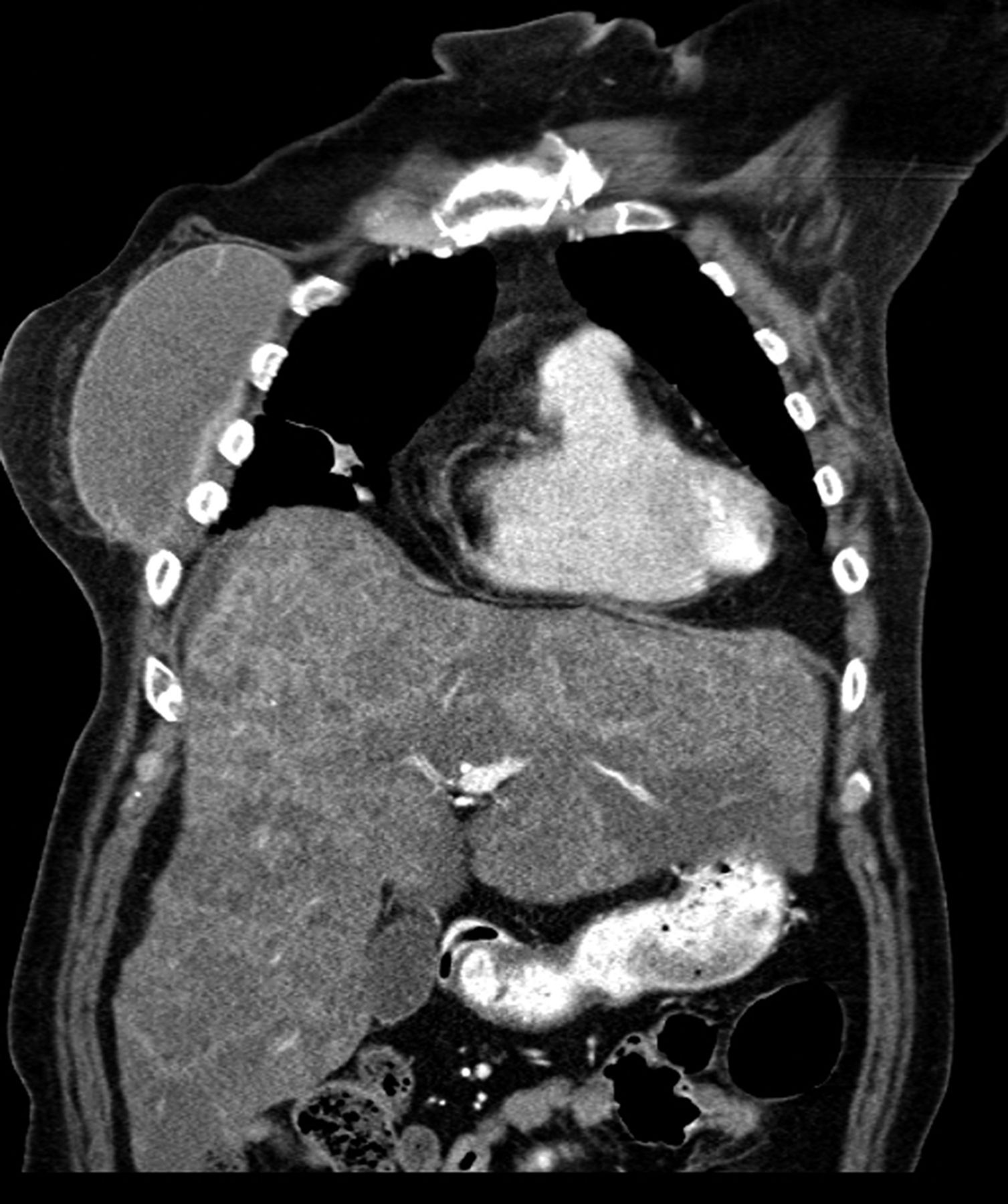
Given the patient’s history of breast cancer and the abnormal results from the liver function studies, concern about metastatic disease to the liver was raised. She underwent further evaluation with contrast-enhanced CT of the chest, abdomen, and pelvis, which confirmed hepatomegaly with extensive metastases (Fig. 3). The patient began to experience mental status changes secondary to hepatic encephalopathy and, less than 2 wk after the bone scan, died.

FIGURE 3.
  • Download figure
  • Open in new tab
  • Download powerpoint
FIGURE 3.

Contrast-enhanced coronal CT scan shows extensive hepatic metastases with hepatomegaly. Right breast prosthesis is also present.

DISCUSSION

In view of the abnormal liver uptake on the bone scan, the need for a regimented interpretation algorithm in this patient was strongly suggested, especially since a detailed history was not available. We recommend the following approach when one is faced with such a case. First, determine whether the patient has undergone a recent nuclear medicine study that would interfere with the bone scan. Examples include a 99mTc-sulfur colloid or 99mTc-labeled white blood cell scan within the last day or an 111In-labeled white blood cell or 67Ga scan within the last week. Additional uptake within the spleen would point toward recent 99mTc-sulfur colloid or 111In-labeled white blood cell injection, whereas activity in the colon would support 67Ga administration. The next step is to determine whether the dose of 99mTc-methylene diphosphonate was degraded by excess alumina chemical impurity, resulting in colloid formation with subsequent liver uptake. The maximum permissible amount of aluminum ion is 10 μg/mL of 99mTc eluate. Diffuse liver uptake with reduced bone uptake supports this conclusion (1). Uptake may also be present in the spleen and bone marrow (2). Finally, one should consider whether the patient has primary or metastatic liver disease. Heterogeneous uptake in the liver is more suggestive of primary and metastatic disease, and the liver may also be enlarged.

CONCLUSION

When faced with unexpected hepatic uptake on a bone scan, the interpreting physician must first exclude technical factors before recommending a work-up for liver disorders. In particular, the presence of alumina and recent administration of various radiopharmaceuticals can mimic liver disease. The interpreting physician should consider all available clues, particularly when a sufficient history is not available. These clues include a high index of suspicion for signs of metastatic disease, a search for additional uptake in the spleen and gastrointestinal tract to suggest prior administration of various radiopharmaceuticals, and scrutiny of the distribution of liver uptake.

DISCLOSURE

No potential conflict of interest relevant to this article was reported.

Footnotes

  • Published online Dec. 23, 2014.

REFERENCES

  1. 1. ↵
    1. Loutfi I ,
    2. Collier D ,
    3. Mohammed AM
    . Nonosseous abnormalities on bone scans. J Nucl Med Technol. 2003;31:149–153.
    OpenUrlAbstract/FREE Full Text
  2. 2. ↵
    1. MacDonald J
    . Idiopathic hepatic uptake of 99mTc methylene diphosphonate: a case report. J Nucl Med Technol. 2001;29:32–36.
    OpenUrlAbstract/FREE Full Text
  • Received for publication July 30, 2014.
  • Accepted for publication October 15, 2014.
View Abstract
PreviousNext
Back to top

In this issue

Journal of Nuclear Medicine Technology: 43 (2)
Journal of Nuclear Medicine Technology
Vol. 43, Issue 2
June 1, 2015
  • Table of Contents
  • About the Cover
  • Index by author
Print
Download PDF
Article Alerts
Sign In to Email Alerts with your Email Address
Email Article

Thank you for your interest in spreading the word on Journal of Nuclear Medicine Technology.

NOTE: We only request your email address so that the person you are recommending the page to knows that you wanted them to see it, and that it is not junk mail. We do not capture any email address.

Enter multiple addresses on separate lines or separate them with commas.
Liver Uptake on Bone Scanning: A Diagnostic Algorithm
(Your Name) has sent you a message from Journal of Nuclear Medicine Technology
(Your Name) thought you would like to see the Journal of Nuclear Medicine Technology web site.
Citation Tools
Liver Uptake on Bone Scanning: A Diagnostic Algorithm
Sayf Al-katib, Zaid Al-faham, Helena Balon
Journal of Nuclear Medicine Technology Jun 2015, 43 (2) 135-136; DOI: 10.2967/jnmt.114.146449

Citation Manager Formats

  • BibTeX
  • Bookends
  • EasyBib
  • EndNote (tagged)
  • EndNote 8 (xml)
  • Medlars
  • Mendeley
  • Papers
  • RefWorks Tagged
  • Ref Manager
  • RIS
  • Zotero
Share
Liver Uptake on Bone Scanning: A Diagnostic Algorithm
Sayf Al-katib, Zaid Al-faham, Helena Balon
Journal of Nuclear Medicine Technology Jun 2015, 43 (2) 135-136; DOI: 10.2967/jnmt.114.146449
Twitter logo Facebook logo LinkedIn logo Mendeley logo
  • Tweet Widget
  • Facebook Like
  • Google Plus One
Bookmark this article

Jump to section

  • Article
    • Abstract
    • CASE REPORT
    • DISCUSSION
    • CONCLUSION
    • DISCLOSURE
    • Footnotes
    • REFERENCES
  • Figures & Data
  • Info & Metrics
  • PDF

Related Articles

  • No related articles found.
  • PubMed
  • Google Scholar

Cited By...

  • No citing articles found.
  • Google Scholar

More in this TOC Section

  • High-Sensitivity Troponin Elevation in a Young Woman with Typical Chest Pain: The Heart of the Matter
  • Pulmonary Adenocarcinoma Revealed by Parathyroid Scintigraphy: An Incidental Case to Remember
  • Prominent Right Ventricular Tracer Uptake: A Harbinger of Multivessel Coronary Artery Disease
Show more Teaching Case Studies

Similar Articles

Keywords

  • bone scan
  • liver metastasis
  • breast carcinoma
SNMMI

© 2025 SNMMI

Powered by HighWire