
Main menu

  • Home
  • Content
    • Current
      • JNMT Supplement
    • Ahead of print
    • Past Issues
    • Continuing Education
    • JNMT Podcast
    • SNMMI Annual Meeting Abstracts
  • Subscriptions
    • Subscribers
    • Rates
    • Journal Claims
    • Institutional and Non-member
  • Authors
    • Submit to JNMT
    • Information for Authors
    • Assignment of Copyright
    • AQARA Requirements
  • Info
    • Reviewers
    • Permissions
    • Advertisers
    • Corporate & Special Sales
  • About
    • About Us
    • Editorial Board
    • Contact Information
  • More
    • Alerts
    • Feedback
    • Help
    • SNMMI Journals
  • SNMMI
    • JNMT
    • JNM
    • SNMMI Journals
    • SNMMI

User menu

  • Subscribe
  • My alerts
  • Log in
  • Log out
  • My Cart

Search

  • Advanced search
Journal of Nuclear Medicine Technology
  • SNMMI
    • JNMT
    • JNM
    • SNMMI Journals
    • SNMMI
  • Subscribe
  • My alerts
  • Log in
  • Log out
  • My Cart
Journal of Nuclear Medicine Technology

Advanced Search

  • Home
  • Content
    • Current
    • Ahead of print
    • Past Issues
    • Continuing Education
    • JNMT Podcast
    • SNMMI Annual Meeting Abstracts
  • Subscriptions
    • Subscribers
    • Rates
    • Journal Claims
    • Institutional and Non-member
  • Authors
    • Submit to JNMT
    • Information for Authors
    • Assignment of Copyright
    • AQARA Requirements
  • Info
    • Reviewers
    • Permissions
    • Advertisers
    • Corporate & Special Sales
  • About
    • About Us
    • Editorial Board
    • Contact Information
  • More
    • Alerts
    • Feedback
    • Help
    • SNMMI Journals
  • Watch or Listen to JNMT Podcast
  • Visit SNMMI on Facebook
  • Join SNMMI on LinkedIn
  • Follow SNMMI on Twitter
  • Subscribe to JNMT RSS feeds
OtherIMAGING

The Day of Contamination

Darcy L. Kasner and Michael E. Spieth
Journal of Nuclear Medicine Technology March 2003, 31 (1) 21-24;
Darcy L. Kasner
  • Find this author on Google Scholar
  • Find this author on PubMed
  • Search for this author on this site
Michael E. Spieth
  • Find this author on Google Scholar
  • Find this author on PubMed
  • Search for this author on this site
  • Article
  • Figures & Data
  • Info & Metrics
  • PDF
Loading

Abstract

Objective:We present 3 cases of suspected radioisotope urinary contamination that occurred on a single day. Because net clearance is via the kidneys, the injected dose is excreted in patients’ urine. It is important to recognize the patterns of urinary contamination to avoid reporting false-positive abnormalities.

Materials and Methods:99mTc-Medronate was administered intravenously and whole-body bone scans and spot views were obtained 2–3 h later.

Results:Two cases of urinary contamination were confirmed, and the third case was false-positive with a urinary collection leg bag seen in an orthogonal view.

Conclusion:We believe that urinary contamination is probably the most common type of contamination. There are distinct urinary contamination patterns among male and female patients. It is unusual to find so many cases of contamination on a single day in our institution. Few statistics are widely available on the number or percentages of contamination for specific radiopharmaceuticals.

  • contamination
  • urine
  • 99mTc-medronate

Radioisotope contamination is a daily occurrence in any nuclear medicine department. Patient contamination is easily identifiable with our sensitive cameras. Contaminated objects (floors, sinks, wheelchairs) around the department that are not routinely imaged are more difficult to identify. This is why daily wipe tests are mandatory. Contamination is an unwanted occurrence for the nuclear technologist, involving cleaning and interruptions in work flow.

On a single day, 3 of 10 (30%) bone scans performed were suspected of contamination. Two cases were proven to be contamination, and the third case was later shown to be a urinary collection leg bag. This exceptional day caused us to search the literature for reports of contamination. Several references were identified in standard textbooks (1–8).

A Medline search also identified references on radionuclide contamination involving 131I (9,10), radiopharmaceutical injections in the cardiology laboratory (11), and during ventilation/perfusion (V/Q) scans (12–13). One review article nicely categorized multiple artifacts, including urinary contamination (14). Urinary contamination on bone scans has been seen at menses with tampon use (15), with a femoral vein Hickman catheter overlying the pelvis (16), and on patient hospital gowns (14).

MATERIALS AND METHODS

All patients received approximately 925 MBq (25 mCi) 99mTc-medronate (technetium-99m MDP; Bracco Diagnostics Inc., Princeton, NJ) and delayed whole-body images and multiple spot images were obtained. In addition, patient 3 had blood flow and blood pool images of the feet and ankles immediately after the radiopharmaceutical administration.

FINDINGS

Case 1

An 85-y-old woman with back pain from possible compression fractures was scheduled for a bone scan to confirm the cause of the pain. Delayed images demonstrated intense activity in the T11 vertebral body, consistent with a recent compression fracture and multiple left upper rib fractures. Abnormal, intense, glove-like activity was noted on the dorsum of the left hand. This proved to be a leaking injection site and the source of gown contamination over the left axilla, abdomen, pelvis, and chest. The contamination was easily demonstrated by pulling the gown up and reimaging (Fig. 1).

FIGURE 1.
  • Download figure
  • Open in new tab
  • Download powerpoint
FIGURE 1.

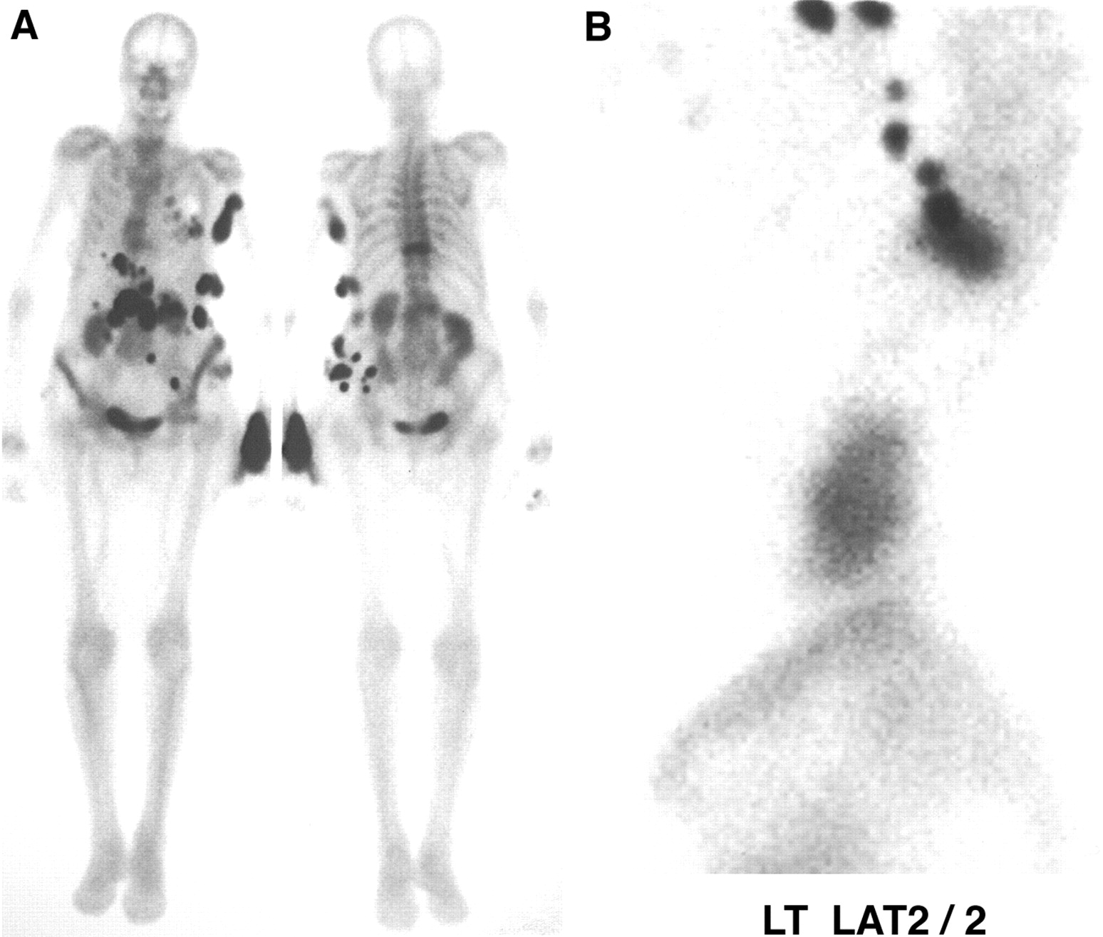
Case 1. (A) Contamination is evident at injection site, but rest of activity could have been mistaken for bone-forming metastases from osteosarcoma or simply leaking intravenous site. (B) As gown was moved, activity also moved, confirming contamination.

Case 2

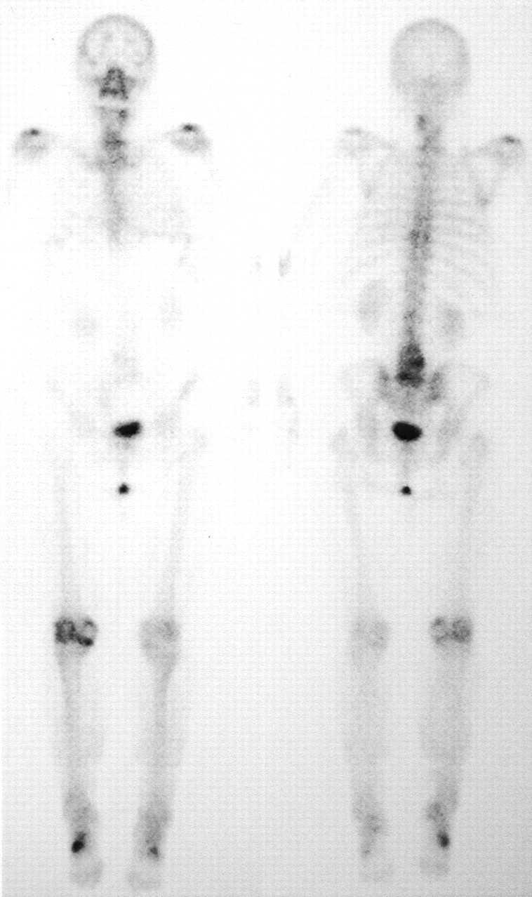
A 22-y-old man had a contusion on the right chest wall and lumbar spine secondary to a work-related fall from a truck. Mild abnormal increased activity was noted in the sternoclavicular joints and shoulders but not the ribs. Both kidneys and bladder were normal. Abnormal activity was noted over both legs, especially on the right leg inferior to the knee, as a result of urinary contamination on a protective sheet (Fig. 2).

FIGURE 2.
  • Download figure
  • Open in new tab
  • Download powerpoint
FIGURE 2.

Case 2. (A) Abnormalities seen only in anterior and posterior views could have been mistaken for sites of trauma. (B) Lateral view shows activity posteriorly on protective sheet.

Case 3

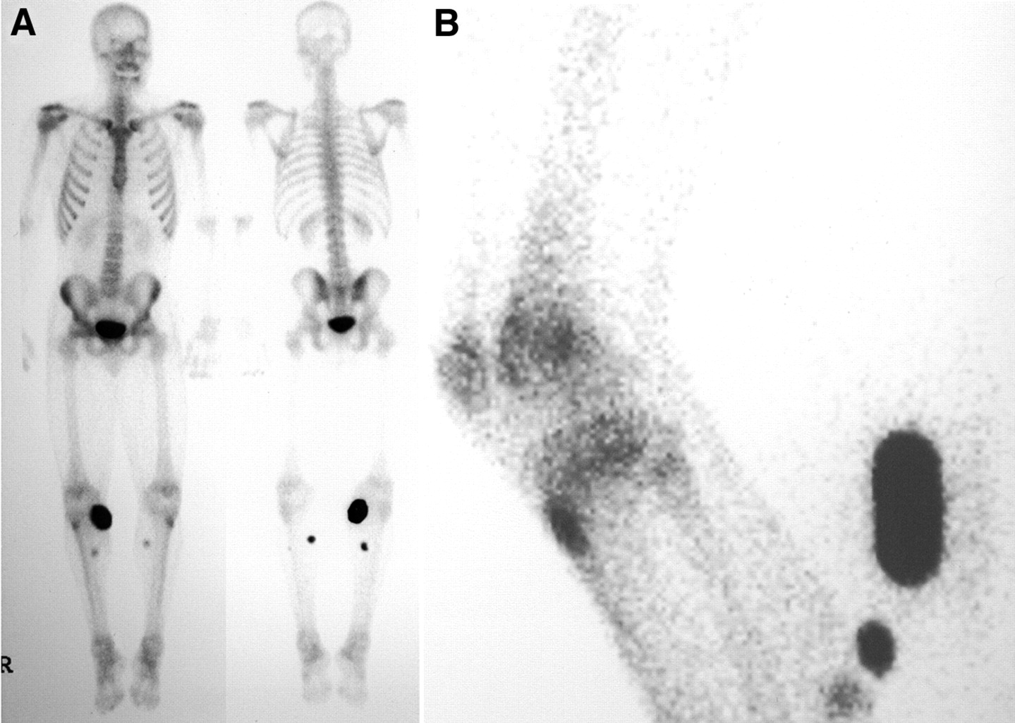
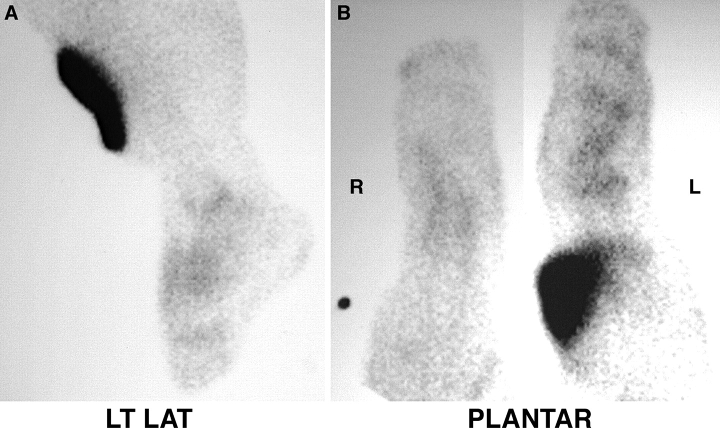
A 33-y-old man presented with persistent left foot cellulitis and was imaged to exclude acute osteomyelitis. The blood pool images showed faint focal activity in the left leg above the ankle. Although the delayed images did not show significant activity in the legs or feet, localized leg activity clearly represented a urine bag best seen on a left lateral view (Fig. 3).

FIGURE 3.
  • Download figure
  • Open in new tab
  • Download powerpoint
FIGURE 3.

Case 3. (A) Originally believed to be urinary contamination, leg bag is clearly seen on lateral view. (B) Posterior view of urinary bag.

RESULTS

A common source of contamination results from the intravenous injection of the radioisotope. Contamination may result from injection site dose infiltration, a leaking intravenous tube, or bleeding from the puncture site because of inadequate hemostasis.

Because all bone agents are excreted through the genitourinary (GU) tract, we obtain an incidental view of the entire GU system from the kidneys to the external genitalia. Some authors report up to 40% of GU abnormalities detected. Not every bone scan detects a GU abnormality. Others may show a urine leak or urinary diversion simulating a leak. Intense abdomen or pelvic activity may represent an unusual case of contamination, bladder rupture, tortuous or dilated ureters, or surgical urinary diversion.

Urinary contamination is a relatively common finding. Some patients are quite fastidious, others are not as careful, and still other patients have renal failure and produce very little urine. Some patients have urinary tract obstructions or Foley catheters.

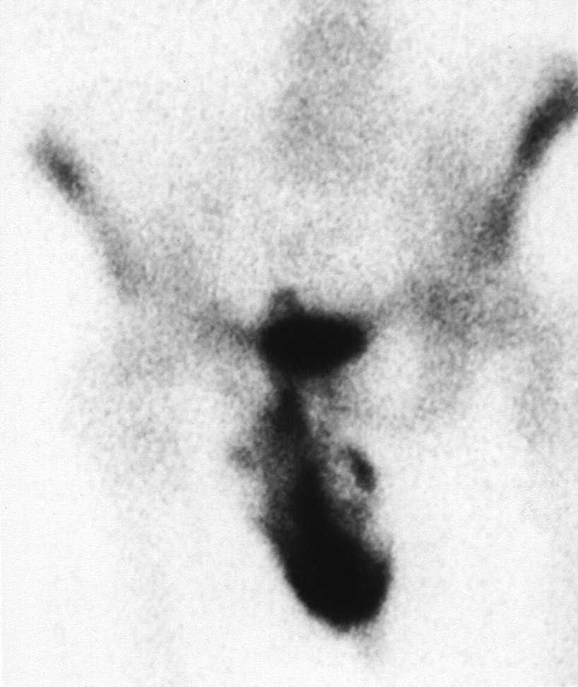
Distinct male (Fig. 4) and female (Fig. 5) patterns of urinary contamination were noted. Among patients with minimal urinary contamination, females produce a short linear area, whereas males have a more random pattern that represents the more mobile external genitalia. In cases of gross contamination, no distinct male or female patterns of contamination were noted.

FIGURE 4.
  • Download figure
  • Open in new tab
  • Download powerpoint
FIGURE 4.

Male urinary contamination pattern.

FIGURE 5.
  • Download figure
  • Open in new tab
  • Download powerpoint
FIGURE 5.

Female urinary contamination pattern.

DISCUSSION

Contamination is seen every day in our department. One author has classified the types of problems associated with radiopharmaceuticals, including contamination (17). We once believed that infiltrated doses were more common with each year’s new students, but we are beginning to rethink that hypothesis. An infiltrated dose can lead to axillary lymph node activity unrelated to possible neoplastic activity.

CONCLUSION

It is important to recognize contamination and differentiate it from a true abnormality. In one of our cases, a leg bag simulated soft-tissue activity that could have been interpreted as cellulitis. A small study looking at 2 different departments for incidental contamination for two 1-mo periods demonstrated our belief that the frequency of contamination varies with the radiopharmaceutical (18). A study dedicated to the topic of contamination would be helpful to establish the frequency of and site location for each radiopharmaceutical. Types and locations of contamination may be radiopharmaceutical dependent.

Footnotes

  • For correspondence or reprints contact: Michael E. Spieth, MD, Department of Radiology, Nuclear Medicine Section, Marshfield Clinic, 1000 North Oak Ave. H-IN-NM, Marshfield, WI 54449.

    E-mail: spieth.michael{at}marshfieldclinic.org

REFERENCES

  1. ↵
    Patton JA. Artifacts. In: Habibian MR, Delbeke D, Martin WH, Sandler MP, eds. Nuclear Medicine Imaging: A Teaching File. Philadelphia, PA: Lippincott Williams & Wilkins; 1999:873–874.
  2. Ryo UY, Bekerman C, Pinsky SM. The skeletal system. In: Ryo UY, Alavi A, Collier BD, Bekerman C, Pinsky SM, eds. Atlas of Nuclear Medicine Artifacts and Variants. 2nd ed. Chicago, IL: Year Book Medical Publishers, Inc.; 1990:155–202.
  3. Wilson MA, Bianco JA, Tu RK, Zager LV. Radiation detection. In: Wilson MA, ed. Textbook of Nuclear Medicine. Philadelphia, PA: Lippincott-Raven; 1998:415–436.
  4. Murray IPC. Bone scintigraphy: the procedure and interpretation. In: Murray IPC, Ell PJ, eds. Nuclear Medicine in Clinical Diagnosis and Treatment. 2nd ed. Edinburgh, U.K.: Churchill Livingstone; 1998:1141–1142.
  5. Campeau RJ. Bone imaging. In: Baum S, Dubovsky EV, Campeau RJ, et al., eds. Atlas of Nuclear Medicine Imaging. 2nd ed. Norwalk, CT: Appleton & Lange; 1993:369–405.
  6. Gray HW. Soft tissue uptake of bone agents. In: Fogelman I, ed. Bone Scanning in Clinical Practice. London, U.K.: Springer-Verlag; 1987:228–229.
  7. Adams KJ, Shuler SE, Witherspoon LR, Neely HR. A retrospective analysis of renal abnormalities detected on bone scans. Clin Nucl Med. 1980;5:1–7.
    OpenUrlPubMed
  8. ↵
    Brill DR. Radionuclide imaging of nonneoplastic soft tissue disorders. Semin Nucl Med. 1981;11:277–288.
    OpenUrlCrossRefPubMed
  9. ↵
    Shapiro B, Rufini V, Jarwan AM, et al. Artifacts, anatomical and physiological variants, and unrelated diseases that may cause false-positive whole-body I-131 scans in patients with thyroid cancer. Semin Nucl Med. 2000;30:115–132.
    OpenUrlCrossRefPubMed
  10. ↵
    Pochis WT, Krasnow AZ, Isitman AT, et al. The radioactive handkerchief sign: a contamination artifact in I-131 imaging for metastatic thyroid carcinoma. Clin Nucl Med. 1990;15:491–494.
    OpenUrlPubMed
  11. ↵
    Mosman EA, Peterson LJ, Hung JC, Gibbons RJ. Practical methods for reducing radioactive contamination incidents in the nuclear cardiology laboratory. J Nucl Med Technol. 1999;27:287–289.
    OpenUrlAbstract
  12. ↵
    Ikehira H, Kinjo M, Yamamoto Y, et al. Hot spots observed on pulmonary perfusion imaging: a case report. J Nucl Med Techol. 1999;27:301–302.
    OpenUrlAbstract
  13. ↵
    Gough J, Spieth ME, Bollech J. Evaluation of room contamination after using Tc-99m-DTPA aerosol ventilation kits [abstract]. J Nucl Med Technol. 2000;28:146.
  14. ↵
    Gentili A, Miron SD, Adler LP. Review of some common artifacts in nuclear medicine. Clin Nucl Med. 1994;19:138–143.
    OpenUrlPubMed
  15. ↵
    Turton DB, Silverman ED. Tampon artifact in bone scintigraphy. Clin Nucl Med. 1994;19:1103–1104.
    OpenUrlPubMed
  16. ↵
    Gandhi SM, Mattox S, Scheiner J, Zuckier LS. Appearance of a common catheter in an uncommon location. Clin Nucl Med. 1996;21:973–974.
    OpenUrlPubMed
  17. ↵
    Hladik WB III, Norenberg JP. Problems associated with the clinical use of radiopharmacueticals: a proposed classification system and trouble shooting guide. Eur J Nucl Med. 1996;23:997–1002.
    OpenUrlPubMed
  18. ↵
    Wiatrowski WA, Cooke EP, Kopp DT, Jordan DW. Radiocontamination in medical centers from diagnostic nuclear medicine departments. Health Phys. 1984;47:297–298.
    OpenUrlPubMed
PreviousNext
Back to top

In this issue

Journal of Nuclear Medicine Technology: 31 (1)
Journal of Nuclear Medicine Technology
Vol. 31, Issue 1
March 1, 2003
  • Table of Contents
  • About the Cover
  • Index by author
Print
Download PDF
Article Alerts
Sign In to Email Alerts with your Email Address
Email Article

Thank you for your interest in spreading the word on Journal of Nuclear Medicine Technology.

NOTE: We only request your email address so that the person you are recommending the page to knows that you wanted them to see it, and that it is not junk mail. We do not capture any email address.

Enter multiple addresses on separate lines or separate them with commas.
The Day of Contamination
(Your Name) has sent you a message from Journal of Nuclear Medicine Technology
(Your Name) thought you would like to see the Journal of Nuclear Medicine Technology web site.
Citation Tools
The Day of Contamination
Darcy L. Kasner, Michael E. Spieth
Journal of Nuclear Medicine Technology Mar 2003, 31 (1) 21-24;

Citation Manager Formats

  • BibTeX
  • Bookends
  • EasyBib
  • EndNote (tagged)
  • EndNote 8 (xml)
  • Medlars
  • Mendeley
  • Papers
  • RefWorks Tagged
  • Ref Manager
  • RIS
  • Zotero
Share
The Day of Contamination
Darcy L. Kasner, Michael E. Spieth
Journal of Nuclear Medicine Technology Mar 2003, 31 (1) 21-24;
Twitter logo Facebook logo LinkedIn logo Mendeley logo
  • Tweet Widget
  • Facebook Like
  • Google Plus One
Bookmark this article

Jump to section

  • Article
    • Abstract
    • MATERIALS AND METHODS
    • FINDINGS
    • RESULTS
    • DISCUSSION
    • CONCLUSION
    • Footnotes
    • REFERENCES
  • Figures & Data
  • Info & Metrics
  • PDF

Related Articles

  • No related articles found.
  • PubMed
  • Google Scholar

Cited By...

  • Contamination, a Major Problem in Nuclear Medicine Imaging: How to Investigate, Handle, and Avoid It
  • Staging of Regional Lymph Nodes in Melanoma Patients by Means of 99mTc-MIBI Scintigraphy
  • Google Scholar

More in this TOC Section

  • Single- Versus Dual-Time-Point Imaging for Transthyretin Cardiac Amyloid Using 99mTc-Pyrophosphate
  • Software Discrepancies in Radionuclide-Derived Left Ventricular Ejection Fraction
  • Monte Carlo Simulation of Characteristics of Discovery NM/CT 670 Pro SPECT System for Routinely Used Diagnostic and Therapeutic Radionuclides
Show more Imaging

Similar Articles

SNMMI

© 2025 SNMMI

Powered by HighWire