  • Main menu
  • User menu
  • Search
  • English ▼
    • English
    • Afrikaans
    • Albanian
    • Amharic
    • Arabic
    • Armenian
    • Azerbaijani
    • Basque
    • Belarusian
    • Bengali
    • Bosnian
    • Bulgarian
    • Catalan
    • Cebuano
    • Chichewa
    • Chinese (Simplified)
    • Chinese (Traditional)
    • Corsican
    • Croatian
    • Czech
    • Danish
    • Dutch
    • Esperanto
    • Estonian
    • Filipino
    • Finnish
    • French
    • Frisian
    • Galician
    • Georgian
    • German
    • Greek
    • Gujarati
    • Haitian Creole
    • Hausa
    • Hawaiian
    • Hebrew
    • Hindi
    • Hmong
    • Hungarian
    • Icelandic
    • Igbo
    • Indonesian
    • Irish
    • Italian
    • Japanese
    • Javanese
    • Kannada
    • Kazakh
    • Khmer
    • Korean
    • Kurdish (Kurmanji)
    • Kyrgyz
    • Lao
    • Latin
    • Latvian
    • Lithuanian
    • Luxembourgish
    • Macedonian
    • Malagasy
    • Malay
    • Malayalam
    • Maltese
    • Maori
    • Marathi
    • Mongolian
    • Myanmar (Burmese)
    • Nepali
    • Norwegian
    • Pashto
    • Persian
    • Polish
    • Portuguese
    • Punjabi
    • Romanian
    • Russian
    • Samoan
    • Scottish Gaelic
    • Serbian
    • Sesotho
    • Shona
    • Sindhi
    • Sinhala
    • Slovak
    • Slovenian
    • Somali
    • Spanish
    • Sudanese
    • Swahili
    • Swedish
    • Tajik
    • Tamil
    • Telugu
    • Thai
    • Turkish
    • Ukrainian
    • Urdu
    • Uzbek
    • Vietnamese
    • Welsh
    • Xhosa
    • Yiddish
    • Yoruba
    • Zulu

Main menu

  • Home
  • Content
    • Current
      • JNMT Supplement
    • Ahead of print
    • Past Issues
    • Continuing Education
    • JNMT Podcast
    • SNMMI Annual Meeting Abstracts
  • Subscriptions
    • Subscribers
    • Rates
    • Journal Claims
    • Institutional and Non-member
  • Authors
    • Submit to JNMT
    • Information for Authors
    • Assignment of Copyright
    • AQARA Requirements
  • Info
    • Reviewers
    • Permissions
    • Advertisers
    • Corporate & Special Sales
  • About
    • About Us
    • Editorial Board
    • Contact Information
  • More
    • Alerts
    • Feedback
    • Help
    • SNMMI Journals
  • SNMMI
    • JNMT
    • JNM
    • SNMMI Journals
    • SNMMI

User menu

  • Subscribe
  • My alerts
  • Log in
  • My Cart

Search

  • Advanced search
Journal of Nuclear Medicine Technology
  • SNMMI
    • JNMT
    • JNM
    • SNMMI Journals
    • SNMMI
  • Subscribe
  • My alerts
  • Log in
  • My Cart
Journal of Nuclear Medicine Technology

Advanced Search

English ▼
  • English
  • Afrikaans
  • Albanian
  • Amharic
  • Arabic
  • Armenian
  • Azerbaijani
  • Basque
  • Belarusian
  • Bengali
  • Bosnian
  • Bulgarian
  • Catalan
  • Cebuano
  • Chichewa
  • Chinese (Simplified)
  • Chinese (Traditional)
  • Corsican
  • Croatian
  • Czech
  • Danish
  • Dutch
  • Esperanto
  • Estonian
  • Filipino
  • Finnish
  • French
  • Frisian
  • Galician
  • Georgian
  • German
  • Greek
  • Gujarati
  • Haitian Creole
  • Hausa
  • Hawaiian
  • Hebrew
  • Hindi
  • Hmong
  • Hungarian
  • Icelandic
  • Igbo
  • Indonesian
  • Irish
  • Italian
  • Japanese
  • Javanese
  • Kannada
  • Kazakh
  • Khmer
  • Korean
  • Kurdish (Kurmanji)
  • Kyrgyz
  • Lao
  • Latin
  • Latvian
  • Lithuanian
  • Luxembourgish
  • Macedonian
  • Malagasy
  • Malay
  • Malayalam
  • Maltese
  • Maori
  • Marathi
  • Mongolian
  • Myanmar (Burmese)
  • Nepali
  • Norwegian
  • Pashto
  • Persian
  • Polish
  • Portuguese
  • Punjabi
  • Romanian
  • Russian
  • Samoan
  • Scottish Gaelic
  • Serbian
  • Sesotho
  • Shona
  • Sindhi
  • Sinhala
  • Slovak
  • Slovenian
  • Somali
  • Spanish
  • Sudanese
  • Swahili
  • Swedish
  • Tajik
  • Tamil
  • Telugu
  • Thai
  • Turkish
  • Ukrainian
  • Urdu
  • Uzbek
  • Vietnamese
  • Welsh
  • Xhosa
  • Yiddish
  • Yoruba
  • Zulu
  • Home
  • Content
    • Current
    • Ahead of print
    • Past Issues
    • Continuing Education
    • JNMT Podcast
    • SNMMI Annual Meeting Abstracts
  • Subscriptions
    • Subscribers
    • Rates
    • Journal Claims
    • Institutional and Non-member
  • Authors
    • Submit to JNMT
    • Information for Authors
    • Assignment of Copyright
    • AQARA Requirements
  • Info
    • Reviewers
    • Permissions
    • Advertisers
    • Corporate & Special Sales
  • About
    • About Us
    • Editorial Board
    • Contact Information
  • More
    • Alerts
    • Feedback
    • Help
    • SNMMI Journals
  • Watch or Listen to JNMT Podcast
  • Visit SNMMI on Facebook
  • Join SNMMI on LinkedIn
  • Follow SNMMI on Twitter
  • Subscribe to JNMT RSS feeds
OtherIMAGING

Lymphoscintigraphy Using 99mTc Filtered Sulfur Colloid in Chylothorax: A Case Report

Bohdan Bybel, Donald R. Neumann, Benedict Y. Kim, Kanak Amin and Thomas Rice
Journal of Nuclear Medicine Technology March 2001, 29 (1) 30-31;
Bohdan Bybel
  • Find this author on Google Scholar
  • Find this author on PubMed
  • Search for this author on this site
Donald R. Neumann
  • Find this author on Google Scholar
  • Find this author on PubMed
  • Search for this author on this site
Benedict Y. Kim
  • Find this author on Google Scholar
  • Find this author on PubMed
  • Search for this author on this site
Kanak Amin
  • Find this author on Google Scholar
  • Find this author on PubMed
  • Search for this author on this site
Thomas Rice
  • Find this author on Google Scholar
  • Find this author on PubMed
  • Search for this author on this site
  • Article
  • Figures & Data
  • Info & Metrics
  • PDF
Loading

Abstract

Objective:A 66-y-old man was diagnosed with esophageal carcinoma and underwent a right thoracotomy and esophagectomy. Postoperatively, a recurring right pleural effusion developed. Because an attempt at lymphangiography failed, lymphoscintigraphy was suggested. Because of the inability to obtain radiolabeled albumin, dextran, or nanocolloid, we used filtered sulfur colloid. (0.1 um). The study confirmed the diagnosis of chylothorax.

  • chylothorax
  • lymphoscintigraphy
  • filtered sulfur colloid

CASE REPORT

A 66-y-old man presented with a 2-mo history of weight loss and dysphagia to solids. A CT scan and subsequent endoscopy demonstrated a 4-cm esophageal mass consistent with esophageal carcinoma. Preoperatively, it was noted that the patient also had severe coronary artery disease and a left upper lung mass. Initially, the patient had coronary bypass grafting and underwent a left upper lobe wedge resection. Approximately 2 weeks later, a right thoracotomy and esophagectomy were performed. The surgery was completed without complication. The patient developed bilateral pleural effusions; the one on the right was worse.

The drainage indicated that the right effusion was chylous, so a radiologic lymphangiogram was ordered to better evaluate the leak. Unfortunately, there was significant difficulty in cannulating the lymphatic channels and the test was not completed. Lymphoscintigraphy was suggested and, after discussion, a lymphoscintigram with 99mTc filtered sulfur colloid was proposed. Radiolabeled human serum albumin, dextran and nanocolloid were not available.

MATERIALS AND METHODS

99mTc sulfur colloid was made per the package insert directions from the kit (CIS); a routine quality control (QC) procedure was performed. The sulfur chloride was then filtered through two 0.1-μm filters (Gelman Science Acrodisc; Ann Arbor, MI) as per our typical protocol for sentinel node lymphoscintigraphy. Approximately 7%–15% of the activity remained after filtering. 99mTc sulfur colloid was divided into 4 insulin syringes. The patient was then injected bilaterally in the first and second web-spaces of the feet. Serial whole-body imaging was performed at 4 and 24 h.

IMAGING RESULTS

Abnormal accumulation of activity is seen in the lower right hemithorax (Fig. 1). Expected activity in inguinal lymph nodes is also noted. No other areas of abnormal accumulation are identified. A region of interest was drawn around the thoracic activity and compared with a region including the total injected activity. There was approximately 1% of injected activity present in the thorax at 4 h.

FIGURE 1.
  • Download figure
  • Open in new tab
  • Download powerpoint
FIGURE 1.

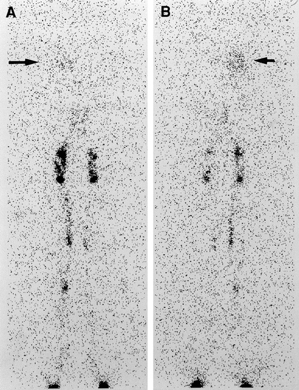
Anterior and posterior whole-body images at 4 h. Radioactivity is seen in the right thorax (arrow) corresponding to the known chylothorax.

Follow-up

The patient had a ligation of the thoracic duct and talc pleurodesis of the left side. Because this procedure did not completely resolve the patient’s problem, a radiographic lymphangiogram with CT imaging was performed; this confirmed an extravasation into the right thorax at the T10 level.

Subsequently, a right thoracotomy, thoracic duct ligation and talc pleurodesis were repeated. Since this procedure, the patient has been stable with no further leakage demonstrated.

DISCUSSION

Chylothorax, a pleural effusion containing chyle, can occur secondary to trauma, neoplasm, malformations of the lymphatic system and surgical procedures involving the pleural space.

Lymphangiography is considered the gold standard in investigating chylothorax, as well as being used to assess other pathologies such as chyloperitoneum, chyluria, thoracic duct patency, abnormal leg lymphatics, and detection of abnormal retroperitoneal lymph nodes.

It is a difficult procedure, requiring cannulation of the lymphatic channels, that can potentially cause adverse effects such as local tissue necrosis, fat embolism to the lungs, hypersensitivity reaction or worsening of lymphedema from the contrast material.

Lymphoscintigraphy has been described previously using radiolabeled human serum albumin, dextran or nanocolloid. It is quick, minimally invasive, and does not have any known side effects. It has become difficult to procure human serum albumin or dextran to do these studies. An attempt was made to use filtered 99mTc sulfur colloid identical to material used in sentinel lymph node lymphoscintigraphy in melanoma or breast cancer.

Our method is similar to what has been described by Hung et al. (3).

Although a portion of the radiopharmaceutical may have been too large and trapped in the inguinal lymph nodes, enough traveled into the thoracic duct to demonstrate the lymphatic leak. A direct comparison with other radiopharmaceuticals was not performed, but if the other materials are not available, adequate studies can be performed with filtered 99mTc sulfur colloid.

Footnotes

  • For correspondence or reprints contact: Dr. Bohdan Bybel, Department of Nuclear Medicine, Cleveland Clinic Foundation, 9500 Euclid Gb3, Cleveland, OH 44195; Phone: 216-444-2099; E-mail: bybelb@ccf.org.

REFERENCES

  1. Pui MH, Yueh TC. Lymphoscintigraphy in chyluria, chyloperitoneum and chylothorax. J Nuc Med 1998:39(7):1292–6.
    OpenUrlAbstract/FREE Full TextGoogle Scholar
  2. Inoue Y, Otake T, Nishikawa J, Sasaki Y. Lymphoscintigraphy using Tc 99m human serum albumin in chylothorax. Clin Nuc Med 1997 22(1):60.
    OpenUrlPubMedGoogle Scholar
  3. ↵
    Hung JC, Wiseman GA, Wahner HW, Mullan BP, Taggart TR and Dunn WL. Filtered Technetium-99m-Sulfur Colloid for Lymphoscintigraphy. J Nuc Med 1995;36(10):1895–1901.
    OpenUrlAbstract/FREE Full TextGoogle Scholar
PreviousNext
Back to top

In this issue

Journal of Nuclear Medicine Technology: 29 (1)
Journal of Nuclear Medicine Technology
Vol. 29, Issue 1
March 1, 2001
  • Table of Contents
  • About the Cover
  • Index by author
Print
Download PDF
Article Alerts
Email Article
Citation Tools
Share
Lymphoscintigraphy Using 99mTc Filtered Sulfur Colloid in Chylothorax: A Case Report
Bohdan Bybel, Donald R. Neumann, Benedict Y. Kim, Kanak Amin, Thomas Rice
Journal of Nuclear Medicine Technology Mar 2001, 29 (1) 30-31;
Twitter logo Facebook logo LinkedIn logo Mendeley logo
  • Tweet Widget
Bookmark this article

Jump to section

  • Article
    • Abstract
    • CASE REPORT
    • MATERIALS AND METHODS
    • IMAGING RESULTS
    • DISCUSSION
    • Footnotes
    • REFERENCES
  • Figures & Data
  • Info & Metrics
  • PDF

Related Articles

  • No related articles found.
  • PubMed
  • Google Scholar

Cited By...

  • No citing articles found.
  • Google Scholar

More in this TOC Section

  • Early 10-Minute Postinjection [18F]F-FAPI-42 uEXPLORER Total-Body PET/CT Scanning Protocol for Staging Lung Cancer Using HYPER Iterative Reconstruction
  • Single- Versus Dual-Time-Point Imaging for Transthyretin Cardiac Amyloid Using 99mTc-Pyrophosphate
  • Software Discrepancies in Radionuclide-Derived Left Ventricular Ejection Fraction
Show more Imaging

Similar Articles

SNMMI

© 2025 SNMMI

Powered by HighWire
Alerts for this Article
Sign In to Email Alerts with your Email Address
Email this Article

Thank you for your interest in spreading the word on Journal of Nuclear Medicine Technology.

NOTE: We only request your email address so that the person you are recommending the page to knows that you wanted them to see it, and that it is not junk mail. We do not capture any email address.

Enter multiple addresses on separate lines or separate them with commas.
Lymphoscintigraphy Using 99mTc Filtered Sulfur Colloid in Chylothorax: A Case Report
(Your Name) has sent you a message from Journal of Nuclear Medicine Technology
(Your Name) thought you would like to see the Journal of Nuclear Medicine Technology web site.
Citation Tools
Lymphoscintigraphy Using 99mTc Filtered Sulfur Colloid in Chylothorax: A Case Report
Bohdan Bybel, Donald R. Neumann, Benedict Y. Kim, Kanak Amin, Thomas Rice
Journal of Nuclear Medicine Technology Mar 2001, 29 (1) 30-31;

Citation Manager Formats

  • BibTeX
  • Bookends
  • EasyBib
  • EndNote (tagged)
  • EndNote 8 (xml)
  • Medlars
  • Mendeley
  • Papers
  • RefWorks Tagged
  • Ref Manager
  • RIS
  • Zotero

We use cookies on this site to enhance your user experience

By clicking any link on this page you are giving your consent for us to set cookies.