
Main menu

  • Home
  • Content
    • Current
      • JNMT Supplement
    • Ahead of print
    • Past Issues
    • Continuing Education
    • JNMT Podcast
    • SNMMI Annual Meeting Abstracts
  • Subscriptions
    • Subscribers
    • Rates
    • Journal Claims
    • Institutional and Non-member
  • Authors
    • Submit to JNMT
    • Information for Authors
    • Assignment of Copyright
    • AQARA Requirements
  • Info
    • Reviewers
    • Permissions
    • Advertisers
    • Corporate & Special Sales
  • About
    • About Us
    • Editorial Board
    • Contact Information
  • More
    • Alerts
    • Feedback
    • Help
    • SNMMI Journals
  • SNMMI
    • JNMT
    • JNM
    • SNMMI Journals
    • SNMMI

User menu

  • Subscribe
  • My alerts
  • Log in
  • Log out
  • My Cart

Search

  • Advanced search
Journal of Nuclear Medicine Technology
  • SNMMI
    • JNMT
    • JNM
    • SNMMI Journals
    • SNMMI
  • Subscribe
  • My alerts
  • Log in
  • Log out
  • My Cart
Journal of Nuclear Medicine Technology

Advanced Search

  • Home
  • Content
    • Current
    • Ahead of print
    • Past Issues
    • Continuing Education
    • JNMT Podcast
    • SNMMI Annual Meeting Abstracts
  • Subscriptions
    • Subscribers
    • Rates
    • Journal Claims
    • Institutional and Non-member
  • Authors
    • Submit to JNMT
    • Information for Authors
    • Assignment of Copyright
    • AQARA Requirements
  • Info
    • Reviewers
    • Permissions
    • Advertisers
    • Corporate & Special Sales
  • About
    • About Us
    • Editorial Board
    • Contact Information
  • More
    • Alerts
    • Feedback
    • Help
    • SNMMI Journals
  • Watch or Listen to JNMT Podcast
  • Visit SNMMI on Facebook
  • Join SNMMI on LinkedIn
  • Follow SNMMI on Twitter
  • Subscribe to JNMT RSS feeds
OtherIMAGING

Duodenogastric Reflux in a Hiatal Hernia Seen as Retrocardiac Activity on 99mTc-Tetrofosmin Cardiac SPECT Raw-Data Images

We-Jen Shih, George L. Shih, Wen-Shen Huang and Primo Milan
Journal of Nuclear Medicine Technology December 2007, 35 (4) 252-254; DOI: https://doi.org/10.2967/jnmt.107.041178
We-Jen Shih
  • Find this author on Google Scholar
  • Find this author on PubMed
  • Search for this author on this site
George L. Shih
  • Find this author on Google Scholar
  • Find this author on PubMed
  • Search for this author on this site
Wen-Shen Huang
  • Find this author on Google Scholar
  • Find this author on PubMed
  • Search for this author on this site
Primo Milan
  • Find this author on Google Scholar
  • Find this author on PubMed
  • Search for this author on this site
  • Article
  • Figures & Data
  • Info & Metrics
  • PDF
Loading

Abstract

We present a patient with a large hiatal hernia resulting in duodenogastric reflux that was seen as abnormal activity below and behind the heart (retrocardiac) on SPECT images and on 99mTc-tetrofosmin cardiac SPECT raw-data projection images. Incidental findings such as extra- and retrocardiac activity in the thorax and abdomen should be included on all comprehensive cardiac SPECT reports.

  • duodenogastric reflux
  • hiatal hernia
  • raw-data images
  • 99mTc-tetrofosmin

Approximately only 4% of 99mTc-labeled perfusion agents such as 99mTc-tetrofosmin or sestamibi is localized in the heart. Because the rest of the pharmaceutical is excreted mainly to the duodenum via the hepatobiliary system, duodenogastric reflux can sometimes be detected on the raw-data images of cardiac SPECT studies (1–3). During acquisition of cardiac SPECT images using 99mTc-labeled perfusion agents such as 99mTc-tetrofosmin and sestamibi, the field of view covers the essential organs of the thorax and abdomen. The abdominal organs such as the upper gastrointestinal tract, liver, gallbladder, stomach, spleen, and small bowel are thus imaged, as on a 99mTc-diisopropyl iminodiacetic acid hepatobiliary scintigraphy study. Therefore, abnormalities in the abdominal organs such as the upper gastrointestinal tract, liver, gallbladder, and spleen can easily be detected.

Abnormal esophageal conditions such as postoperative esophageal carcinoma or Barrett's esophagus may constitute an abnormal area of retrocardiac activity (4,5). We present a patient with a large hiatal hernia that caused duodenogastric reflux to be exhibited on vertical and horizontal longitudinal SPECT images and on the raw-data projection images of a 2-d 99mTc-tetrofosmin cardiac SPECT study. Although the incidental findings in the extra- and retrocardiac regions of the thorax and abdomen did not affect the study, a comprehensive interpretation of any cardiac SPECT study should note these relevant findings.

CASE REPORT

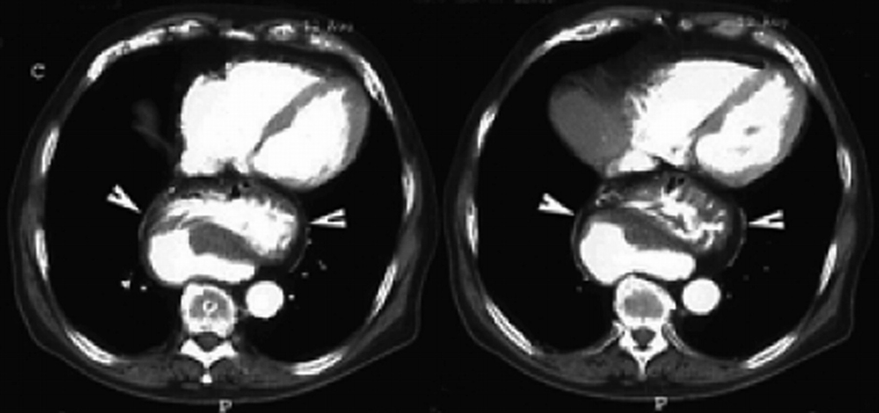
An 83-y-old male smoker with a history of cardiovascular disease and prostate cancer complained of atypical chest pain and was referred for a cardiac SPECT study. Two-day 99mTc-tetrofosmin cardiac gated images showed a fixed defect involving the apical and the apical anterior wall of the left ventricle, and the left ventricular ejection fraction was 54%. A rather large area of radioactivity was seen posterior to the basal heart on horizontal and vertical longitudinal SPECT images and was consistent with duodenogastric reflux on short-axis SPECT images (Fig. 1). Raw-data projection images showed duodenogastric reflux (Fig. 2A): a large area of abnormally increased activity in the area behind the heart (Fig. 2B). Concurrent CT of the chest showed a large intrathoracic hiatal hernia containing part of the stomach between the heart and the thoracic vertebrae, in the retrocardiac space (Fig. 3). The patient underwent an esophagogastroduodenoscopic examination, which showed a hiatal hernia, a small diverticulum in the lower third of the esophagus, and multiple linear superficial erosions in the gastric body and fundus.

FIGURE 1. 
  • Download figure
  • Open in new tab
  • Download powerpoint
FIGURE 1. 

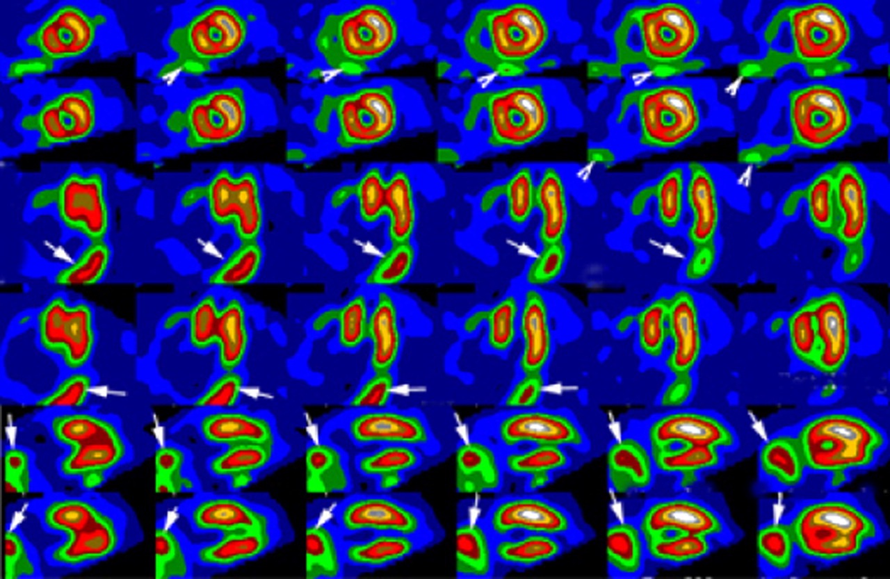
Two-day 99mTc-tetrofosmin SPECT images (top 2 rows, stress and rest short-axis slices; middle 2 rows, stress and rest horizontal slices; bottom 2 rows, stress and rest vertical slices) show absence of activity in apical wall and decreased activity in apical anterior wall. Increased activity in inferior to the inferior wall is noted on short-axis slices (arrowheads); large areas of activity (arrows) are noted in basal region on vertical and horizontal images.

FIGURE 2. 
  • Download figure
  • Open in new tab
  • Download powerpoint
FIGURE 2. 

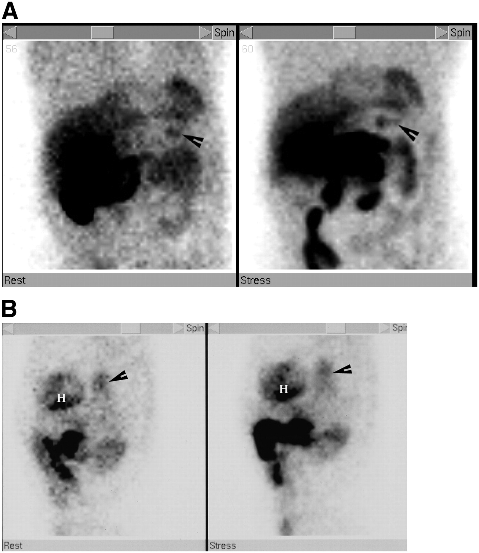
(A) Raw-data images in anterior projections at stress (right) and rest (left) on 2-d 99mTc-tetrofosmin SPECT study show duodenogastric reflux (arrowheads). (B) Raw-data images in left lateral projections on 2-d 99mTc-tetrofosmin SPECT study show extracardiac activity (arrows) in posterior aspect of heart (H) at rest (left) and stress (right).

FIGURE 3. 
  • Download figure
  • Open in new tab
  • Download powerpoint
FIGURE 3. 

Contrast-enhanced CT images of chest show large part of stomach and its omentum (arrowheads) between heart and thoracic vertebrae.

DISCUSSION

Postoperative esophageal carcinoma (1) or Barrett's esophagus (2) may cause extra- and retrocardiac activity on raw-data projection images on 99mTc-tetrofosmin cardiac SPECT studies. These findings are due to duodenogastroesophageal reflux. Because 99mTc-myocardial perfusion imaging agents such as 99mTc-tetrofosmin are excreted by the hepatobiliary system, duodenogastric reflux can be incidentally detected on a 99mTc-tetrofosmin cardiac SPECT study. The duodenogastric reflux demonstrated by our patient's SPECT study appeared to be due to a hiatal hernia, which exhibited duodenogastric reflux on short-axis SPECT images and bile regurgitation to the hiatal hernia on longitudinal SPECT images; these abnormalities were seen as retrocardiac activity on raw-data projection images (Fig. 2B). The large hiatal hernia was confirmed by CT of the chest as shown in Figure 3. Although the hiatal hernia could have been diagnosed on CT of the chest, CT images could not depict the presence or absence of duodenogastric reflux, which was visualized in this patient.

Cases of extracardiac activity posterior to the heart in hiatal hernia have been reported (6,7); however, in these reports, retrocardiac activity was described only on raw-data projection images; the reports did not describe retrocardiac activity or duodenogastric reflux on SPECT images. In our case report, we described 2 abnormal findings, namely duodenogastric reflux on short-axis SPECT images (Fig. 1) and retrocardiac activity from the hiatal hernia on long-axis images (Fig. 2B).

Interpretation of raw-data projection images on gated cardiac SPECT studies is essential both for quality control and for evaluating incidental abnormal findings in the thorax and abdomen. To further evaluate the possibility of a tumor or benign lesion in the thorax, other examinations such as chest radiography, chest CT, or upper gastrointestinal tract imaging may be helpful. Our patient's cardiac SPECT study concomitantly exhibited duodenogastric reflux and retrocardiac activity. The cause of the duodenogastric reflux appeared to be hiatal hernia, and the retrocardiac activity might have been either benign or malignant. The hiatal hernia seen on chest CT yielded the cause of the duodenogastric reflux and excluded the diagnosis of malignancy.

Footnotes

  • COPYRIGHT © 2007 by the Society of Nuclear Medicine, Inc.

References

  1. 1.↵
    Middleton GW, Williams JH. Interference from duodeno-gastric reflux of 99Tcm radiopharmaceuticals in SPET myocardial perfusion imaging. Nucl Med Commun. 1996;17:114–118.
    OpenUrlPubMed
  2. 2.↵
    Shih WJ, Donovan P, Kiffer V. Significant entero-gastric reflux secondary to Billroth II gastrectomy leads to reverse redistribution of inferior wall of the left ventricle on dipyridamole myocardial SPECT. Clin Nucl Med. 2001;26:543–545.
    OpenUrlCrossRefPubMed
  3. 3.↵
    Middleton GW, Williams JH. Significant gastric reflux of Tc-99m sestamibi cardiac imaging. J Nucl Med. 1994;35:619–620.
    OpenUrlAbstract/FREE Full Text
  4. 4.↵
    Shih WJ, Milan PP. Duodenogastroesophageal reflux in a patient with postoperative esophageal cancer shown on Tc-99m tetrofosmin raw data images of dual-isotope gated cardiac SPECT. J Nucl Cardiol. 2004;11:512–514.
    OpenUrlCrossRefPubMed
  5. 5.↵
    Shih WJ, Milan PP, Shih LG. Duodenogastric reflux shown on raw data images on dual-isotope gated cardiac Tc-99m tetrofosmin SPECT in a patient with esophagectomy for Barrett esophagus. Clin Nucl Med. 2005;30:30–31.
    OpenUrlCrossRefPubMed
  6. 6.↵
    Zoghbi GJ, Heo J, Iskandrian AE. Hiatal hernia detected by Tc-99m tetrofosmin SPECT. J Nucl Cardiol. 2003;10:712–713.
    OpenUrlCrossRefPubMed
  7. 7.↵
    Slavin JD, Engin IO, Spencer RP. Retrocardiac uptake of Tc-99m sestamibi: manifestation of a hiatus hernia. Clin Nucl Med. 1998;23:239–240.
    OpenUrlCrossRefPubMed
  • Received for publication February 26, 2007.
  • Accepted for publication May 15, 2007.
PreviousNext
Back to top

In this issue

Journal of Nuclear Medicine Technology: 35 (4)
Journal of Nuclear Medicine Technology
Vol. 35, Issue 4
December 2007
  • Table of Contents
  • About the Cover
  • Index by author
Print
Download PDF
Article Alerts
Sign In to Email Alerts with your Email Address
Email Article

Thank you for your interest in spreading the word on Journal of Nuclear Medicine Technology.

NOTE: We only request your email address so that the person you are recommending the page to knows that you wanted them to see it, and that it is not junk mail. We do not capture any email address.

Enter multiple addresses on separate lines or separate them with commas.
Duodenogastric Reflux in a Hiatal Hernia Seen as Retrocardiac Activity on 99mTc-Tetrofosmin Cardiac SPECT Raw-Data Images
(Your Name) has sent you a message from Journal of Nuclear Medicine Technology
(Your Name) thought you would like to see the Journal of Nuclear Medicine Technology web site.
Citation Tools
Duodenogastric Reflux in a Hiatal Hernia Seen as Retrocardiac Activity on 99mTc-Tetrofosmin Cardiac SPECT Raw-Data Images
We-Jen Shih, George L. Shih, Wen-Shen Huang, Primo Milan
Journal of Nuclear Medicine Technology Dec 2007, 35 (4) 252-254; DOI: 10.2967/jnmt.107.041178

Citation Manager Formats

  • BibTeX
  • Bookends
  • EasyBib
  • EndNote (tagged)
  • EndNote 8 (xml)
  • Medlars
  • Mendeley
  • Papers
  • RefWorks Tagged
  • Ref Manager
  • RIS
  • Zotero
Share
Duodenogastric Reflux in a Hiatal Hernia Seen as Retrocardiac Activity on 99mTc-Tetrofosmin Cardiac SPECT Raw-Data Images
We-Jen Shih, George L. Shih, Wen-Shen Huang, Primo Milan
Journal of Nuclear Medicine Technology Dec 2007, 35 (4) 252-254; DOI: 10.2967/jnmt.107.041178
Twitter logo Facebook logo LinkedIn logo Mendeley logo
  • Tweet Widget
  • Facebook Like
  • Google Plus One
Bookmark this article

Jump to section

  • Article
    • Abstract
    • CASE REPORT
    • DISCUSSION
    • Footnotes
    • References
  • Figures & Data
  • Info & Metrics
  • PDF

Related Articles

  • No related articles found.
  • PubMed
  • Google Scholar

Cited By...

  • No citing articles found.
  • Google Scholar

More in this TOC Section

  • Single- Versus Dual-Time-Point Imaging for Transthyretin Cardiac Amyloid Using 99mTc-Pyrophosphate
  • Software Discrepancies in Radionuclide-Derived Left Ventricular Ejection Fraction
  • Monte Carlo Simulation of Characteristics of Discovery NM/CT 670 Pro SPECT System for Routinely Used Diagnostic and Therapeutic Radionuclides
Show more Imaging

Similar Articles

SNMMI

© 2025 SNMMI

Powered by HighWire