
Main menu

  • Home
  • Content
    • Current
      • JNMT Supplement
    • Ahead of print
    • Past Issues
    • Continuing Education
    • JNMT Podcast
    • SNMMI Annual Meeting Abstracts
  • Subscriptions
    • Subscribers
    • Rates
    • Journal Claims
    • Institutional and Non-member
  • Authors
    • Submit to JNMT
    • Information for Authors
    • Assignment of Copyright
    • AQARA Requirements
  • Info
    • Reviewers
    • Permissions
    • Advertisers
    • Corporate & Special Sales
  • About
    • About Us
    • Editorial Board
    • Contact Information
  • More
    • Alerts
    • Feedback
    • Help
    • SNMMI Journals
  • SNMMI
    • JNMT
    • JNM
    • SNMMI Journals
    • SNMMI

User menu

  • Subscribe
  • My alerts
  • Log in
  • Log out
  • My Cart

Search

  • Advanced search
Journal of Nuclear Medicine Technology
  • SNMMI
    • JNMT
    • JNM
    • SNMMI Journals
    • SNMMI
  • Subscribe
  • My alerts
  • Log in
  • Log out
  • My Cart
Journal of Nuclear Medicine Technology

Advanced Search

  • Home
  • Content
    • Current
    • Ahead of print
    • Past Issues
    • Continuing Education
    • JNMT Podcast
    • SNMMI Annual Meeting Abstracts
  • Subscriptions
    • Subscribers
    • Rates
    • Journal Claims
    • Institutional and Non-member
  • Authors
    • Submit to JNMT
    • Information for Authors
    • Assignment of Copyright
    • AQARA Requirements
  • Info
    • Reviewers
    • Permissions
    • Advertisers
    • Corporate & Special Sales
  • About
    • About Us
    • Editorial Board
    • Contact Information
  • More
    • Alerts
    • Feedback
    • Help
    • SNMMI Journals
  • Watch or Listen to JNMT Podcast
  • Visit SNMMI on Facebook
  • Join SNMMI on LinkedIn
  • Follow SNMMI on Twitter
  • Subscribe to JNMT RSS feeds
Review ArticleTeaching Case Studies

PET/CT: First-Line Examination to Assess Disease Extent of Disseminated Coccidioidomycosis

Jason Foerter, John Sundell and Penny Vroman
Journal of Nuclear Medicine Technology September 2016, 44 (3) 212-213; DOI: https://doi.org/10.2967/jnmt.115.170449
Jason Foerter
Department of Radiology, San Antonio Military Medical Center, Fort Sam Houston, Texas
  • Find this author on Google Scholar
  • Find this author on PubMed
  • Search for this author on this site
John Sundell
Department of Radiology, San Antonio Military Medical Center, Fort Sam Houston, Texas
  • Find this author on Google Scholar
  • Find this author on PubMed
  • Search for this author on this site
Penny Vroman
Department of Radiology, San Antonio Military Medical Center, Fort Sam Houston, Texas
  • Find this author on Google Scholar
  • Find this author on PubMed
  • Search for this author on this site
  • Article
  • Figures & Data
  • Info & Metrics
  • PDF
Loading

Abstract

Coccidioidomycosis is an infection caused by inhalation of the Coccidioides fungus. Most infections remain subclinical or are confined to the pulmonary system. Disseminated disease is rare. Traditionally, a combination of imaging modalities has been used to determine disease extent. We suggest 18F-FDG PET/CT as a single first-line imaging examination to assess disease extent. We present a case of disseminated coccidioidomycosis to the lung, mediastinum, soft tissues, and skeletal system. To our knowledge, no prior case reports demonstrate such widespread disease using PET/CT.

  • PET/CT
  • coccidioidomycosis
  • osteolytic lesions

Less than 1% of coccidioidomycosis infections result in extrapulmonary disseminated disease, and less than half of those involve the skeletal system (1). A single imaging examination with 18F-FDG PET/CT provides comprehensive assessment of disease extent.

CASE REPORT

An immunocompetent, 22-y-old African-American man was admitted for work-up and treatment of presumed pneumonia, which was refractory to empiric therapy. The patient reported 3 mo of fever, night sweats, weight loss, and difficulty breathing. He was born in Liberia, had moved to Texas 15 y earlier, and recently had traveled to Ohio. CT of the chest, abdomen, and pelvis demonstrated multiple osteolytic lesions. Left iliac lesion biopsy demonstrated coccidioidomycosis.

PET/CT was performed to determine disease extent (Figs. 1–3). Pertinent findings included hypermetabolic mediastinal, hilar, and mesenteric/retroperitoneal lymphadenopathy with an SUVmax of 30.8 g/mL; nonhypermetabolic pulmonary nodules; and widespread hypermetabolic osteolytic lesions with an SUVmax of 30.6 g/mL.

FIGURE 1.
  • Download figure
  • Open in new tab
  • Download powerpoint
FIGURE 1.

Maximum-intensity-projection images demonstrate too numerous to count foci of increased 18F-FDG activity representing disseminated disease.

FIGURE 2.
  • Download figure
  • Open in new tab
  • Download powerpoint
FIGURE 2.

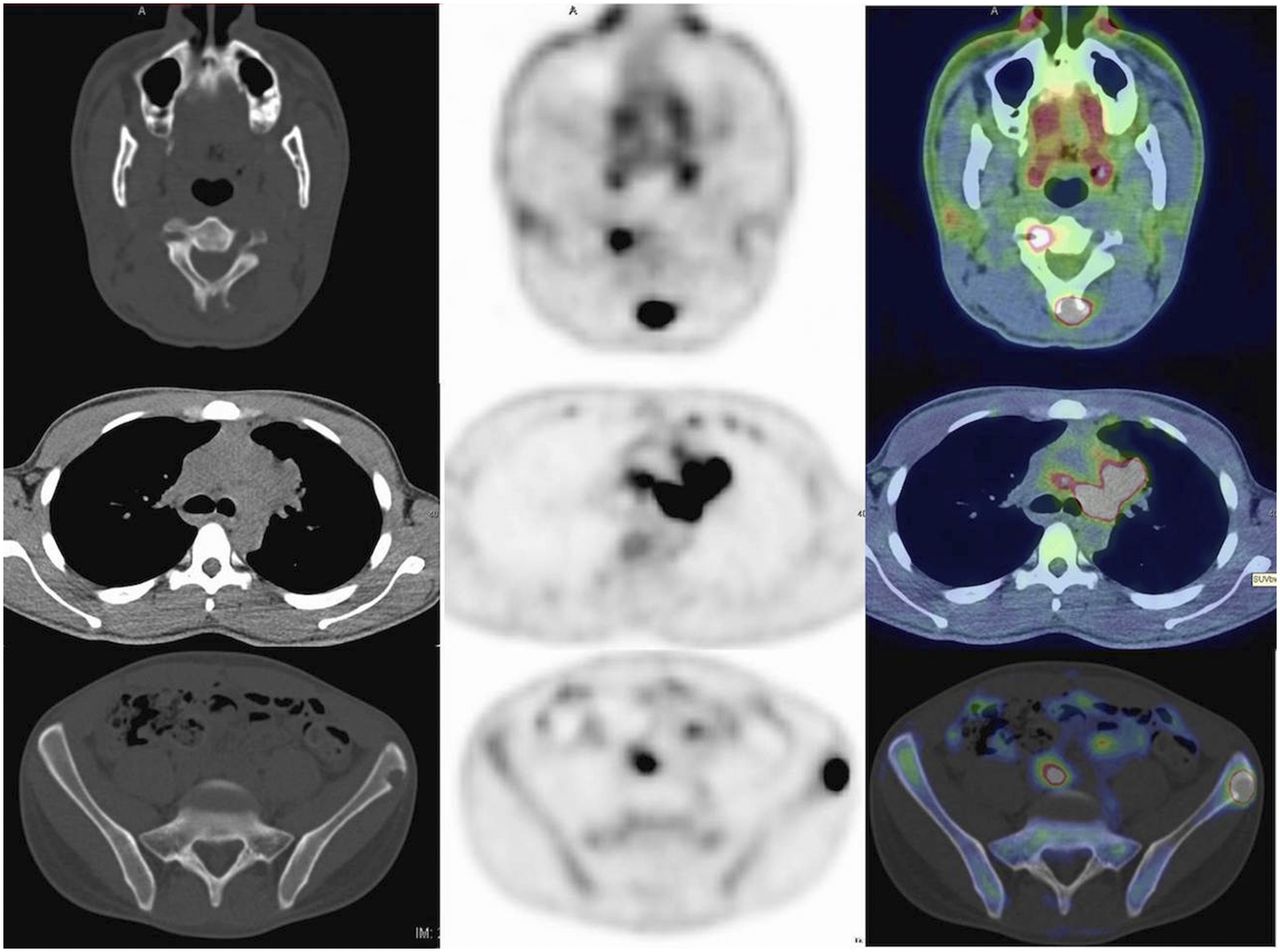
Axial CT (left), corrected PET (center), and fused PET/CT (right) images through neck (top), mid thorax (center), and pelvis (bottom) demonstrate hypermetabolic mediastinal lymph nodes and lytic posterior C2 and left iliac lesions.

FIGURE 3.
  • Download figure
  • Open in new tab
  • Download powerpoint
FIGURE 3.

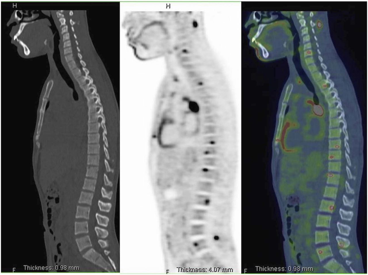
Sagittal CT (left), PET (center), and fused PET/CT (right) images demonstrate disseminated disease scattered throughout vertebral bodies and posterior spinal elements at multiple levels.

He was subsequently treated with a combination of high-dose fluconazole, itraconazole, and amphotericin. He responded well clinically and was discharged home.

DISCUSSION

18F-FDG is a glucose analog positron emitter, whose primary imaging use has traditionally been staging and monitoring of malignancy. Infectious and inflammatory cells, however, also preferentially metabolize glucose (2). PET/CT imaging of infection and inflammation is gaining greater acceptance, to include evaluation of joint prosthesis, fever of unknown origin, AIDS-related disorders, and vascular graft infections (3). Previously, multiple imaging modalities were used to assess infectious and inflammatory disease extent, to include bone scintigraphy, radiography, CT, and MRI (4). Now, whole-body PET/CT can provide a single comprehensive assessment of anatomic and metabolic extent of infection and inflammation involving both the soft tissues and the osseous structures.

Risk factors for disseminated coccidioidomycosis include African-American or Filipino ancestry, immunocompromised state, and male sex (4). The nonspecific symptomatology and indolent nature can lead to delayed diagnosis, with recurrence or spread possible 2 or more years after the initial infection. Fever of unknown origin work-up is then pursued and frequently uses multiple imaging examinations to determine disease extent.

Skeletal involvement of disseminated coccidioidomyocosis predominantly causes osteolytic lesions, which are 18F-FDG–avid on PET/CT. Additionally, soft-tissue assessment with PET/CT can demonstrate clinically occult soft-tissue infection/abscesses, which may need surgical debridement. Thus, 18F-FDG PET/CT can provide whole-body comprehensive assessment of disease extent and guide biopsy targets and therapy (3).

CONCLUSION

Though rare, disseminated coccidioidomycosis is associated with significant morbidity and mortality to include clinically occult abscesses. Previously, a combination of multiple imaging examinations was used to determine disease extent. PET/CT, however, provides whole-body metabolic and anatomic assessment of disease and should be considered a first-line imaging examination in these patients.

DISCLOSURE

No potential conflict of interest relevant to this article was reported.

Footnotes

  • Published online Apr. 21, 2016.

REFERENCES

  1. 1.↵
    1. Arora NP,
    2. Taneja V
    . ReyesSacin C, Bhanot R, Natesan SK. Coccidioidomycosis masquerading as malignancy. BMJ Case Rep. February 25, 2012 [Epub ahead of print].
  2. 2.↵
    1. Zhuang H,
    2. Alavi A
    . 18-fluorodeoxyglucose positron emission tomographic imaging in the detection and monitoring of infection and inflammation. Semin Nucl Med. 2002;32:47–59.
    OpenUrlCrossRefPubMed
  3. 3.↵
    1. Basu S,
    2. Chryssikos T,
    3. Moghadam-Kia S,
    4. Zhuang H,
    5. Torigian DA,
    6. Alavi A
    . Positron emission tomography as a diagnostic tool in infection: present role and future possibilities. Semin Nucl Med. 2009;39:36–51.
    OpenUrlCrossRefPubMed
  4. 4.↵
    1. Taljanovic MS,
    2. Adam RD
    . Musculoskeletal coccidioidomycosis. Semin Musculoskelet Radiol. 2011;15:511–526.
    OpenUrlCrossRefPubMed
  • Received for publication November 25, 2015.
  • Accepted for publication February 29, 2016.
PreviousNext
Back to top

In this issue

Journal of Nuclear Medicine Technology: 44 (3)
Journal of Nuclear Medicine Technology
Vol. 44, Issue 3
September 1, 2016
  • Table of Contents
  • About the Cover
  • Index by author
Print
Download PDF
Article Alerts
Sign In to Email Alerts with your Email Address
Email Article

Thank you for your interest in spreading the word on Journal of Nuclear Medicine Technology.

NOTE: We only request your email address so that the person you are recommending the page to knows that you wanted them to see it, and that it is not junk mail. We do not capture any email address.

Enter multiple addresses on separate lines or separate them with commas.
PET/CT: First-Line Examination to Assess Disease Extent of Disseminated Coccidioidomycosis
(Your Name) has sent you a message from Journal of Nuclear Medicine Technology
(Your Name) thought you would like to see the Journal of Nuclear Medicine Technology web site.
Citation Tools
PET/CT: First-Line Examination to Assess Disease Extent of Disseminated Coccidioidomycosis
Jason Foerter, John Sundell, Penny Vroman
Journal of Nuclear Medicine Technology Sep 2016, 44 (3) 212-213; DOI: 10.2967/jnmt.115.170449

Citation Manager Formats

  • BibTeX
  • Bookends
  • EasyBib
  • EndNote (tagged)
  • EndNote 8 (xml)
  • Medlars
  • Mendeley
  • Papers
  • RefWorks Tagged
  • Ref Manager
  • RIS
  • Zotero
Share
PET/CT: First-Line Examination to Assess Disease Extent of Disseminated Coccidioidomycosis
Jason Foerter, John Sundell, Penny Vroman
Journal of Nuclear Medicine Technology Sep 2016, 44 (3) 212-213; DOI: 10.2967/jnmt.115.170449
Twitter logo Facebook logo LinkedIn logo Mendeley logo
  • Tweet Widget
  • Facebook Like
  • Google Plus One
Bookmark this article

Jump to section

  • Article
    • Abstract
    • CASE REPORT
    • DISCUSSION
    • CONCLUSION
    • DISCLOSURE
    • Footnotes
    • REFERENCES
  • Figures & Data
  • Info & Metrics
  • PDF

Related Articles

  • No related articles found.
  • PubMed
  • Google Scholar

Cited By...

  • No citing articles found.
  • Google Scholar

More in this TOC Section

  • High-Sensitivity Troponin Elevation in a Young Woman with Typical Chest Pain: The Heart of the Matter
  • Prominent Right Ventricular Tracer Uptake: A Harbinger of Multivessel Coronary Artery Disease
  • SPECT/CT for Discrimination Between Active and Inactive Os Trigonum in Posterior Ankle Syndrome
Show more Teaching Case Studies

Similar Articles

Keywords

  • PET/CT
  • coccidioidomycosis
  • osteolytic lesions
SNMMI

© 2025 SNMMI

Powered by HighWire