


Abstract
This multicenter study aimed to determine the reproducibility of quantitative SPECT images reconstructed using a commercially available method of ordered-subset conjugate-gradient minimization. Methods: A common cylindric phantom containing a 100 kBq/mL concentration of 99mTc-pertechnetate solution in a volume of 7 L was scanned under standard imaging conditions at 6 institutions using the local clinical protocol of each. Interinstitutional variation among the quantitative SPECT images was evaluated using the coefficient of variation. Dose calibrator accuracy was also investigated by measuring the same lot of commercially available 99mTc vials at each institution. Results: The respective radioactivity concentrations under standard and clinical conditions ranged from 95.71 ± 0.60 (mean ± SD) to 108.35 ± 0.36 kBq/mL and from 96.78 ± 0.64 to 108.49 ± 0.11 kBq/mL, respectively. Interinstitutional variation in radioactivity concentration was 4.20%. The bias in the radioactivity concentrations in SPECT images was associated with the accuracy of the dose calibrator at each institution. Conclusion: The reproducibility of the commercially available quantitative SPECT reconstruction method is high and comparable to that of PET, for comparatively large (∼7 L), homogeneous objects.
SPECT imaging has been considered less quantitatively accurate than PET because of issues with sensitivity, spatial resolution, and various corrections, including photon attenuation and scatter (1–4). The recent introduction of hybrid SPECT devices equipped with CT capability has allowed not only lesion localization but also more accurate quantitative assessment by correcting image-degradation factors (1,5). Several studies have suggested that the SUV in SPECT/CT images is sufficiently accurate to have clinical value (1,5–13), and Bailey et al. (1) reported that SPECT/CT quantitative accuracy is comparable to that of PET/CT.
Siemens Healthcare introduced the xSPECT Quant reconstruction engine to apply SPECT quantitation to clinical practice (14,15). For accuracy, xSPECT Quant uses the CT coordinate system to improve alignment between SPECT and CT images. The change in image-formation space from a SPECT image to a CT image increases data volumes and prolongs calculation time. To address this change, xSPECT Quant uses a unique algorithm, namely ordered-subset conjugate-gradient minimization (OSCGM), which has faster convergence than conventional ordered-subset expectation maximization. The projection data are processed as count rates in OSCGM reconstruction. This concept differs from conventional count-based SPECT reconstruction and is similar to PET, with voxel units of Bq/mL. Moreover, by calibrating the scanner to a reference source, xSPECT Quant reconstructs SPECT voxels in units of Bq/mL. Calibration is performed once every 30 d using a 57Co standard point source (or 99mTc) to maintain quantitative accuracy. Kuji et al. reported that the quantitative indices generated by xSPECT Quant are helpful for bone SPECT/CT (16). According to previous studies that used a uniform phantom, the quantitative accuracy of xSPECT Quant is 3%–6% (17). The clinical value and quantitative accuracy of xSPECT Quant have been reported as above, but reproducibility at several institutions has not yet been reported, to our knowledge. The present multicenter study aimed to determine this reproducibility. To determine whether there is any interinstitutional bias associated with the radioactivity measurements, the accuracy of the dose calibrator at each institution was also evaluated.
MATERIALS AND METHODS
Participating Institutions
The Symbia Intevo (Siemens Medical Solutions USA Inc.) SPECT/CT system with xSPECT Quant was installed at the 6 institutions (institutions A–F) that participated in the present phantom study. Table 1 summarizes the calibration sources and the sensitivity calibration factors (SCFs) used at these institutions during phantom image acquisition. Two institutions measured the SCF using a 57Co standard point source within 3% National Institute of Standards and Technology–traceable accuracy. Others used 99mTc point sources created in house, whose radioactivity were measured with the individual dose calibrator certified by each manufacturer within a year for the SCF measurements.
Details of System Sensitivity Calibration at Participating Institutions
Phantom Measurements
A uniform cylindric phantom with a diameter of 21 cm (volume, 6,810 mL) was set up by removing all the internal features (cold spheres and cold rods) of a Jaszczak phantom (Data Spectrum Corp.) and was scanned at each institution. The concentrations of aqueous 99mTc solution in the phantoms were adjusted to 100 kBq/mL using the dose calibrator at each institution, with the pure water volume determined according to the measured radioactivity (∼800 MBq). We used a graduated cylinder with a total volume of 1,000 mL and accuracy of 2.0 mL to adjust solution volumes. SPECT scans were started immediately after phantom filling. The radioactive decay of 99mTc was corrected in the SPECT quantification process; the radioactivity concentration of the output images was referenced to the radioactivity measured during the middle of the SPECT acquisition.
We conducted tests under 2 imaging conditions. First, standardized study conditions were created to minimize variables when acquiring phantom imaging data at each site. Then, the routine acquisition and reconstruction conditions for bone SPECT imaging at each institution were adopted to assess potential interinstitutional variability in daily clinical practice. Under the standardized study conditions, the images were acquired using a low-energy high-resolution collimator, a 256 × 256 matrix, and a 2.4-mm pixel size. The energy window setting for 99mTc was 129.5–150.5 keV, and the scatter window setting was 108.5–129.5 keV. The cylindric phantom was carefully located at the center of the field of view using CT positioning lasers. Phantom images were acquired from 72 projections over a 360° circular orbit with step-and-shoot mode, and the rotation radius of the detector was 260 mm. The time taken for each projection was adjusted to 20 s, corresponding to a total acquisition duration of 12 min. CT images were then acquired using 130 keV, 50 mA, a tube rotation duration of 0.6 s, and a pitch of 1.0. The CT data were reconstructed with a slice thickness of 2.0 mm and a display field of view of 500 mm. Under the clinical conditions, the modes of acquisition, projection numbers, and amount of time per view varied among institutions (Table 2). The CT acquisition parameters were not standardized, and interinstitutional variability existed with respect to mA, slice thickness, and field-of-view settings.
Bone SPECT Image Acquisition and Reconstruction Conditions Used for Clinical Conditions at Participating Institutions
Images were reconstructed using the OSCGM algorithm, integrating scatter correction with energy window–based scatter estimation and attenuation correction according to an attenuation map derived from the CT data. The scatter estimation is modeled in OSCGM as part of the forward-projection step in the reconstruction iteration. Details on OSCGM reconstruction have been described elsewhere (14,15). Under the standardized study conditions, the OSCGM reconstruction was set to 30 iterations per subset, as based on the previous study (17), to optimize balance between the convergence for accurate quantification and the degradation of image uniformity. A gaussian filter with a full width at half maximum of 6 mm was used for postacquisition smoothing. Table 2 shows that these reconstruction parameters varied among the institutions under the clinical conditions. To assess further variabilities introduced by differences in clinical routine, an optional reconstruction application adapted for bone SPECT, namely xSPECT Bone, was used at institutions A, C, E, and F. All reconstructed data units were generated in Bq/mL using the SCF measured at each institution.
Intrainstitutional reproducibility was examined at institution A, in which 2 Symbia Intevo SPECT/CT systems were installed. To get 6 datasets from 1 institution to complement the 6 datasets from 6 institutions, phantom filling and data acquisition were repeated 3 times on separate days for each of the 2 systems.
Dose Calibrator Accuracy
The accuracy of the dose calibrators was investigated using a commercially available 99mTc source; its manufacturer (Nihon Medi-Physics Co. Ltd.) delivered the same lot number to each institution. Because the manufacturer’s dose calibrator is certified regularly with National Institute of Standards and Technology–traceable standards, it was assumed that variations in the radioactivity and volume of the 99mTc solution in the same lot were negligible, and we defined the operational true activity in the reference source as the value measured at the manufacturer’s factory (410.6 MBq in 1.13 mL at the assay date and time, with variation of ±2%). The diameter of each glass vial containing 99mTc solution was 17.0 mm.
The 99mTc vial was measured at 5 time points over 3 d, with the theoretic activity ranging from 615 to 2 MBq, using a dose calibrator available at each institution. To minimize background radioactivity, each measurement was taken in an environment that had no other radioactive sources or radiowave-emitting devices, after the dose calibrators had been warmed up.
Data Analysis
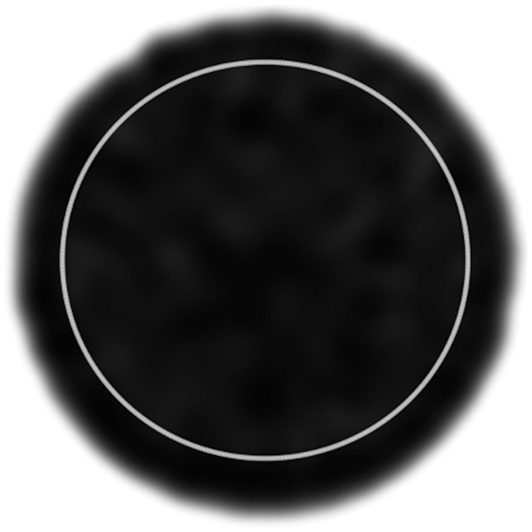
All SPECT images, acquired and reconstructed at the individual institutions, were transferred to the central institution (Tottori University Hospital) in DICOM format and analyzed using the OsiriX DICOM viewer, version 5.6 (Pixmeo). The mean radioactivity concentration (kBq/mL) in 5 circular regions of interest, drawn on consecutive slices in the center of the cylinder phantom, was calculated. Each region of interest encompassed about 80% of the interior diameter of the phantom (Fig. 1). The results were expressed as mean ± SD.

Representative slice of cylindric phantom. Gray circle indicates placement of region of interest on phantom.
We evaluated reproducibility as the interinstitutional variation in radioactivity concentrations in SPECT images, calculated using the following formula:
where mean represents the mean radioactivity concentration of acquired SPECT images and SD represents the SD of the radioactivity concentrations of the participating institutions.
Measurement accuracy in the dose calibrator test was calculated as the difference in radioactivity from the reference value as follows:
where A meas is the activity measured at each of the participating institutions and A ref is the activity measured at the manufacturer’s factory (410.6 MBq at the assay date and time).
Statistical Analysis
Differences in reproducibility were compared between the 2 imaging conditions using Wilcoxon signed-rank tests and F tests. P values of less than 0.05 were considered to indicate a statistically significant difference. All data were statistically analyzed using MATLAB, version R2013a (The MathWorks Inc.).
RESULTS
The radioactivity concentrations in SPECT images acquired under the standardized and clinical conditions were 95.71 ± 0.60 to 108.35 ± 0.36 and 96.78 ± 0.64 to 108.49 ± 0.11 kBq/mL, respectively (Table 3). Interinstitutional variation under these 2 conditions was, respectively, 4.20% and 3.89%. Reproducibility did not significantly differ between the 2 imaging conditions (P = 0.394, Wilcoxon signed-rank test; P = 0.893, F test). The results of the intrainstitutional examination at institution A are summarized in Table 4. The radioactivity concentrations in SPECT images acquired under the standardized conditions, as tested with the 2 scanners repeatedly, were 98.96 ± 0.07 to 101.81 ± 0.30 kBq/mL.
Radioactivity Concentrations in SPECT Images Under 2 Conditions at Participating Institutions
Radioactivity Concentrations in SPECT Images in Repetitive Experiment at Institution A
In Table 5, we show that at most institutions, the measurement accuracy of the dose calibrators was within ±5% of the manufacturer’s measurement. However, the measurement error at institution F was relatively high (6.13% ± 0.44%). The measured values at institutions A, B, C, D, and F tended to be higher than the reference value, whereas the measured value at institution E was an underestimation.
Measurement Accuracy for Each Institutional Dose Calibrator
DISCUSSION
Quantitative SPECT images can be generated using a commercially available OSCGM application, xSPECT Quant, the clinical use of which is becoming more prevalent. The present multicenter study investigated the reproducibility of quantitative SPECT images generated using this application. The interinstitutional variation in radioactivity concentrations was 4% under the 2 study conditions. PET studies have been reported to show a similar level of variability (18), suggesting that the quantitative reproducibility of xSPECT Quant for a homogeneous distribution of radiotracer throughout a relatively large (∼7 L) volume is good and comparable to that of PET (1,13).
Scanner variability and reconstruction parameters have been considered technologic factors affecting the accuracy of quantitative measurements in PET studies (19,20). The reproducibility of SPECT quantitation in the present study was good regardless of variability in imaging parameters. This good result might be associated with the cylindric phantom. We selected this phantom to avoid errors due to the technical difficulties involved in phantom preparation at the participating institutions. One study that used a body phantom with spheric inserts found a larger variation in quantitative values for small spheres (21). The large object size and the difference in the radiotracer are the major limitations in this study, since absolute measurements are often of most interest when applied to much smaller foci of radiotracer uptake and are of special interest for the dosimetry of therapy agents (22). Because partial-volume effects are influenced by imaging conditions (23–25), the quantitative accuracy of xSPECT Quant requires further evaluation for smaller regions of interest that might be representative of focal uptake in a lesion, for example. Moreover, previous studies mentioned that the quantitative values might be influenced by the noise characteristics in the xSPECT Bone algorithm (26,27). xSPECT Bone incorporated a weighted correction according to zone classification based on CT data. Our study design did not assess the effect of tissues zones used during reconstruction, and the 3.89% variation in clinical protocols that we found between institutions does not address the effect of zoning during reconstruction.
Dose calibrator accuracy and scanner calibration are also considerable factors in quantitative measurements (19,20). Because the filled radioactivity in the phantom was measured with each institution’s own dose calibrator, dose calibrator bias had to be considered. A comparison of Tables 3 and 5 shows that the measurement accuracy of each institution’s dose calibrator had a direct impact on bias in the radioactivity concentration in SPECT images. For example, because the dose calibrator at institution E was shown to underestimate the radioactivity, compared with the value calibrated by the manufacturer, a higher radioactivity might have been used in the cylinder phantom prepared at this site. In short, the interinstitutional variation in the phantom study can be attributed mainly to the variability in dose calibrator accuracy at the participating sites. This attribution is strengthened by the excellent intrainstitutional repeatability at institution A, as tested with identical dose calibrators. In addition to intrinsic factors such as device calibration and electronic response, the radioactivity measurement of the dose calibrator depends on source shape, material, volume, and surroundings (28–31). Each institution in our study used a commercially available 99mTc source with the same lot number to minimize variables. However, slight individual differences cannot be denied. Our study showed some general differences in measurement of radioactivity among the 6 institutions, including differences in manipulation (e.g., shielding with lead material) and in environment (e.g., temperature and humidity), not only in the intrinsic error of the devices.
Lack of a common source for calibrating detector sensitivity created potential for variability in our study. Miyaji et al. reported that the SCF of the 99mTc source depended on the preparation method whereas calibration using the 57Co standard source was stable over a long period (32). Anizan et al. also mentioned that precise preparation and careful measurement of the calibration-source activity and acquisition on a background of negligible radiation are required for stable planar-sensitivity–based calibration (33). The effects of differences among calibration sources were not assessed in this study; hence, we have no details about variability.
Although the types of dose calibrators, calibration sources, and other items differed among the participating institutions, the reproducibility of SPECT quantitation was sufficient to discuss quantitative uptake equally among multiple centers. Our results indicated that xSPECT Quant harmonizes variability in a multicenter setting. However, the present phantom measurements were limited to a single radioactivity concentration, and only 1 measurement was conducted at each institute. The interinstitutional variability and accuracy of SPECT quantitation await future evaluation.
CONCLUSION
A commercially available quantitative SPECT application reproduced radioactivity concentrations with an interinstitutional variation of 4.2%, which is comparable to the variation for PET in a comparatively large (∼7 L), homogeneous object. This multicenter study was the first step toward verification of SPECT quantitation, and further investigation of accuracy is desirable. Nonetheless, our findings are significant in terms of clinical assessments of SUV using SPECT/CT.
DISCLOSURE
No potential conflict of interest relevant to this article was reported.
Acknowledgments
We thank Nihon Medi-Physics Co., Ltd., for technical support. We also thank Takeshi Shimizu (Siemens Healthcare K.K.) for assistance.
Footnotes
Published online Jan. 8, 2021.
REFERENCES
- Received for publication September 17, 2020.
- Accepted for publication January 3, 2021.


Jump to section
Related Articles
Cited By...
- No citing articles found.