
Main menu

  • Home
  • Content
    • Current
      • JNMT Supplement
    • Ahead of print
    • Past Issues
    • Continuing Education
    • JNMT Podcast
    • SNMMI Annual Meeting Abstracts
  • Subscriptions
    • Subscribers
    • Rates
    • Journal Claims
    • Institutional and Non-member
  • Authors
    • Submit to JNMT
    • Information for Authors
    • Assignment of Copyright
    • AQARA Requirements
  • Info
    • Reviewers
    • Permissions
    • Advertisers
    • Corporate & Special Sales
  • About
    • About Us
    • Editorial Board
    • Contact Information
  • More
    • Alerts
    • Feedback
    • Help
    • SNMMI Journals
  • SNMMI
    • JNMT
    • JNM
    • SNMMI Journals
    • SNMMI

User menu

  • Subscribe
  • My alerts
  • Log in
  • My Cart

Search

  • Advanced search
Journal of Nuclear Medicine Technology
  • SNMMI
    • JNMT
    • JNM
    • SNMMI Journals
    • SNMMI
  • Subscribe
  • My alerts
  • Log in
  • My Cart
Journal of Nuclear Medicine Technology

Advanced Search

  • Home
  • Content
    • Current
    • Ahead of print
    • Past Issues
    • Continuing Education
    • JNMT Podcast
    • SNMMI Annual Meeting Abstracts
  • Subscriptions
    • Subscribers
    • Rates
    • Journal Claims
    • Institutional and Non-member
  • Authors
    • Submit to JNMT
    • Information for Authors
    • Assignment of Copyright
    • AQARA Requirements
  • Info
    • Reviewers
    • Permissions
    • Advertisers
    • Corporate & Special Sales
  • About
    • About Us
    • Editorial Board
    • Contact Information
  • More
    • Alerts
    • Feedback
    • Help
    • SNMMI Journals
  • Watch or Listen to JNMT Podcast
  • Visit SNMMI on Facebook
  • Join SNMMI on LinkedIn
  • Follow SNMMI on Twitter
  • Subscribe to JNMT RSS feeds
Research ArticleTeaching Case Studies

Isolated Malignant Portal Vein Thrombus in Colon Carcinoma: A Rare Finding on 18F-FDG PET/CECT

Tarun Kumar Jain, Ajay Yadav, Hemant Malhotra, Nitin Khunteta and Guman Singh
Journal of Nuclear Medicine Technology June 2021, 49 (2) 190-192; DOI: https://doi.org/10.2967/jnmt.120.249532
Tarun Kumar Jain
1Department of Nuclear Medicine, Mahatma Gandhi Medical College and Hospital, Jaipur, India;
  • Find this author on Google Scholar
  • Find this author on PubMed
  • Search for this author on this site
Ajay Yadav
2Department of Medical Oncology, Mahatma Gandhi Medical College and Hospital, Jaipur, India;
  • Find this author on Google Scholar
  • Find this author on PubMed
  • Search for this author on this site
Hemant Malhotra
2Department of Medical Oncology, Mahatma Gandhi Medical College and Hospital, Jaipur, India;
  • Find this author on Google Scholar
  • Find this author on PubMed
  • Search for this author on this site
Nitin Khunteta
3Department of Surgical Oncology, Mahatma Gandhi Medical College and Hospital, Jaipur, India; and
  • Find this author on Google Scholar
  • Find this author on PubMed
  • Search for this author on this site
Guman Singh
4Department of Radiation Oncology, Mahatma Gandhi Medical College and Hospital, Jaipur, India
  • Find this author on Google Scholar
  • Find this author on PubMed
  • Search for this author on this site
  • Article
  • Figures & Data
  • Info & Metrics
  • PDF
Loading

Abstract

In the absence of hepatic metastasis in abdominal cancers, an isolated malignant portal vein thrombus is very rare. The presence of a malignant thrombus has clinical significance for determining the stage, treatment, and prognosis. 18F-FDG PET/CECT is a noninvasive modality for discriminating between malignant and benign thrombi. We present a case of primary sigmoid colon carcinoma for which 18F-FDG PET/CT showed, in addition to the 18F-FDG–avid primary lesion, an 18F-FDG–avid filling defect in the portal vein, likely malignant thrombus.

  • malignant portal vein thrombus
  • colon carcinoma
  • 18F FDG PET-contrast enhanced CT

In the absence of hepatic metastasis in abdominal cancers, an isolated malignant portal vein thrombus is very rare. The presence of a malignant thrombus has clinical significance for determining the stage, treatment, and prognosis. 18F-FDG PET/CECT in which the CT portion is performed with contrast enhancement is a noninvasive modality for determining the vessel thrombus/embolus. We present a case of primary sigmoid colon carcinoma for which 18F-FDG PET/CECT showed, in addition to the 18F-FDG–avid primary lesion, an 18F-FDG–avid filling defect in the portal vein, likely malignant thrombus.

CASE REPORT

A 67-y-old man presented with a history of black stools for the last 20 days. He had severe anemia, with a hemoglobin level of 5.0 mg/dL. There was no past history of previous surgery, tuberculosis, hepatitis, or any other chronic illness. On physical examination, there was no specific finding other than severe pallor. On ultrasonography, a long segmental wall thickening was seen in the sigmoid colon and a filling defect in the portal vein. The patient underwent sigmoidoscopy and biopsy of the lesion, histopathologic examination of which revealed moderately differentiated adenocarcinoma.

18F-FDG PET/CECT was performed for staging and to assess the nature of the filling defect in the portal venous system. Fifty minutes after intravenous injection of 370 MBq (10 mCi) of 18F-FDG, PET/CECT was performed on a dedicated hybrid scanner (Discovery STE 16; GE Healthcare). Before the PET/CECT scan, a low-dose scout scan (120 kV, 10 mA) was acquired from the base of the skull to the mid thigh. For the PET/CECT scan, a portal-venous–phase contrast-enhanced CT scan (120 kV, 340 mA) was first obtained from the base of the skull to the mid thigh, 55 s after the beginning of 90 mL of isoosmolar contrast medium had been intravenously injected. After the CECT acquisition, a 3-dimensional PET acquisition was done in the caudocranial direction with an acquisition time of 2 min per bed position.

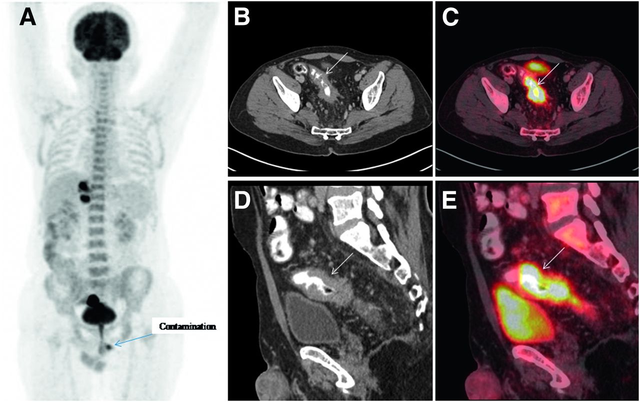
A maximum-intensity-projection PET image (Fig. 1A) revealed a few hot spots in the paramedian plane of the right upper abdominal region and the pelvic region. Transaxial and sagittal CECT and PET/CECT images of the pelvis (Figs. 1B–1E) revealed 18F-FDG–avid (SUVmax, ∼21.7) long segmental asymmetrical wall thickening in the sigmoid colon.

FIGURE 1.
  • Download figure
  • Open in new tab
  • Download powerpoint
FIGURE 1.

18F-FDG PET maximum-intensity-projection image (A) reveals few hot spots in paramedian plane of right upper abdominal region and in pelvic region. Transaxial (B and C) and sagittal (D and E) CT images (B and D) and PET/CECT images (C and E) of pelvis reveal 18F-FDG–avid (SUVmax, ∼21.7) long segmental asymmetric wall thickening (arrows) in sigmoid colon, with perilesional stranding and nodularity.

Transaxial (Fig. 2) and sagittal and coronal (Fig. 3) CECT and PET/CECT images of the upper abdomen revealed an 18F-FDG–avid filling defect in the main portal vein (SUVmax, 18.3) and left portal vein (SUVmax, 16.9), suggestive of a malignant thrombus. There was no 18F-FDG–avid lesion in the liver parenchyma.

FIGURE 2.
  • Download figure
  • Open in new tab
  • Download powerpoint
FIGURE 2.

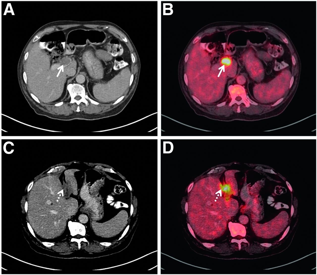
Transaxial CT images (A and C) and PET/CECT images (B and D) of upper abdomen reveal 18F-FDG–avid filling defect in main portal vein (solid arrows; SUVmax, 18.3) and left portal vein (dashed arrows; SUVmax, 16.9).

FIGURE 3.
  • Download figure
  • Open in new tab
  • Download powerpoint
FIGURE 3.

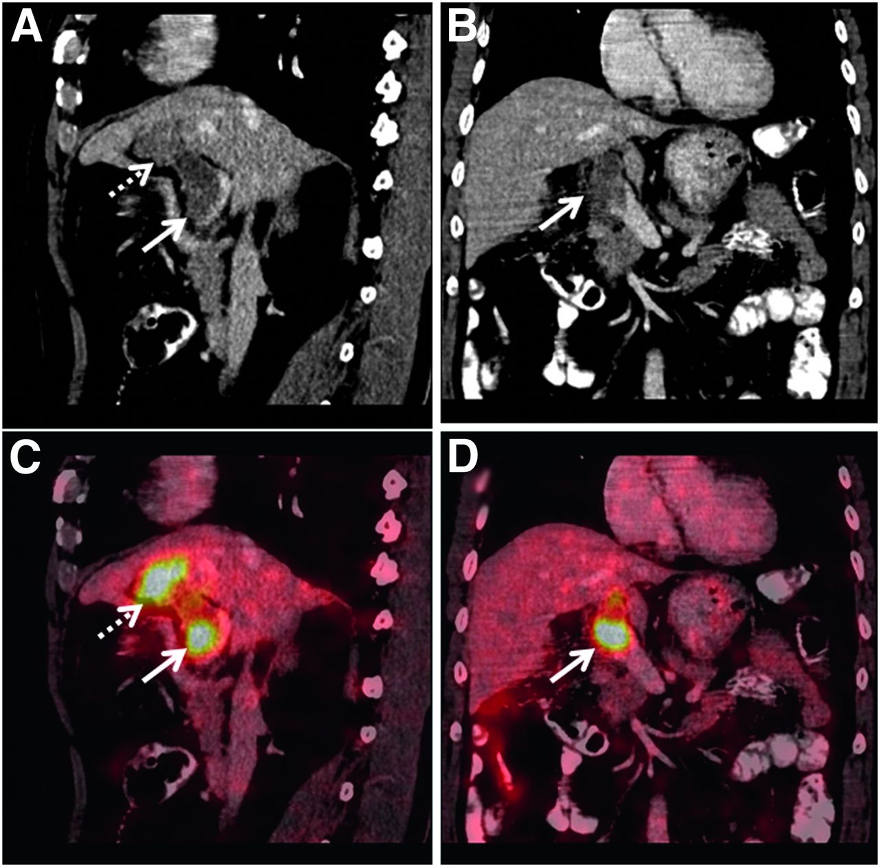
Sagittal (A and C) and coronal (B and D) CT images (A and B) and PET/CECT images (C and D) of abdomen reveal 18F-FDG–avid filling defect in main portal vein (solid arrows) and left portal vein (dashed arrows).

After PET/CECT, it was decided to give the patient initial chemotherapy with the capecitabine-plus-oxaliplatin regimen along with oral anticoagulant medication (dabigatran, 150 mg twice daily) rather than surgery. After 3 cycles of chemotherapy, the patient died at his home after a few hours of sudden respiratory distress, possibly from pulmonary thromboembolism (unconfirmed), before a response evaluation could be done.

DISCUSSION

Malignant portal vein tumor thrombosis is a rare entity, seen in approximately only 2%–3% of patients with either primary or secondary malignant liver lesions (1). Possible mechanisms for malignant portal vein tumor thrombosis that have been mentioned in the literature are shunt formation between the hepatic artery and the portal vein and microscopic invasion of the portal vein from an adjacent hepatic lobule (2,3). An isolated malignant portal vein tumor thrombus without liver involvement is very rare, and only a few cases have been reported in the literature (2,3). The possible pathogenesis is a hypercoagulable state induced by the primary malignancy (4). During routine contrast-enhanced CT imaging, venous thrombosis—whether malignant or benign—appears as a low-attenuation area or filling defect in the vein. Therefore, it is difficult to differentiate between tumor thrombosis and blood clot thrombosis on the basis of the CECT data alone (5). 18F-FDG PET/CECT helps distinguish tumor thrombosis from blood clot thrombosis on the basis of the tracer accumulation (6). In the present case, 18F-FDG PET/CECT showed tracer-avid malignancy in the sigmoid colon (primary) and portal vein, with no avid lesions in the liver.

A possible approach to management of such patients is resection of the primary tumor and of the portal venous thrombus followed by chemotherapy along with oral anticoagulants or subcutaneous low-molecular-weight heparin. As per the limited literature available, the prognosis of these patients is poor; our patient died after 3 cycles of chemotherapy, before the disease response could be evaluated (7).

CONCLUSION

The presence of malignant venous thrombi is a poor prognostic factor in malignancies. 18F-FDG PET/CECT is a reliable, noninvasive modality to detect a malignant venous thrombus.

DISCLOSURE

No potential conflict of interest relevant to this article was reported.

Footnotes

  • Published online Nov. 20, 2020.

REFERENCES

  1. 1.↵
    1. Oh SD,
    2. Oh SJ,
    3. Suh BJ,
    4. Shin JY,
    5. Park JK
    . Long-term survival of a patient with adenocarcinoma of the esophagogastric junction with a portal vein tumor thrombosis who underwent palliative total gastrectomy: a case report. Case Rep Oncol. 2017;10:916–922.
    OpenUrl
  2. 2.↵
    1. Yamamoto N,
    2. Sugano N,
    3. Morinaga S,
    4. et al.
    Massive portal vein tumor thrombus from colorectal cancer without any metastatic nodules in the liver parenchyma. Rare Tumors. 2011;3:e47.
    OpenUrlPubMed
  3. 3.↵
    1. Matsumoto J,
    2. Kojima T,
    3. Hiraguchi E,
    4. Abe M
    . Portal vein tumor thrombus from colorectal cancer with no definite metastatic nodules in liver parenchyma. J Hepatobiliary Pancreat Surg. 2009;16:688–691.
    OpenUrlPubMed
  4. 4.↵
    1. Rees PA,
    2. Clouston HW,
    3. Duff S,
    4. Kirwan CC
    . Colorectal cancer and thrombosis. Int J Colorectal Dis. 2018;33:105–108.
    OpenUrl
  5. 5.↵
    1. Kim SE,
    2. Lee SJ,
    3. Cha JY,
    4. et al.
    Ascending colon cancer with pathologically confirmed tumor thrombosis of superior mesenteric vein: a case report. Clin Endosc. 2019;52:506–509.
    OpenUrl
  6. 6.↵
    1. Hu S,
    2. Zhang J,
    3. Cheng C,
    4. Liu Q,
    5. Sun G,
    6. Zuo C
    . The role of 18F-FDG PET/CT in differentiating malignant from benign portal vein thrombosis. Abdom Imaging. 2014;39:1221–1227.
    OpenUrl
  7. 7.↵
    1. Tada K,
    2. Kokudo N,
    3. Seki M,
    4. et al.
    Hepatic resection for colorectal metastasis with macroscopic tumor thrombus in the portal vein. World J Surg. 2003;27:299–303.
    OpenUrlPubMed
  • Received for publication June 11, 2020.
  • Accepted for publication October 19, 2020.
PreviousNext
Back to top

In this issue

Journal of Nuclear Medicine Technology: 49 (2)
Journal of Nuclear Medicine Technology
Vol. 49, Issue 2
June 1, 2021
  • Table of Contents
  • About the Cover
  • Index by author
Print
Download PDF
Article Alerts
Sign In to Email Alerts with your Email Address
Email Article

Thank you for your interest in spreading the word on Journal of Nuclear Medicine Technology.

NOTE: We only request your email address so that the person you are recommending the page to knows that you wanted them to see it, and that it is not junk mail. We do not capture any email address.

Enter multiple addresses on separate lines or separate them with commas.
Isolated Malignant Portal Vein Thrombus in Colon Carcinoma: A Rare Finding on 18F-FDG PET/CECT
(Your Name) has sent you a message from Journal of Nuclear Medicine Technology
(Your Name) thought you would like to see the Journal of Nuclear Medicine Technology web site.
Citation Tools
Isolated Malignant Portal Vein Thrombus in Colon Carcinoma: A Rare Finding on 18F-FDG PET/CECT
Tarun Kumar Jain, Ajay Yadav, Hemant Malhotra, Nitin Khunteta, Guman Singh
Journal of Nuclear Medicine Technology Jun 2021, 49 (2) 190-192; DOI: 10.2967/jnmt.120.249532

Citation Manager Formats

  • BibTeX
  • Bookends
  • EasyBib
  • EndNote (tagged)
  • EndNote 8 (xml)
  • Medlars
  • Mendeley
  • Papers
  • RefWorks Tagged
  • Ref Manager
  • RIS
  • Zotero
Share
Isolated Malignant Portal Vein Thrombus in Colon Carcinoma: A Rare Finding on 18F-FDG PET/CECT
Tarun Kumar Jain, Ajay Yadav, Hemant Malhotra, Nitin Khunteta, Guman Singh
Journal of Nuclear Medicine Technology Jun 2021, 49 (2) 190-192; DOI: 10.2967/jnmt.120.249532
Twitter logo Facebook logo LinkedIn logo Mendeley logo
  • Tweet Widget
  • Facebook Like
  • Google Plus One
Bookmark this article

Jump to section

  • Article
    • Abstract
    • CASE REPORT
    • DISCUSSION
    • CONCLUSION
    • DISCLOSURE
    • Footnotes
    • REFERENCES
  • Figures & Data
  • Info & Metrics
  • PDF

Related Articles

  • No related articles found.
  • PubMed
  • Google Scholar

Cited By...

  • No citing articles found.
  • Google Scholar

More in this TOC Section

  • High-Sensitivity Troponin Elevation in a Young Woman with Typical Chest Pain: The Heart of the Matter
  • Pulmonary Adenocarcinoma Revealed by Parathyroid Scintigraphy: An Incidental Case to Remember
  • Prominent Right Ventricular Tracer Uptake: A Harbinger of Multivessel Coronary Artery Disease
Show more Teaching Case Studies

Similar Articles

Keywords

  • malignant portal vein thrombus
  • colon carcinoma
  • 18F FDG PET-contrast enhanced CT
SNMMI

© 2025 SNMMI

Powered by HighWire