
Main menu

  • Home
  • Content
    • Current
      • JNMT Supplement
    • Ahead of print
    • Past Issues
    • Continuing Education
    • JNMT Podcast
    • SNMMI Annual Meeting Abstracts
  • Subscriptions
    • Subscribers
    • Rates
    • Journal Claims
    • Institutional and Non-member
  • Authors
    • Submit to JNMT
    • Information for Authors
    • Assignment of Copyright
    • AQARA Requirements
  • Info
    • Reviewers
    • Permissions
    • Advertisers
    • Corporate & Special Sales
  • About
    • About Us
    • Editorial Board
    • Contact Information
  • More
    • Alerts
    • Feedback
    • Help
    • SNMMI Journals
  • SNMMI
    • JNMT
    • JNM
    • SNMMI Journals
    • SNMMI

User menu

  • Subscribe
  • My alerts
  • Log in
  • My Cart

Search

  • Advanced search
Journal of Nuclear Medicine Technology
  • SNMMI
    • JNMT
    • JNM
    • SNMMI Journals
    • SNMMI
  • Subscribe
  • My alerts
  • Log in
  • My Cart
Journal of Nuclear Medicine Technology

Advanced Search

  • Home
  • Content
    • Current
    • Ahead of print
    • Past Issues
    • Continuing Education
    • JNMT Podcast
    • SNMMI Annual Meeting Abstracts
  • Subscriptions
    • Subscribers
    • Rates
    • Journal Claims
    • Institutional and Non-member
  • Authors
    • Submit to JNMT
    • Information for Authors
    • Assignment of Copyright
    • AQARA Requirements
  • Info
    • Reviewers
    • Permissions
    • Advertisers
    • Corporate & Special Sales
  • About
    • About Us
    • Editorial Board
    • Contact Information
  • More
    • Alerts
    • Feedback
    • Help
    • SNMMI Journals
  • Watch or Listen to JNMT Podcast
  • Visit SNMMI on Facebook
  • Join SNMMI on LinkedIn
  • Follow SNMMI on Twitter
  • Subscribe to JNMT RSS feeds
Review ArticleTeaching Case Studies

Intracranial Leptomeningeal Carcinomatosis from Breast Cancer Detected on 18F-FDG PET

Bradley J. Carra and Michael N. Clemenshaw
Journal of Nuclear Medicine Technology September 2015, 43 (3) 227-228; DOI: https://doi.org/10.2967/jnmt.114.148791
Bradley J. Carra
Department of Nuclear Medicine, San Antonio Military Medical Center, Fort Sam Houston, Texas
  • Find this author on Google Scholar
  • Find this author on PubMed
  • Search for this author on this site
Michael N. Clemenshaw
Department of Nuclear Medicine, San Antonio Military Medical Center, Fort Sam Houston, Texas
  • Find this author on Google Scholar
  • Find this author on PubMed
  • Search for this author on this site
  • Article
  • Figures & Data
  • Info & Metrics
  • PDF
Loading

Abstract

Leptomeningeal carcinomatosis is an uncommon manifestation of non–central nervous system (CNS) metastatic disease. Diagnosis, however, has important prognostic and treatment implications. We present a case in which intracranial leptomeningeal carcinomatosis from a primary breast cancer was detected with 18F-FDG PET/CT, despite its low sensitivity for detection of CNS metastases from non-CNS primary tumors.

  • 18F-FDG PET/CT
  • leptomeningeal carcinomatosis
  • breast cancer

Leptomeningeal carcinomatosis (LC) is an uncommon late manifestation of non–central nervous system (CNS) solid malignancies. 18F-FDG PET/CT has a low sensitivity for detection of CNS metastases from non-CNS primary tumors (1). 18F-FDG PET/CT detection of LC from other non-CNS primary tumors has been described (1), and a few cases of 18F-FDG PET/CT detection of spinal LC from breast cancer (2,3) have been reported. To our knowledge, however, this is the first reported case of intracranial LC from breast cancer detected with 18F-FDG PET/CT.

CASE REPORT

A 38-y-old woman with stage IV inflammatory ductal carcinoma of the breast metastatic to the bones presented with ataxia 2 wk after completion of palliative chemotherapy. Contrast-enhanced brain MR imaging demonstrated nodular leptomeningeal enhancement primarily of the cerebellar folia (Fig. 1). A differential diagnosis of LC versus infectious meningitis was provided. A total of 4 cerebrospinal fluid (CSF) samples were negative for infection and metastatic disease.

FIGURE 1.
  • Download figure
  • Open in new tab
  • Download powerpoint
FIGURE 1.

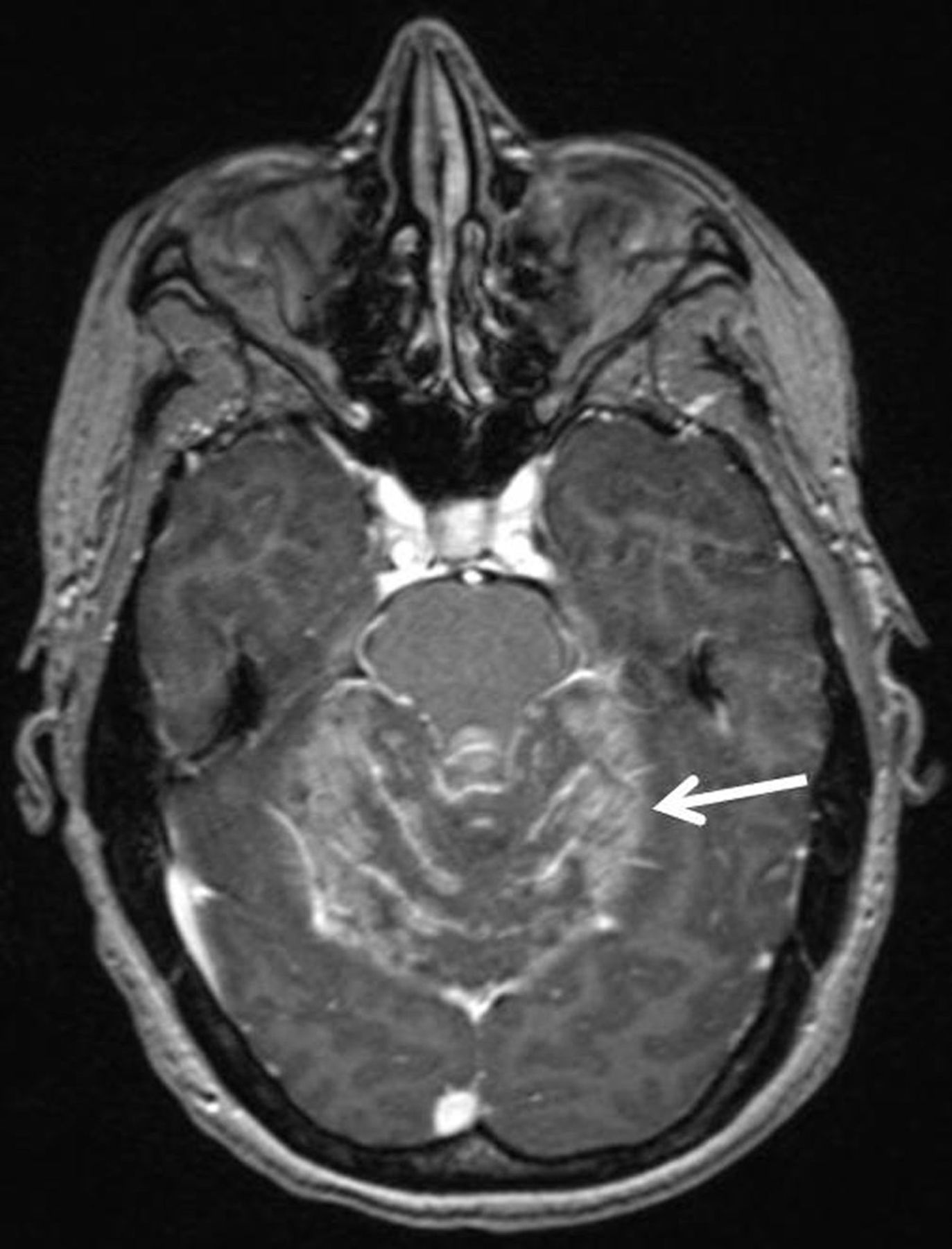
Postcontrast axial T1-weighted image of brain demonstrating leptomeningeal enhancement of cerebellar folia (arrow).

18F-FDG PET/CT was performed for restaging purposes. Imaging from the skull vertex through the mid femora was begun 90 min after intravenous injection of 384 MBq of 18F-FDG. Imaging revealed increased 18F-FDG avidity conforming to the sites of leptomeningeal enhancement on brain MR imaging, as well as progression of osseous metastatic disease (Fig. 2). Despite negative CSF cytology results, the presence of other metastatic disease detected on 18F-FDG PET/CT and a lack of infectious symptoms supported a diagnosis of LC. Neurologic symptoms rapidly regressed after placement of a ventriculostomy and initiation of high-dose steroids and intrathecal chemotherapy.

FIGURE 2.
  • Download figure
  • Open in new tab
  • Download powerpoint
FIGURE 2.

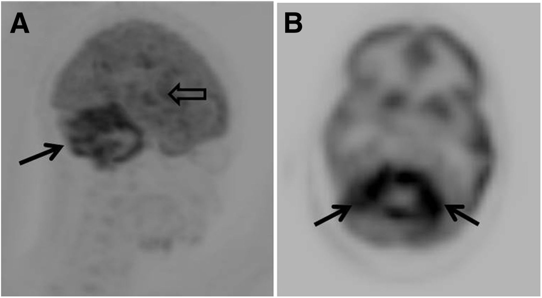
Maximum intensity projection (A) and axial image (B) from 18F-FDG PET showing increased leptomeningeal radiotracer uptake in cerebellum (solid arrows) and temporal lobe (open arrow).

DISCUSSION

The most common intracranial locations of CSF tumor seeding are the basilar cisterns and cerebellum. Although breast cancer is the most common nonhematologic tumor with leptomeningeal spread, the incidence remains low at only 1%–5% (4,5). LC has a poor prognosis, with median survival of only 2–4 mo (2).

Detection of metastatic cells within the CSF remains the gold standard for diagnosis, but the yield is poor, with detection rates of only 50% on initial CSF sampling (4). Contrast-enhanced MR imaging is the imaging modality of choice for diagnosis showing leptomeningeal enhancement. Despite its low sensitivity, 18F-FDG PET/CT may show increased leptomeningeal metabolic activity (1).

Leptomeningeal enhancement and increased 18F-FDG avidity are both nonspecific findings and can be seen in infectious meningitis, LC, or neurosarcoidosis. In conjunction with clinical information, however, they can suggest the diagnosis of LC. Although MR imaging is likely more sensitive in detecting LC, whole-body 18F-FDG PET/CT has the advantage of demonstrating other metastatic disease, an important finding because LC is usually a late manifestation of metastatic disease and less commonly seen in isolation (5).

CONCLUSION

Although the sensitivity of 18F-FDG PET/CT for detection of CNS metastases is poor, routine staging examinations cover the most common locations of intracranial leptomeningeal metastases. Detection is therefore possible and has important prognostic and treatment implications. Although MR imaging is the imaging modality of choice for diagnosing leptomeningeal metastasis, whole-body 18F-FDG PET/CT is uniquely able to detect other systemic metastatic disease, important information supporting a diagnosis of LC.

DISCLOSURE

No potential conflict of interest relevant to this article was reported.

Footnotes

  • Published online Feb. 5, 2015.

REFERENCES

  1. 1.↵
    1. Rohren EM,
    2. Provenzale JM,
    3. Barboriak DP,
    4. Coleman RE
    . Screening for cerebral metastases with FDG PET in patients undergoing whole-body staging of non-central nervous system malignancy. Radiology. 2003;226:181–187.
    OpenUrlCrossRefPubMed
  2. 2.↵
    1. Shah S,
    2. Rangarajan V,
    3. Purandare N,
    4. Luthra K,
    5. Medhi S
    . 18F-FDG uptakes in leptomeningeal metastases from carcinoma of the breast on a PET/CT study. Indian J Cancer. 2007;44:115–118.
    OpenUrlCrossRefPubMed
  3. 3.↵
    1. Grande ML,
    2. Rayo J,
    3. Serrano J,
    4. Infante J,
    5. Garcia L,
    6. Duran C
    . Leptomeningeal carcinomatosis as only pathological finding at FDG-PET/CT in case of tumor marker elevation in breast cancer. Indian J Nucl Med. 2014;29:53–54.
    OpenUrlCrossRefPubMed
  4. 4.↵
    1. Wasserstrom WR,
    2. Glass JP,
    3. Posner JB
    . Diagnosis and treatment of leptomeningeal metastases from solid tumors. Cancer. 1982;49:759–772.
    OpenUrlCrossRefPubMed
  5. 5.↵
    1. Boogerd W,
    2. Hart AAM,
    3. vanderSande JJ,
    4. Engelsman E
    . Meningeal carcinomatosis in breast cancer. Cancer. 1991;67:1685–1695.
    OpenUrlCrossRefPubMed
  • Received for publication September 19, 2014.
  • Accepted for publication November 13, 2014.
PreviousNext
Back to top

In this issue

Journal of Nuclear Medicine Technology: 43 (3)
Journal of Nuclear Medicine Technology
Vol. 43, Issue 3
September 1, 2015
  • Table of Contents
  • About the Cover
  • Index by author
Print
Download PDF
Article Alerts
Sign In to Email Alerts with your Email Address
Email Article

Thank you for your interest in spreading the word on Journal of Nuclear Medicine Technology.

NOTE: We only request your email address so that the person you are recommending the page to knows that you wanted them to see it, and that it is not junk mail. We do not capture any email address.

Enter multiple addresses on separate lines or separate them with commas.
Intracranial Leptomeningeal Carcinomatosis from Breast Cancer Detected on 18F-FDG PET
(Your Name) has sent you a message from Journal of Nuclear Medicine Technology
(Your Name) thought you would like to see the Journal of Nuclear Medicine Technology web site.
Citation Tools
Intracranial Leptomeningeal Carcinomatosis from Breast Cancer Detected on 18F-FDG PET
Bradley J. Carra, Michael N. Clemenshaw
Journal of Nuclear Medicine Technology Sep 2015, 43 (3) 227-228; DOI: 10.2967/jnmt.114.148791

Citation Manager Formats

  • BibTeX
  • Bookends
  • EasyBib
  • EndNote (tagged)
  • EndNote 8 (xml)
  • Medlars
  • Mendeley
  • Papers
  • RefWorks Tagged
  • Ref Manager
  • RIS
  • Zotero
Share
Intracranial Leptomeningeal Carcinomatosis from Breast Cancer Detected on 18F-FDG PET
Bradley J. Carra, Michael N. Clemenshaw
Journal of Nuclear Medicine Technology Sep 2015, 43 (3) 227-228; DOI: 10.2967/jnmt.114.148791
Twitter logo Facebook logo LinkedIn logo Mendeley logo
  • Tweet Widget
  • Facebook Like
  • Google Plus One
Bookmark this article

Jump to section

  • Article
    • Abstract
    • CASE REPORT
    • DISCUSSION
    • CONCLUSION
    • DISCLOSURE
    • Footnotes
    • REFERENCES
  • Figures & Data
  • Info & Metrics
  • PDF

Related Articles

  • No related articles found.
  • PubMed
  • Google Scholar

Cited By...

  • Diagnostic challenges in neoplastic meningitis presenting as intracranial hypertension
  • Google Scholar

More in this TOC Section

  • High-Sensitivity Troponin Elevation in a Young Woman with Typical Chest Pain: The Heart of the Matter
  • Pulmonary Adenocarcinoma Revealed by Parathyroid Scintigraphy: An Incidental Case to Remember
  • Prominent Right Ventricular Tracer Uptake: A Harbinger of Multivessel Coronary Artery Disease
Show more Teaching Case Studies

Similar Articles

Keywords

  • 18F-FDG PET/CT
  • leptomeningeal carcinomatosis
  • breast cancer
SNMMI

© 2025 SNMMI

Powered by HighWire