
Main menu

  • Home
  • Content
    • Current
      • JNMT Supplement
    • Ahead of print
    • Past Issues
    • Continuing Education
    • JNMT Podcast
    • SNMMI Annual Meeting Abstracts
  • Subscriptions
    • Subscribers
    • Rates
    • Journal Claims
    • Institutional and Non-member
  • Authors
    • Submit to JNMT
    • Information for Authors
    • Assignment of Copyright
    • AQARA Requirements
  • Info
    • Reviewers
    • Permissions
    • Advertisers
    • Corporate & Special Sales
  • About
    • About Us
    • Editorial Board
    • Contact Information
  • More
    • Alerts
    • Feedback
    • Help
    • SNMMI Journals
  • SNMMI
    • JNMT
    • JNM
    • SNMMI Journals
    • SNMMI

User menu

  • Subscribe
  • My alerts
  • Log in
  • My Cart

Search

  • Advanced search
Journal of Nuclear Medicine Technology
  • SNMMI
    • JNMT
    • JNM
    • SNMMI Journals
    • SNMMI
  • Subscribe
  • My alerts
  • Log in
  • My Cart
Journal of Nuclear Medicine Technology

Advanced Search

  • Home
  • Content
    • Current
    • Ahead of print
    • Past Issues
    • Continuing Education
    • JNMT Podcast
    • SNMMI Annual Meeting Abstracts
  • Subscriptions
    • Subscribers
    • Rates
    • Journal Claims
    • Institutional and Non-member
  • Authors
    • Submit to JNMT
    • Information for Authors
    • Assignment of Copyright
    • AQARA Requirements
  • Info
    • Reviewers
    • Permissions
    • Advertisers
    • Corporate & Special Sales
  • About
    • About Us
    • Editorial Board
    • Contact Information
  • More
    • Alerts
    • Feedback
    • Help
    • SNMMI Journals
  • Watch or Listen to JNMT Podcast
  • Visit SNMMI on Facebook
  • Join SNMMI on LinkedIn
  • Follow SNMMI on Twitter
  • Subscribe to JNMT RSS feeds
Review ArticleTEACHING FILES

Increased Sacral Uptake on a Bone Scan with SPECT/CT in a Patient with Achondroplasia: Normal or Abnormal?

Alexander Copelan, Kristin Booth and Helena Balon
Journal of Nuclear Medicine Technology September 2013, 41 (3) 234-235; DOI: https://doi.org/10.2967/jnmt.113.126417
Alexander Copelan
1Beaumont Health System, Royal Oak, Michigan; and
  • Find this author on Google Scholar
  • Find this author on PubMed
  • Search for this author on this site
Kristin Booth
2McLaren Oakland Medical Center, Pontiac, Michigan
  • Find this author on Google Scholar
  • Find this author on PubMed
  • Search for this author on this site
Helena Balon
1Beaumont Health System, Royal Oak, Michigan; and
  • Find this author on Google Scholar
  • Find this author on PubMed
  • Search for this author on this site
  • Article
  • Figures & Data
  • Info & Metrics
  • PDF
Loading

A 26-y-old man with achondroplasia presented with chronic lower back pain with no history of trauma. MR imaging performed 5 mo previously demonstrated a congenitally small lumbar canal causing multilevel stenosis.

After intravenous administration of 814 MBq (22 mCi) of 99mTc-methylene diphosphonate, delayed anterior and posterior whole-body bone scans were performed, along with spot views of the pelvis. On review of the planar images, additional SPECT/CT imaging of the pelvis was performed to clarify the findings.

Limited-field-of-view, low-dose CT imaging was performed for anatomic localization and attenuation correction of the SPECT data.

QUESTION 1

After reviewing Figures 1–4⇓⇓⇓, what is the most likely cause of the focal increased uptake in the sacral region seen only on the planar images?

  1. Sacral fracture.

  2. Osteomyelitis.

  3. Horizontal orientation of the sacrum.

  4. Osteoid osteoma.

FIGURE 1.
  • Download figure
  • Open in new tab
  • Download powerpoint
FIGURE 1.

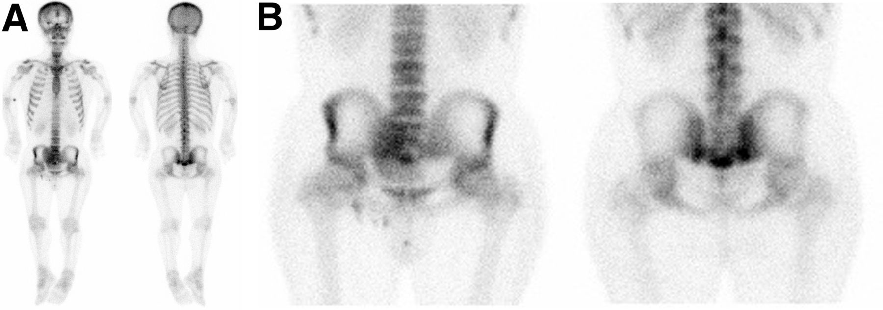
Whole-body bone scan and spot views of pelvis demonstrate mild to moderate increased radiotracer activity in right and left sacroiliac joints inferiorly and in sacrococcygeal area, raising possibility of fracture. Mild focal increased uptake is also noted bilaterally along proximal humeral cortices.

FIGURE 2.
  • Download figure
  • Open in new tab
  • Download powerpoint
FIGURE 2.

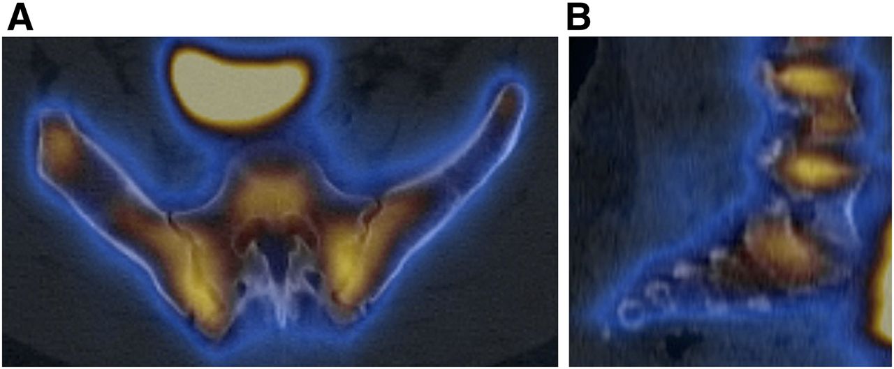
Axial (A) and sagittal (B) SPECT/CT images of pelvis and sacroiliac joints demonstrate mild increased radiotracer activity in sacroiliac joints bilaterally. Sacrococcygeal uptake seen on planar images is not apparent on SPECT images.

FIGURE 3.
  • Download figure
  • Open in new tab
  • Download powerpoint
FIGURE 3.

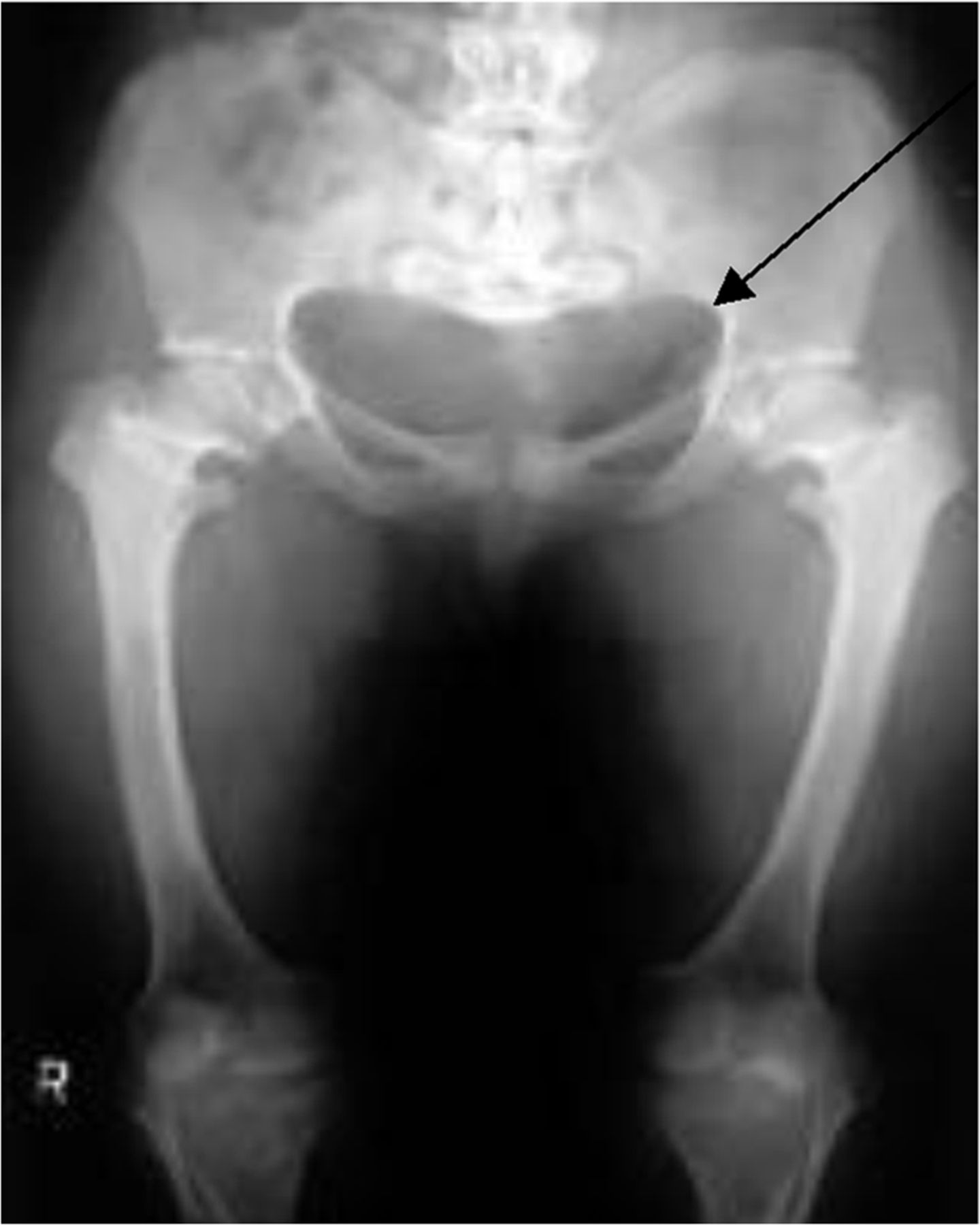
Champagne glass appearance of pelvis (arrow), which is due to anteversion of pelvis, squaring-off of ilia, lower lying anterior superior iliac spine, and higher posterior superior iliac spine.

FIGURE 4.
  • Download figure
  • Open in new tab
  • Download powerpoint
FIGURE 4.

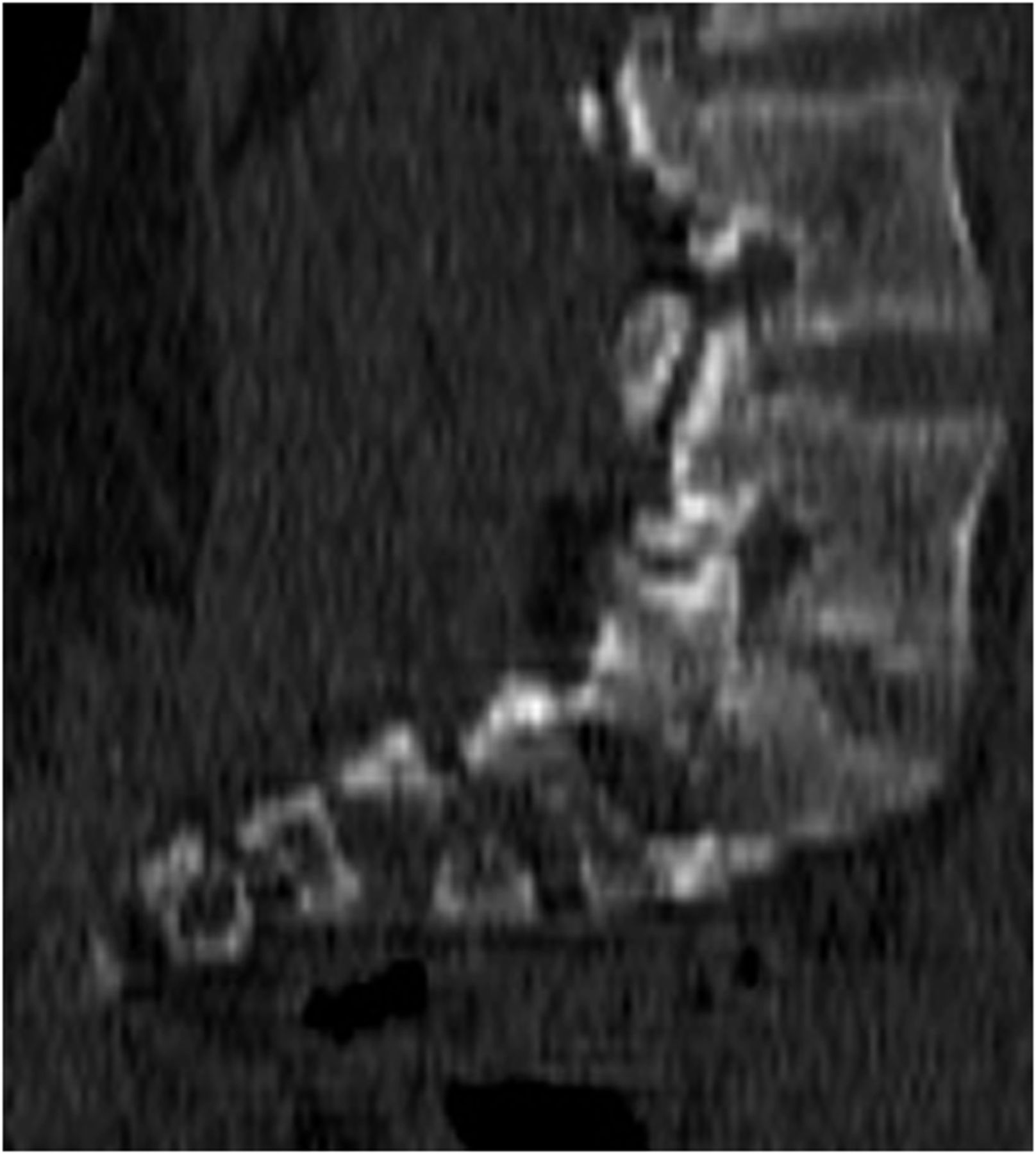
Sagittal slice (from SPECT/CT) demonstrating horizontal configuration of sacrum.

QUESTION 2

Which of the following is true regarding achondroplasia?

  1. It can be diagnosed on prenatal ultrasound.

  2. It is the second most common cause of dwarfism.

  3. It is typically diagnosed with genetic testing.

  4. It affects the axial skeleton more than the appendicular skeleton.

QUESTION 3

Which of the following is not associated with achondroplasia?

  1. Narrowing of the spinal canal.

  2. Below average IQ.

  3. “Champagne glass” pelvis.

  4. Depressed nasal bridge

QUESTION 4

Which of the following is a major complication later in life in achondroplasia patients?

  1. Genu valgus of the knee.

  2. Pulmonary hypertension.

  3. Infertility.

  4. Foramen magnum stenosis.

DISCUSSION

Achondroplasia is the most common cause of dwarfism. About 75% of cases are due to sporadic mutations, often associated with advanced maternal age, but it can also be inherited as an autosomal dominant genetic disorder. Achondroplasia occurs because of an abnormality of cartilage formation secondary to a mutation in the gene for fibroblast growth factor receptor 3 (1). The condition can be diagnosed with prenatal ultrasound or on the basis of clinical and radiographic findings; genetic testing is not typically needed (2). The main radiographic findings involve the skull, extremities, spine, and pelvis. Narrowing of the spinal canal is the pathologic hallmark (3). Platyspondyly (flattened vertebral bodies) can be seen, and decreasing interpedicular spaces caudally from L1 to L5 can lead to spinal stenosis and lower back pain. The skull often demonstrates a prominent forehead and depressed nasal bridge. The skull base is disproportionately small because it forms by endochondral ossification, whereas the skull vault is normal because it forms by membranous ossification. This disproportionate growth can lead to a small cisterna magna, empty sella, and a narrowed foramen magnum, which may necessitate cervicomedullary decompression (4,5). The extremities are disproportionately shortened, especially proximally—known as rhizomelic shortening—and are often widened because of unopposed appositional bone growth. This is the most likely explanation for the faint uptake in the proximal humeri in Figure 1. The pelvis can demonstrate a champagne glass appearance secondary to anteversion, squared-off and shortened ilia, a low anterior superior iliac spine, and a high posterior superior iliac spine. The pelvis itself is typically small, and horizontally oriented acetabula are frequently seen.

Footnotes

  • Published online Jul. 18, 2013.

  • ↵* For the answers, see page 241.

REFERENCES

  1. 1.↵
    1. Horton WA,
    2. Hall JG,
    3. Hecht JT
    . Achondroplasia. Lancet. 2007;370:162–172.
    OpenUrlCrossRefPubMed
  2. 2.↵
    1. Modaff P,
    2. Horton VK,
    3. Pauli RM
    . Errors in the prenatal diagnosis of children with achondroplasia. Prenat Diagn. 1996;16:525–530.
    OpenUrlCrossRefPubMed
  3. 3.↵
    1. Lonstein JE
    . Treatment of kyphosis and lumbar stenosis in achondroplasia. Basic Life Sci. 1988;48:283–292.
    OpenUrlPubMed
  4. 4.↵
    1. Smoker WR,
    2. Khanna G
    . Imaging the craniocervical junction. Childs Nerv Syst. 2008;24:1123–1145.
    OpenUrlCrossRefPubMed
  5. 5.↵
    1. Morgan DF,
    2. Young RF
    . Spinal neurological complications of achondroplasia: results of surgical treatment. J Neurosurg. 1980;52:463–472.
    OpenUrlPubMed
PreviousNext
Back to top

In this issue

Journal of Nuclear Medicine Technology: 41 (3)
Journal of Nuclear Medicine Technology
Vol. 41, Issue 3
September 1, 2013
  • Table of Contents
  • About the Cover
  • Index by author
Print
Download PDF
Article Alerts
Sign In to Email Alerts with your Email Address
Email Article

Thank you for your interest in spreading the word on Journal of Nuclear Medicine Technology.

NOTE: We only request your email address so that the person you are recommending the page to knows that you wanted them to see it, and that it is not junk mail. We do not capture any email address.

Enter multiple addresses on separate lines or separate them with commas.
Increased Sacral Uptake on a Bone Scan with SPECT/CT in a Patient with Achondroplasia: Normal or Abnormal?
(Your Name) has sent you a message from Journal of Nuclear Medicine Technology
(Your Name) thought you would like to see the Journal of Nuclear Medicine Technology web site.
Citation Tools
Increased Sacral Uptake on a Bone Scan with SPECT/CT in a Patient with Achondroplasia: Normal or Abnormal?
Alexander Copelan, Kristin Booth, Helena Balon
Journal of Nuclear Medicine Technology Sep 2013, 41 (3) 234-235; DOI: 10.2967/jnmt.113.126417

Citation Manager Formats

  • BibTeX
  • Bookends
  • EasyBib
  • EndNote (tagged)
  • EndNote 8 (xml)
  • Medlars
  • Mendeley
  • Papers
  • RefWorks Tagged
  • Ref Manager
  • RIS
  • Zotero
Share
Increased Sacral Uptake on a Bone Scan with SPECT/CT in a Patient with Achondroplasia: Normal or Abnormal?
Alexander Copelan, Kristin Booth, Helena Balon
Journal of Nuclear Medicine Technology Sep 2013, 41 (3) 234-235; DOI: 10.2967/jnmt.113.126417
Twitter logo Facebook logo LinkedIn logo Mendeley logo
  • Tweet Widget
  • Facebook Like
  • Google Plus One
Bookmark this article

Jump to section

  • Article
    • QUESTION 1
    • QUESTION 2
    • QUESTION 3
    • QUESTION 4
    • DISCUSSION
    • Footnotes
    • REFERENCES
  • Figures & Data
  • Info & Metrics
  • PDF

Related Articles

  • No related articles found.
  • PubMed
  • Google Scholar

Cited By...

  • No citing articles found.
  • Google Scholar

More in this TOC Section

  • Patient with Acute Shortness of Breath, Elevated D-Dimer, and a Previous History of Cancer
  • Bronchopleural Fistula Mimicking Chronic Obstructive Lung Disease on 133Xe Scan
Show more TEACHING FILES

Similar Articles

SNMMI

© 2025 SNMMI

Powered by HighWire