


Article Figures & Data
Figures
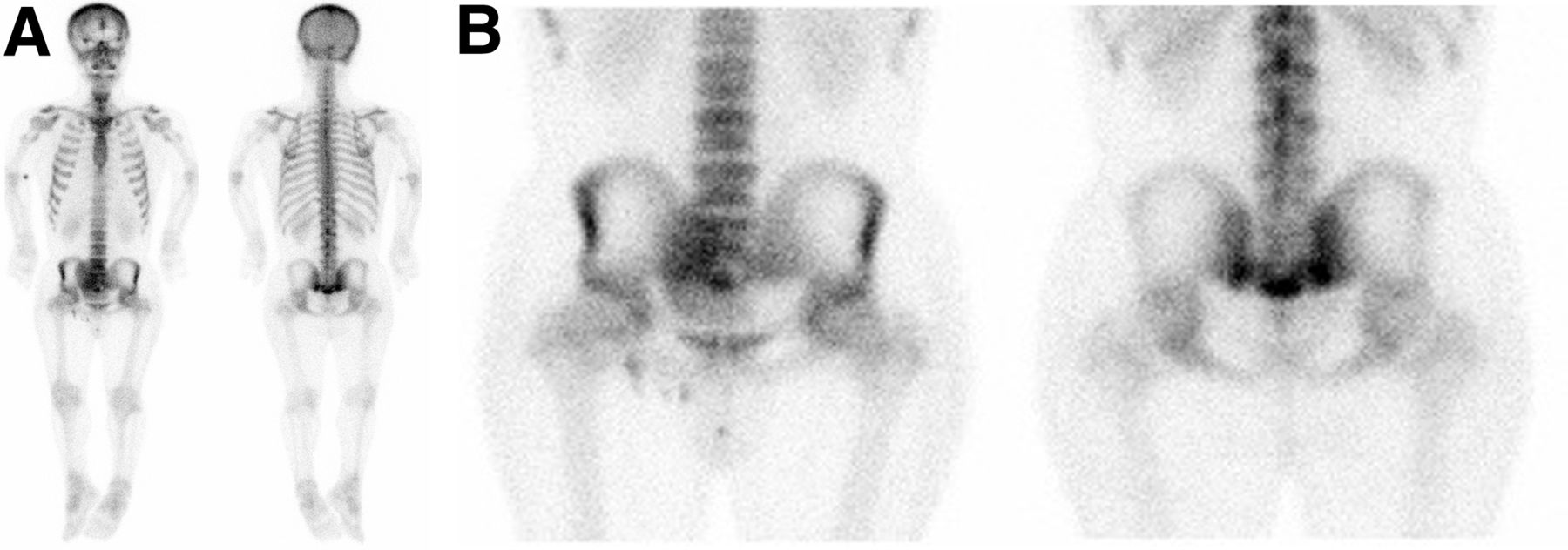
- FIGURE 1.
Whole-body bone scan and spot views of pelvis demonstrate mild to moderate increased radiotracer activity in right and left sacroiliac joints inferiorly and in sacrococcygeal area, raising possibility of fracture. Mild focal increased uptake is also noted bilaterally along proximal humeral cortices.
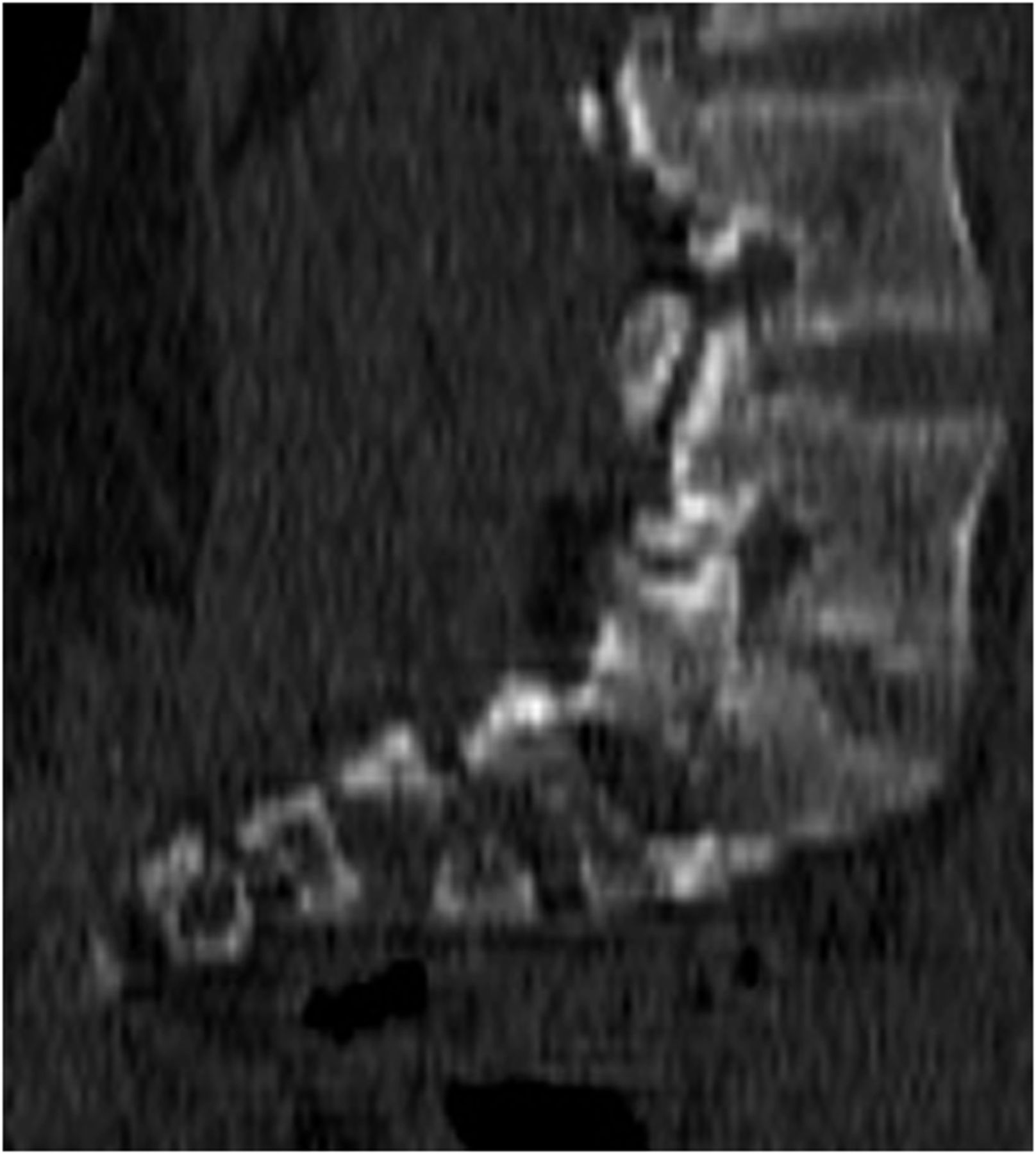
- FIGURE 2.
Axial (A) and sagittal (B) SPECT/CT images of pelvis and sacroiliac joints demonstrate mild increased radiotracer activity in sacroiliac joints bilaterally. Sacrococcygeal uptake seen on planar images is not apparent on SPECT images.
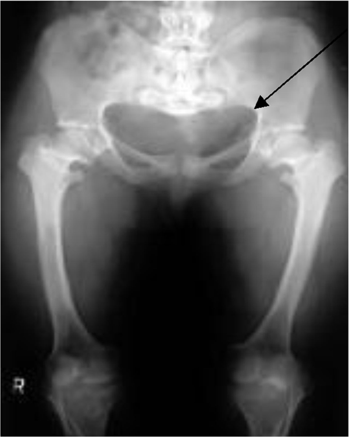
- FIGURE 3.
Champagne glass appearance of pelvis (arrow), which is due to anteversion of pelvis, squaring-off of ilia, lower lying anterior superior iliac spine, and higher posterior superior iliac spine.
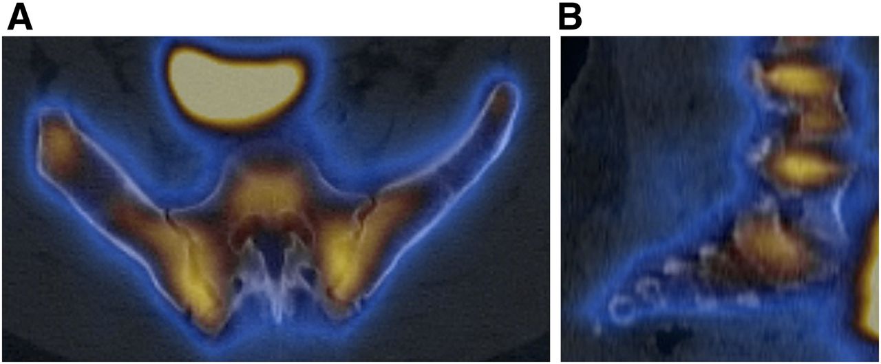
- FIGURE 4.
Sagittal slice (from SPECT/CT) demonstrating horizontal configuration of sacrum.


