
Main menu

  • Home
  • Content
    • Current
      • JNMT Supplement
    • Ahead of print
    • Past Issues
    • Continuing Education
    • JNMT Podcast
    • SNMMI Annual Meeting Abstracts
  • Subscriptions
    • Subscribers
    • Rates
    • Journal Claims
    • Institutional and Non-member
  • Authors
    • Submit to JNMT
    • Information for Authors
    • Assignment of Copyright
    • AQARA Requirements
  • Info
    • Reviewers
    • Permissions
    • Advertisers
    • Corporate & Special Sales
  • About
    • About Us
    • Editorial Board
    • Contact Information
  • More
    • Alerts
    • Feedback
    • Help
    • SNMMI Journals
  • SNMMI
    • JNMT
    • JNM
    • SNMMI Journals
    • SNMMI

User menu

  • Subscribe
  • My alerts
  • Log in
  • My Cart

Search

  • Advanced search
Journal of Nuclear Medicine Technology
  • SNMMI
    • JNMT
    • JNM
    • SNMMI Journals
    • SNMMI
  • Subscribe
  • My alerts
  • Log in
  • My Cart
Journal of Nuclear Medicine Technology

Advanced Search

  • Home
  • Content
    • Current
    • Ahead of print
    • Past Issues
    • Continuing Education
    • JNMT Podcast
    • SNMMI Annual Meeting Abstracts
  • Subscriptions
    • Subscribers
    • Rates
    • Journal Claims
    • Institutional and Non-member
  • Authors
    • Submit to JNMT
    • Information for Authors
    • Assignment of Copyright
    • AQARA Requirements
  • Info
    • Reviewers
    • Permissions
    • Advertisers
    • Corporate & Special Sales
  • About
    • About Us
    • Editorial Board
    • Contact Information
  • More
    • Alerts
    • Feedback
    • Help
    • SNMMI Journals
  • Watch or Listen to JNMT Podcast
  • Visit SNMMI on Facebook
  • Join SNMMI on LinkedIn
  • Follow SNMMI on Twitter
  • Subscribe to JNMT RSS feeds
Research ArticleTeaching Case Studies

Evaluation of Hepatopulmonary Syndrome with 99mTc-Macroaggregated Albumin Scintigraphy

Fathima Fijula Palot Manzil, Iqbal Haq and Xiaofei Wang
Journal of Nuclear Medicine Technology December 2022, 50 (4) 377-378; DOI: https://doi.org/10.2967/jnmt.122.264190
Fathima Fijula Palot Manzil
Department of Radiology, University of Arkansas for Medical Sciences, Little Rock, Arkansas
  • Find this author on Google Scholar
  • Find this author on PubMed
  • Search for this author on this site
Iqbal Haq
Department of Radiology, University of Arkansas for Medical Sciences, Little Rock, Arkansas
  • Find this author on Google Scholar
  • Find this author on PubMed
  • Search for this author on this site
Xiaofei Wang
Department of Radiology, University of Arkansas for Medical Sciences, Little Rock, Arkansas
  • Find this author on Google Scholar
  • Find this author on PubMed
  • Search for this author on this site
  • Article
  • Figures & Data
  • Info & Metrics
  • PDF
Loading

Abstract

Hepatopulmonary syndrome is characterized by intrapulmonary vascular dilatation causing hypoxemia in patients with liver disease. 99mTc-macroaggregated albumin (99mTc-MAA) scintigraphy has diagnostic value in suspected hepatopulmonary syndrome by detecting a clinically significant right-to-left shunt. In the presence of cirrhosis, 99mTc-MAA scanning with extrapulmonary organ visualization is specific for intrapulmonary shunting. 99mTc-MAA scintigraphy also provides the added value of quantification of the shunt.

  • hepatopulmonary syndrome
  • 99mTc-MAA scintigraphy
  • intrapulmonary shunt

We report a case of hepatopulmonary syndrome (HPS) confirmed by 99mTc-macroaggregated albumin (99mTc-MAA) scintigraphy. HPS is an uncommon condition in which there is hypoxemia due to intrapulmonary vascular dilatation in the context of liver disease.

CASE REPORT

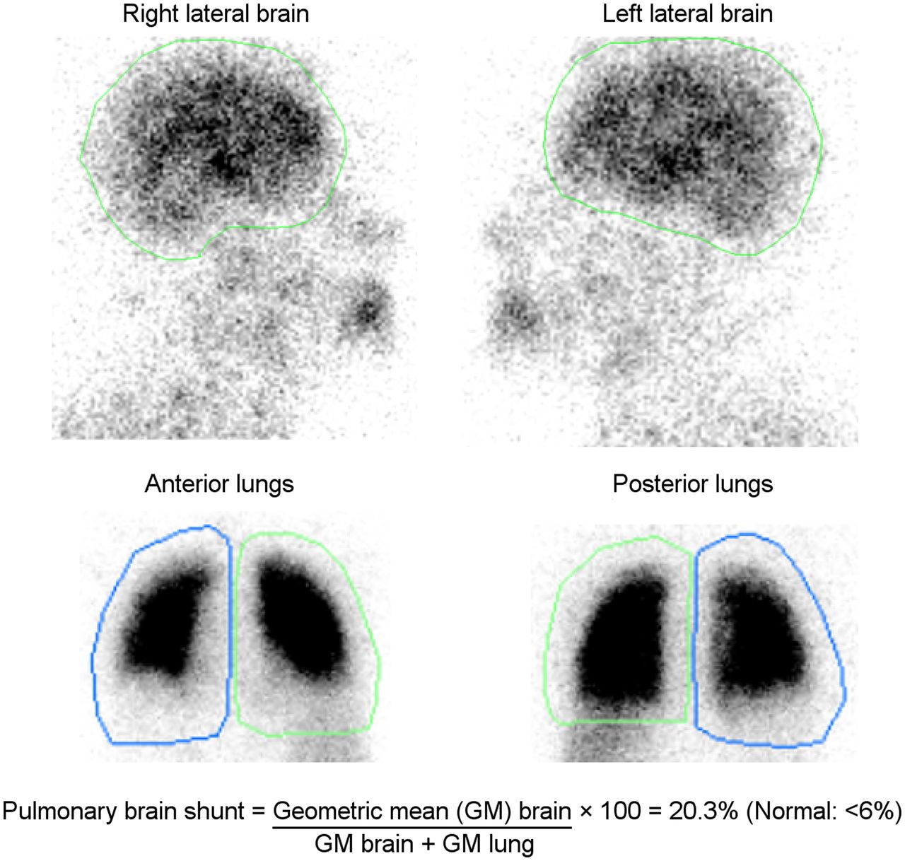
A 38-y-old man presented with dyspnea on exertion and thrombocytopenia. The patient was extensively evaluated, including MRI and liver biopsy, which demonstrated cirrhosis. The patient had a consistently low partial pressure of oxygen (≈55 mm Hg). The alveolar arterial oxygen gradient was 25 mm Hg. A transthoracic echocardiogram with saline showed microbubbles in the left heart chambers 4 cardiac cycles after contrast appearance in the right heart, suggestive of an extracardiac shunt—likely pulmonary arteriovenous malformation. 99mTc-MAA lung scintigraphy after injection of 144.3 MBq (3.9 mCi) of radiotracer showed increased tracer uptake in the brain, kidneys, spleen, and subcutaneous tissues, indicating a right-to-left shunt—likely an intrapulmonary shunt in the setting of cirrhosis (Fig. 1). On quantification, the brain shunt fraction was 20.3% (Fig. 2). An elevated alveolar–arterial gradient with a partial pressure of oxygen of less than 60, echocardiographic and scintigraphic evidence of intrapulmonary shunting, and no known chronic lung disease in the setting of cirrhosis were consistent with HPS in our patient. The patient is currently undergoing evaluation for a liver transplant.

FIGURE 1.
  • Download figure
  • Open in new tab
  • Download powerpoint
FIGURE 1.

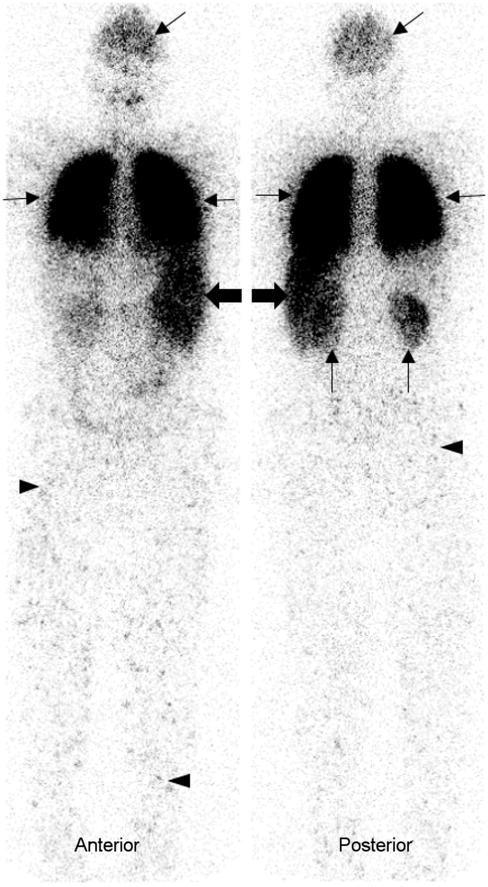
99mTc-MAA planar whole-body image in anterior and posterior projections show intense radiotracer uptake in lungs (thin transverse arrows) and shunted activity in brain (oblique arrows), spleen (thick transverse arrows), kidneys (vertical arrows), and subcutaneous tissues (arrowheads).

FIGURE 2.
  • Download figure
  • Open in new tab
  • Download powerpoint
FIGURE 2.

Planar images of brain in right and left lateral projections and of lungs in anterior and posterior projections show areas of interest drawn to calculate lung–brain shunt.

DISCUSSION

The pathognomonic intrapulmonary vascular dilatations in HPS cause impaired oxygen transfer from alveoli to red blood cells, inducing an intrapulmonary right-to-left shunt. In HPS, hypoxemia and dyspnea may increase in the upright position because of preferential perfusion of dilated vessels in the lung bases (1). The severity of HPS based on the degree of hypoxemia is described as mild, moderate, severe, and very severe if PaO2 is 80 or more, 60–79, 50–59, and less than 50 mm Hg, respectively. HPS is frequently underdiagnosed (2). Currently, the only effective treatment for HPS is liver transplantation. It is important to diagnose HPS as early as possible to expedite treatment for a better outcome. Hypoxemia can be distinguished from HPS and other etiologies through bubble echocardiography, with arrival of bubbles in the left heart at least 3 cardiac cycles after contrast appearance in the right heart, or through 99mTc-MAA scintigraphy showing uptake in the brain (3). 99mTc-MAA scintigraphy is more specific than echocardiography and also can quantify and measure the degree of the shunt. Under good quality control measures and absence of a shunt, no extrapulmonary organs should be visualized, as the injected 99mTc-MAA particles are trapped in the pulmonary microvasculature. However, when there is an intrapulmonary shunt, a fraction of the particles enters the systemic circulation, leading to visualization of other organs and systems. The standard technique of calculating the percentage of lung–brain shunting is done by drawing regions of interest around the brain and lungs and determining the geometric mean of the brain and lung counts (4,5). HPS is suggested if the alveolar arterial oxygen gradient is at least 15 mm Hg or at least 20 mm Hg for patients more than 64 y old and the pulmonary brain shunt quantitative index for 99mTc-MAA is at least 6%. A whole-body 99mTc-MAA uptake calculation is another method for detecting intrapulmonary vascular dilatation (6).

CONCLUSION

Patients with a history of chronic liver disease along with dyspnea should be further evaluated for possible HPS. In patients with an elevated alveolar arterial gradient, bubble echocardiography or 99mTc scintigraphy aids in diagnosis of an intrapulmonary shunt. Though echocardiography is sensitive, it lacks specificity by providing false-positive results in patients with concomitant lung diseases. A positive 99mTc-MAA scan with tracer uptake in extrapulmonary organs in a cirrhotic patient is specific for HPS. 99mTc-MAA scintigraphy also quantifies the extent of a shunt.

DISCLOSURE

No potential conflict of interest relevant to this article was reported.

Footnotes

  • Published online May. 24, 2022.

REFERENCES

  1. 1.↵
    1. Koch DG,
    2. Fallon MB
    . Hepatopulmonary syndrome. Clin Liver Dis. 2014;18:407–420.
    OpenUrl
  2. 2.↵
    1. Hoeper MM,
    2. Krowka MJ,
    3. Strassburg CP
    . Portopulmonary hypertension and hepatopulmonary syndrome. Lancet. 2004;363:1461–1468.
    OpenUrlCrossRefPubMed
  3. 3.↵
    1. Rodríguez-Roisin R,
    2. Krowka MJ
    . Hepatopulmonary syndrome: a liver-induced lung vascular disorder. N Engl J Med. 2008;358:2378–2387.
    OpenUrlCrossRefPubMed
  4. 4.↵
    1. Abrams GA,
    2. Nanda NC,
    3. Dubovsky EV,
    4. et al
    . Use of macroaggregated albumin lung perfusion scan to diagnose hepatopulmonary syndrome: a new approach. Gastroenterology. 1998;114:305–310.
    OpenUrlCrossRefPubMed
  5. 5.↵
    1. Surasi DS,
    2. Manapragada P,
    3. Bhambhvani P
    . Lung perfusion imaging in hepatopulmonary syndrome using 99mTc macroaggregated albumin. J Nucl Cardiol. 2015;22:586–588.
    OpenUrl
  6. 6.↵
    1. Zhao H,
    2. Tsauo J,
    3. Zhang XW,
    4. et al
    . Technetium-99m-labeled macroaggregated albumin lung perfusion scan for diagnosis of hepatopulmonary syndrome: a prospective study comparing brain uptake and whole-body uptake. World J Gastroenterol. 2020;26:1088–1097.
    OpenUrl
  • Received for publication March 28, 2022.
  • Revision received April 27, 2022.
PreviousNext
Back to top

In this issue

Journal of Nuclear Medicine Technology: 50 (4)
Journal of Nuclear Medicine Technology
Vol. 50, Issue 4
December 1, 2022
  • Table of Contents
  • About the Cover
  • Index by author
  • Complete Issue (PDF)
Print
Download PDF
Article Alerts
Sign In to Email Alerts with your Email Address
Email Article

Thank you for your interest in spreading the word on Journal of Nuclear Medicine Technology.

NOTE: We only request your email address so that the person you are recommending the page to knows that you wanted them to see it, and that it is not junk mail. We do not capture any email address.

Enter multiple addresses on separate lines or separate them with commas.
Evaluation of Hepatopulmonary Syndrome with 99mTc-Macroaggregated Albumin Scintigraphy
(Your Name) has sent you a message from Journal of Nuclear Medicine Technology
(Your Name) thought you would like to see the Journal of Nuclear Medicine Technology web site.
Citation Tools
Evaluation of Hepatopulmonary Syndrome with 99mTc-Macroaggregated Albumin Scintigraphy
Fathima Fijula Palot Manzil, Iqbal Haq, Xiaofei Wang
Journal of Nuclear Medicine Technology Dec 2022, 50 (4) 377-378; DOI: 10.2967/jnmt.122.264190

Citation Manager Formats

  • BibTeX
  • Bookends
  • EasyBib
  • EndNote (tagged)
  • EndNote 8 (xml)
  • Medlars
  • Mendeley
  • Papers
  • RefWorks Tagged
  • Ref Manager
  • RIS
  • Zotero
Share
Evaluation of Hepatopulmonary Syndrome with 99mTc-Macroaggregated Albumin Scintigraphy
Fathima Fijula Palot Manzil, Iqbal Haq, Xiaofei Wang
Journal of Nuclear Medicine Technology Dec 2022, 50 (4) 377-378; DOI: 10.2967/jnmt.122.264190
Twitter logo Facebook logo LinkedIn logo Mendeley logo
  • Tweet Widget
  • Facebook Like
  • Google Plus One
Bookmark this article

Jump to section

  • Article
    • Abstract
    • CASE REPORT
    • DISCUSSION
    • CONCLUSION
    • DISCLOSURE
    • Footnotes
    • REFERENCES
  • Figures & Data
  • Info & Metrics
  • PDF

Related Articles

  • No related articles found.
  • PubMed
  • Google Scholar

Cited By...

  • No citing articles found.
  • Google Scholar

More in this TOC Section

  • High-Sensitivity Troponin Elevation in a Young Woman with Typical Chest Pain: The Heart of Matter
  • Prominent Right Ventricular Tracer Uptake: A Harbinger of Multivessel Coronary Artery Disease
  • SPECT/CT for Discrimination Between Active and Inactive Os Trigonum in Posterior Ankle Syndrome
Show more Teaching Case Studies

Similar Articles

Keywords

  • hepatopulmonary syndrome
  • 99mTc-MAA scintigraphy
  • intrapulmonary shunt
SNMMI

© 2025 SNMMI

Powered by HighWire