
Main menu

  • Home
  • Content
    • Current
      • JNMT Supplement
    • Ahead of print
    • Past Issues
    • Continuing Education
    • JNMT Podcast
    • SNMMI Annual Meeting Abstracts
  • Subscriptions
    • Subscribers
    • Rates
    • Journal Claims
    • Institutional and Non-member
  • Authors
    • Submit to JNMT
    • Information for Authors
    • Assignment of Copyright
    • AQARA Requirements
  • Info
    • Reviewers
    • Permissions
    • Advertisers
    • Corporate & Special Sales
  • About
    • About Us
    • Editorial Board
    • Contact Information
  • More
    • Alerts
    • Feedback
    • Help
    • SNMMI Journals
  • SNMMI
    • JNMT
    • JNM
    • SNMMI Journals
    • SNMMI

User menu

  • Subscribe
  • My alerts
  • Log in
  • Log out
  • My Cart

Search

  • Advanced search
Journal of Nuclear Medicine Technology
  • SNMMI
    • JNMT
    • JNM
    • SNMMI Journals
    • SNMMI
  • Subscribe
  • My alerts
  • Log in
  • Log out
  • My Cart
Journal of Nuclear Medicine Technology

Advanced Search

  • Home
  • Content
    • Current
    • Ahead of print
    • Past Issues
    • Continuing Education
    • JNMT Podcast
    • SNMMI Annual Meeting Abstracts
  • Subscriptions
    • Subscribers
    • Rates
    • Journal Claims
    • Institutional and Non-member
  • Authors
    • Submit to JNMT
    • Information for Authors
    • Assignment of Copyright
    • AQARA Requirements
  • Info
    • Reviewers
    • Permissions
    • Advertisers
    • Corporate & Special Sales
  • About
    • About Us
    • Editorial Board
    • Contact Information
  • More
    • Alerts
    • Feedback
    • Help
    • SNMMI Journals
  • Watch or Listen to JNMT Podcast
  • Visit SNMMI on Facebook
  • Join SNMMI on LinkedIn
  • Follow SNMMI on Twitter
  • Subscribe to JNMT RSS feeds
Research ArticleImaging

The Semicolon Sign: Dopamine Transporter Imaging Artifact from Head Tilt

Matthew F. Covington, Natalie A. McMillan, Ryan J. Avery and Phillip H. Kuo
Journal of Nuclear Medicine Technology June 2013, 41 (2) 105-107; DOI: https://doi.org/10.2967/jnmt.112.117184
Matthew F. Covington
Department of Medical Imaging, University of Arizona, Tucson, Arizona
  • Find this author on Google Scholar
  • Find this author on PubMed
  • Search for this author on this site
Natalie A. McMillan
Department of Medical Imaging, University of Arizona, Tucson, Arizona
  • Find this author on Google Scholar
  • Find this author on PubMed
  • Search for this author on this site
Ryan J. Avery
Department of Medical Imaging, University of Arizona, Tucson, Arizona
  • Find this author on Google Scholar
  • Find this author on PubMed
  • Search for this author on this site
Phillip H. Kuo
Department of Medical Imaging, University of Arizona, Tucson, Arizona
  • Find this author on Google Scholar
  • Find this author on PubMed
  • Search for this author on this site
  • Article
  • Figures & Data
  • Info & Metrics
  • PDF
Loading

Abstract

Dopamine transporter (DAT) imaging is a valuable tool to aid in the diagnosis of Parkinson disease and other Parkinsonian syndromes. DAT imaging is special among clinical nuclear medicine scans in that the already small caudate and putamen are presented in multiple thin axial cuts. Because the imaged basal ganglia are small, slight differences in head tilt may result in a significant artifact that we have termed the semicolon sign. The semicolon sign occurs when forward head tilt creates select images that show the caudate nuclei separate from the putamen. This gives the false impression that DAT activity in the putamen is decreased or absent. To avoid falsely attributing this artifact to loss of putaminal activity, it is imperative that the interpreting physician first recognize the artifact and then mentally integrate all provided images to identify normal activity in the putamen on subsequent levels. Furthermore, quantitative software packages for automated DAT scan interpretation are now available. If images demonstrating the semicolon sign are used for automated interpretation, loss of activity in the putamen may be falsely calculated, thereby contributing to erroneous results. Quality control measures are essential to ensure that technologists correctly position each patient’s head to minimize head tilt artifact on DAT scan images. A protocol to obtain optimal head positioning is presented.

  • dopamine transporter (DAT) imaging
  • imaging artifact
  • basal ganglia
  • Parkinson disease

Dopamine transporter (DAT) imaging is a valuable tool to aid in the diagnosis of Parkinson disease and other Parkinsonian syndromes such as Lewy body dementia and progressive supranuclear palsy (1,2). DAT imaging is special among clinical nuclear medicine scans in that the already small caudate and putamen, with average volumes of 3.4 and 4.3 cm3, respectively (3), are presented in multiple thin axial cuts. Because the imaged basal ganglia are small, slight differences in head tilt may result in a significant artifact (Fig. 1).

FIGURE 1.
  • Download figure
  • Open in new tab
  • Download powerpoint
FIGURE 1.

Effect of head positioning on axial imaging of caudate (gold) and putamen (blue). These images represent rough approximation of basal ganglia anatomy. (A) Abnormal forward head tilt results in slices (red lines) through caudate head that do not include putamen. Subsequent slices (white lines) demonstrate both caudate and putamen. (B) With correct head positioning, all slices through caudate head also include putamen (white lines).

We have coined the term semicolon sign for the characteristic artifact from frontal head tilt. This nomenclature is in keeping with the “commas” and “periods” commonly used to describe the normal and abnormal appearance of the basal ganglia in Parkinsonian syndromes, respectively. The semicolon sign occurs when forward head tilt creates select images that show the caudate nuclei separate from the putamen (Fig. 2). This may give the false impression that DAT activity in the putamen is decreased or absent. Therefore, the physician may provide an incorrect diagnosis of dopaminergic deficiency, with potential negative clinical consequences.

FIGURE 2.
  • Download figure
  • Open in new tab
  • Download powerpoint
FIGURE 2.

Semicolon sign. Abnormal forward head tilt creates images in which caudate head is imaged in separate axial slices from putamen. (Images are obtained with filtered backprojection using Butterworth filter with cutoff of 0.6 and order of 8. No attenuation correction was applied. Matrix size of 128 × 128 was acquired at 1.23 zoom and was manually adjusted to zoom of 1.6. Axial images are presented at slice thickness of 2.5 mm. Images were acquired at 30 s/frame over 30-min acquisition time using dual-head camera. Circular orbit was used with radius of 13 cm. Stop-and-shoot setting was 3°. Bottom edge of field of view is positioned at level of patient’s nose). (A) Top 2 rows demonstrate normal appearance of properly aligned caudate heads and putamen with comma appearance. Bottom 2 rows demonstrate semicolon artifact in which caudate heads appear separate from putamen on isolated slices. (B) This graphic depicts separate axial images of caudate heads and putamen that have been superimposed to illustrate semicolon appearance.

To avoid falsely attributing this artifact to loss of putaminal activity, the interpreting physician must first recognize the artifact and then mentally integrate all provided images to identify normal activity in the putamen on subsequent levels. Quality control measures are essential to ensure that technologists correctly position each patient’s head to minimize head tilt artifact on DAT scan images. If technologists readily recognize this artifact, they can potentially rescan the patient in a more optimal position. According to consensus practice guidelines (4), the technologist may alternatively use widely available software packages to correct the head tilt artifact by manipulating the orientation of acquired axial images. Such head tilt correction may be particularly useful for patients who are unable to maintain proper head positioning because of discomfort or illness. Software packages are also available for quantitative automated DAT scan interpretation (5,6). These rely on axial images for automated analysis. If images demonstrating the semicolon sign are used for automated interpretation, loss of activity in the putamen may be falsely calculated, thereby contributing to erroneous results.

Proper head positioning may be obtained by the following protocol. A head and neck holder should be used with the head resting entirely within the corresponding concavity of the holder and the vertex of the head reaching the head holder’s superior edge (Fig. 3A). Failure to position the head properly within the head holder may cause forward head tilt (Fig. 3B). Next, the canthomeatal line (imaginary line from the lateral corner of the eye to the ear canal) should be identified and oriented as vertically as possible. Even small deflections of the canthomeatal line from the vertical position may signify abnormal head tilt. Chin position is also a useful measure of head tilt. The chin should lie in a neutral position when viewed from the side. Deflection of the chin toward the neck indicates abnormal forward head tilt. Finally, the technologist should stand at the head of the patient’s bed and examine the vertex, ears, and nose to correct any signs of lateral head rotation.

FIGURE 3.
  • Download figure
  • Open in new tab
  • Download powerpoint
FIGURE 3.

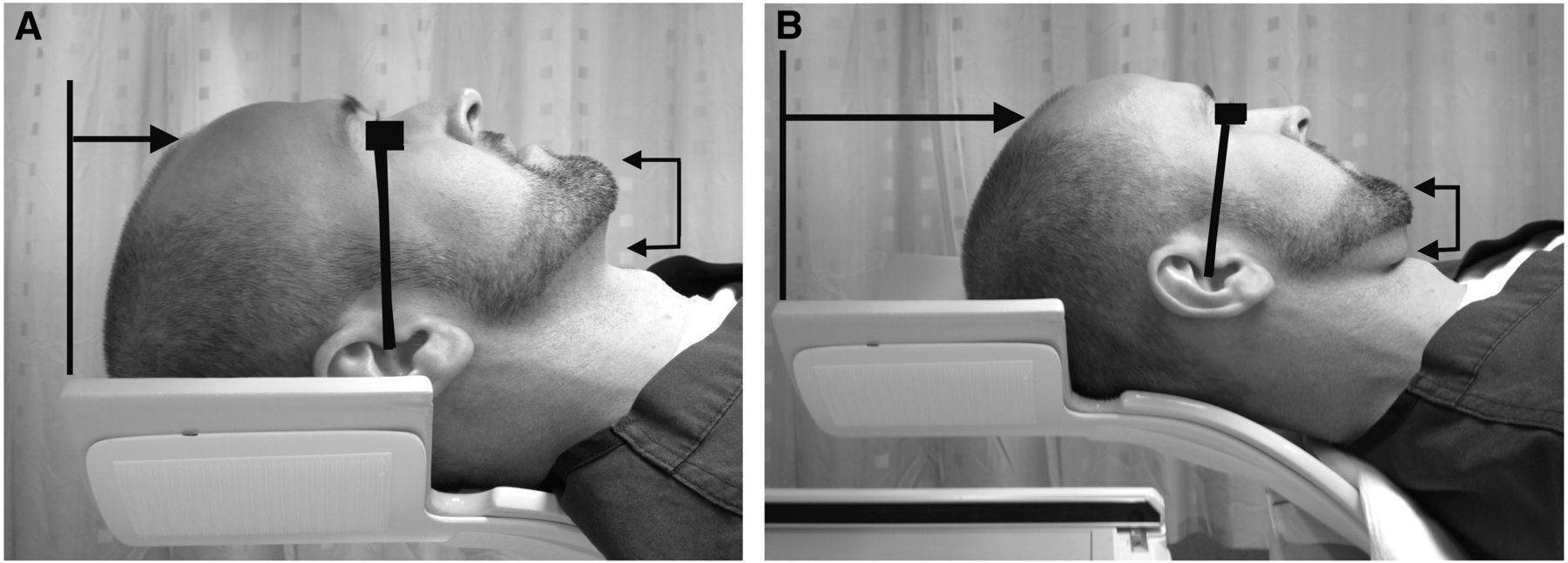
Correct head positioning using head holder (Composites Horizons, Inc.). (A) Vertex of head should reach head holder’s superior edge (black line with arrow). Canthomeatal line should be oriented as vertically as possible (black line). Chin should rest in neutral position (connected arrowheads). (B) Incorrect positioning of head within head holder may result in head tilt artifact. Vertex of head does not reach head holder’s superior edge (black line with arrow). Canthomeatal line (black line) is slightly deflected forward, indicating abnormal forward head tilt. Chin is also deflected toward neck (connected arrowheads).

Footnotes

  • Published online Mar. 21, 2013.

REFERENCES

  1. 1.↵
    1. Ravina B,
    2. Marek K,
    3. Eberly S,
    4. et al
    . Dopamine transporter imaging is associated with long-term outcomes in Parkinson’s disease. Mov Disord. 2012;27:1392–1397.
    OpenUrlCrossRefPubMed
  2. 2.↵
    1. Varrone A,
    2. Marek KL,
    3. Jennings D,
    4. Innis RB,
    5. Seibyl JP
    . [123I]beta-CIT SPECT imaging demonstrates reduced density of striatal dopamine transporters in Parkinson’s disease and multiple system atrophy. Mov Disord. 2001;16:1023–1032.
    OpenUrlCrossRefPubMed
  3. 3.↵
    1. Gunning-Dixon FM,
    2. Head D,
    3. McQuain J,
    4. Acker JD,
    5. Raz N
    . Differential aging of the human striatum: a prospective MR imaging study. AJNR. 1998;19:1501–1507.
    OpenUrlAbstract
  4. 4.↵
    1. Djang DSW,
    2. Janssen MJR,
    3. Bohnen N,
    4. et al
    . SNM practice guideline for dopamine transporter imaging with 123 I-ioflupane SPECT 1.0. J Nucl Med. 2012;53:154–163.
    OpenUrlFREE Full Text
  5. 5.↵
    1. Tatsch K,
    2. Poepperl G
    . Quantitative approaches to dopaminergic brain imaging. Q J Nucl Med Mol Imaging. 2012;56:27–38.
    OpenUrlPubMed
  6. 6.↵
    1. Zubal IG,
    2. Early M,
    3. Yuan O,
    4. et al
    . Optimized, automated striatal uptake analysis applied to SPECT brain scans of Parkinson’s disease patients. J Nucl Med. 2007;48:857–864.
    OpenUrlAbstract/FREE Full Text
PreviousNext
Back to top

In this issue

Journal of Nuclear Medicine Technology: 41 (2)
Journal of Nuclear Medicine Technology
Vol. 41, Issue 2
June 1, 2013
  • Table of Contents
  • About the Cover
  • Index by author
Print
Download PDF
Article Alerts
Sign In to Email Alerts with your Email Address
Email Article

Thank you for your interest in spreading the word on Journal of Nuclear Medicine Technology.

NOTE: We only request your email address so that the person you are recommending the page to knows that you wanted them to see it, and that it is not junk mail. We do not capture any email address.

Enter multiple addresses on separate lines or separate them with commas.
The Semicolon Sign: Dopamine Transporter Imaging Artifact from Head Tilt
(Your Name) has sent you a message from Journal of Nuclear Medicine Technology
(Your Name) thought you would like to see the Journal of Nuclear Medicine Technology web site.
Citation Tools
The Semicolon Sign: Dopamine Transporter Imaging Artifact from Head Tilt
Matthew F. Covington, Natalie A. McMillan, Ryan J. Avery, Phillip H. Kuo
Journal of Nuclear Medicine Technology Jun 2013, 41 (2) 105-107; DOI: 10.2967/jnmt.112.117184

Citation Manager Formats

  • BibTeX
  • Bookends
  • EasyBib
  • EndNote (tagged)
  • EndNote 8 (xml)
  • Medlars
  • Mendeley
  • Papers
  • RefWorks Tagged
  • Ref Manager
  • RIS
  • Zotero
Share
The Semicolon Sign: Dopamine Transporter Imaging Artifact from Head Tilt
Matthew F. Covington, Natalie A. McMillan, Ryan J. Avery, Phillip H. Kuo
Journal of Nuclear Medicine Technology Jun 2013, 41 (2) 105-107; DOI: 10.2967/jnmt.112.117184
Twitter logo Facebook logo LinkedIn logo Mendeley logo
  • Tweet Widget
  • Facebook Like
  • Google Plus One
Bookmark this article

Jump to section

  • Article
    • Abstract
    • Footnotes
    • REFERENCES
  • Figures & Data
  • Info & Metrics
  • PDF

Related Articles

  • No related articles found.
  • PubMed
  • Google Scholar

Cited By...

  • Amyloid Imaging Update: How the Amyloid Landscape Is Changing in Light of the Recent Food and Drug Administration Approval of Antiamyloid Therapeutics
  • Brain Imaging Techniques: Improving the Quality
  • Pitfalls and Artifacts of 123I-Ioflupane SPECT in Parkinsonian Syndromes: A Quality Improvement Teaching Tool
  • DaTQUANT: The Future of Diagnosing Parkinson Disease
  • Brain Imaging Quality Assurance: How to Acquire the Best Brain Images Possible
  • False-Positive Findings on Dopamine Transporter SPECT Due to Therapeutic Dextroamphetamine and Amphetamine
  • Google Scholar

More in this TOC Section

  • Early 10-Minute Postinjection [18F]F-FAPI-42 uEXPLORER Total-Body PET/CT Scanning Protocol for Staging Lung Cancer Using HYPER Iterative Reconstruction
  • Single- Versus Dual-Time-Point Imaging for Transthyretin Cardiac Amyloid Using 99mTc-Pyrophosphate
  • Does Arthrography Improve Accuracy of SPECT/CT for Diagnosis of Aseptic Loosening in Patients with Painful Knee Arthroplasty: A Systematic Review and Metaanalysis
Show more Imaging

Similar Articles

Keywords

  • dopamine transporter (DAT) imaging
  • imaging artifact
  • basal ganglia
  • Parkinson disease
SNMMI

© 2025 SNMMI

Powered by HighWire