


Visual Abstract

Abstract
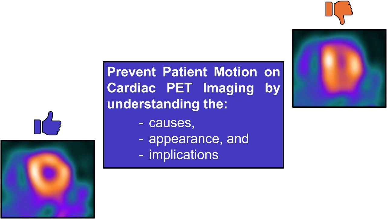
Cardiac PET imaging is increasingly used for myocardial perfusion studies because of its high diagnostic accuracy and low radiation exposure to the patient. However, patient motion can be challenging, affecting a large number of studies. Motion artifacts can lead to inconclusive or false-positive results, complicating clinical interpretation. This article explores the causes of motion artifacts and their characteristic appearance in cardiac PET imaging, highlighting their distinction from true perfusion abnormalities. Strategies for minimizing motion through effective patient positioning and communication are discussed. Understanding and addressing motion artifacts are crucial for optimizing diagnostic accuracy and ensuring the full benefit of cardiac PET imaging.
Footnotes
Published online Apr. 22, 2025.
This article requires a subscription to view the full text. If you have a subscription you may use the login form below to view the article. Access to this article can also be purchased.
SNMMI members
Login to the site using your SNMMI member credentials
Individuals
Login as an individual user
In this issue


Jump to section
- Article
- Visual Abstract
- Abstract
- PATIENT MOTION CAUSE AND PREVENTION
- CARDIAC SPECT VERSUS PET MOTION ARTIFACT
- RECOGNIZING PET MOTION ARTIFACT
- CT ATTENUATION CORRECTION (TRANSMISSION)/EMISSION MISALIGNMENT
- EFFECT OF MOTION ON MYOCARDIAL BLOOD FLOW QUANTIFICATION
- INTERPRETATION STRATEGIES FOR STUDIES WITH MOTION ARTIFACT
- CONCLUSION
- Footnotes
- REFERENCES
- Figures & Data
- Info & Metrics
Related Articles
Cited By...
- No citing articles found.