


Article Figures & Data
Figures
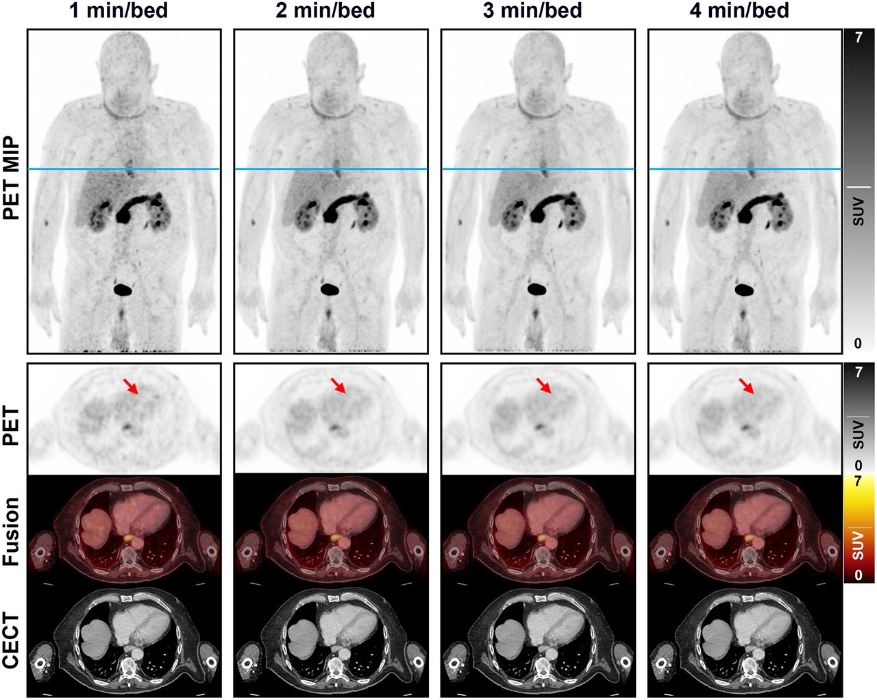
- FIGURE 1.
Maximum-intensity projection and axial PET images with corresponding contrast-enhanced CT and fused images presenting image quality in relation to acquisition duration of [68Ga]FAPI-46 imaging. Reference line (blue) for axial images is presented on maximum-intensity projection images. Arrows indicate interventricular septum of heart (image quality criterion B). CECT = contrast-enhanced CT; MIP = maximum-intensity projection.
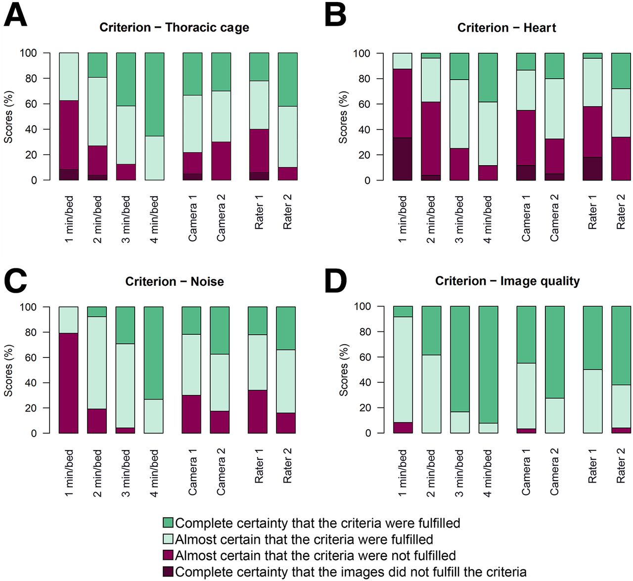
- FIGURE 2.
Results from scoring of criteria A–D presented with histogram for each camera, each rater, and each acquisition duration.
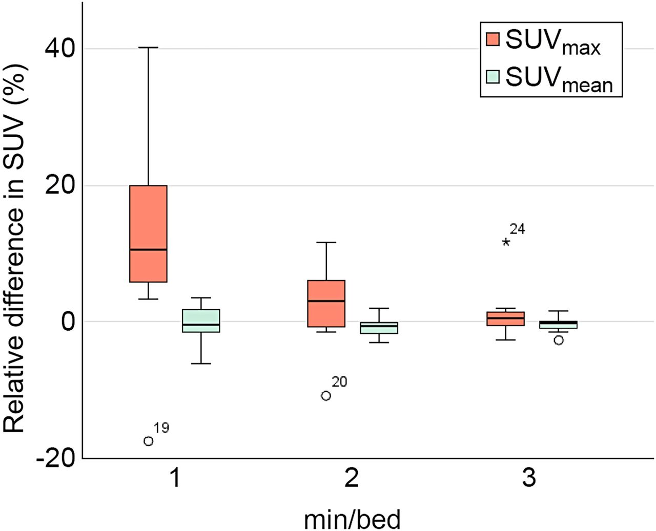
- FIGURE 3.
Box plots of differences in SUVmax and SUVmean for 1, 2, and 3 min/bed position relative to 4 min/bed position. Asterisk represents outlier plotted as individual point.
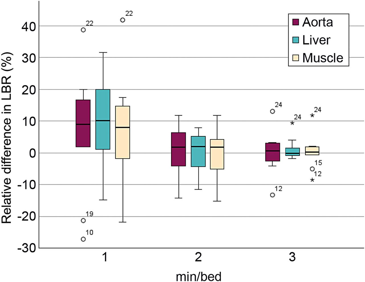
- FIGURE 4.
Box plots of LBR relative to 4 min/bed position using background liver, aorta, and musculature. Asterisks represent outliers plotted as individual points.
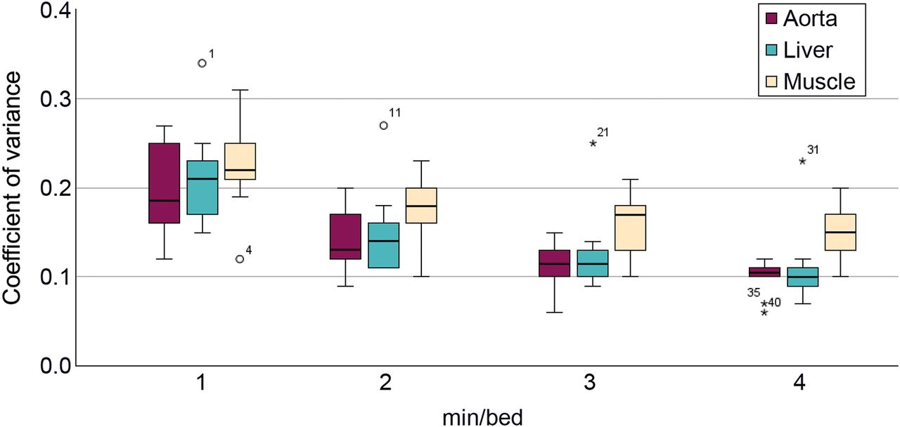
- FIGURE 5.
Box plots of COV measured in background liver, aorta, and musculature at 1, 2, 3, and 4 min/bed position. Asterisks represent outliers plotted as individual points.


Tables
Criterion Definition A Intercostal spaces are distinguishable from ribs, and structures such as costochondral cartilage and sternum are clearly discernible and distinct B Interventricular septum of heart is clearly discernible and distinct C Noise level does not disrupt evaluation D Overall image quality is good enough to make clinical diagnosis Patient no. Sex Body weight (kg) Age (y) BMI Injected activity (MBq) Activity by body weight (MBq/kg) Camera Histologic diagnosis SUVmax SUVmean LBRmax, blood Lesion volume (cm3) 1 F 57 85 22 213.2 3.7 1 PDAC 23.8 14.2 20.6 10 2 F 62 72 20 248.8 4.0 1 PDAC 25.0 13.0 19.3 41 3 F 68 49 28 257.1 3.8 1 PDAC 18.4 12.0 16.1 4 4 M 64 83 22 239.8 3.7 1 PDAC 15.4 8.1 10.6 14 5 M 72 57 23 268.3 3.7 2 PanIN 3.7 3.0 2.8 1 6 M 76 74 25 261.2 3.4 2 PDAC 18.9 12.4 10.6 21 7 M 82 65 25 292.1 3.6 2 PDAC 15.6 8.0 12.1 11 8 M 77 69 24 265.5 3.4 1 PanIN 1.1 0.7 1.1 4 9 M 65 66 21 218.2 3.4 1 IPMN 5.3 2.9 5.2 10 10 F 78 79 24 285.3 3.7 2 PDAC 9.9 6.3 7.4 8 BMI = body mass index; LBRmax,blood = SUVmax divided by background measurement in aorta; PDAC = pancreatic ductal adenocarcinoma; PanIN = pancreatic intraepithelial neoplasia; IPMN = intraductal papillary mucinous neoplasm.
Criterion A Criterion B Criterion C Criterion D Condition Regression coefficient P Regression coefficient P Regression coefficient P Regression coefficient P 1 min/bed −8.12 <0.0001 −7.19 <0.0001 −8.89 <0.0001 −6.61 <0.0001 2 min/bed −4.58 <0.0001 −3.99 <0.0001 −4.95 <0.0001 −3.77 0.0001 3 min/bed −2.63 0.0022 −1.36 0.0419 −2.71 0.0007 −1.02 0.2881 4 min/bed — — — — — — — — Camera 2 −0.35 0.8284 1.97 0.0450 2.04 0.0131 2.94 0.0005 Camera 1 — — — — — — — — Rater 2 3.30 <0.0001 3.20 <0.0001 1.94 0.0010 0.69 0.2495 Rater 1 — — — — — — — — Reference categories are 4 min/bed, camera 1, and rater 1.
- TABLE 4.
Comparison of SUVmax, SUVmean, and COV Measurements Using Paired Wilcoxon Signed-Rank Test
Parameter Measurement z P Mean SUVmax 1 min/bed 14.92 −1.99 <0.05 2 min/bed 13.98 −1.48 0.14 3 min/bed 13.79 −1.12 0.26 4 min/bed 13.71 — — Mean SUVmean 1 min/bed 4.64 −1.22 0.22 2 min/bed 4.77 −1.79 0.07 3 min/bed 4.90 −1.13 0.26 4 min/bed 4.78 — — Mean liver COV 1 min/bed 0.21 −2.81 <0.01 2 min/bed 0.15 −2.81 <0.01 3 min/bed 0.14 −2.70 <0.01 4 min/bed 0.11 — — Mean aorta COV 1 min/bed 0.19 −2.81 <0.01 2 min/bed 0.14 −2.81 <0.01 3 min/bed 0.12 −2.26 <0.05 4 min/bed 0.10 — — Mean muscle COV 1 min/bed 0.23 −2.81 <0.01 2 min/bed 0.18 −2.68 <0.01 3 min/bed 0.16 −2.23 <0.05 4 min/bed 0.15 — —
Supplemental Data
Files in this Data Supplement:


