


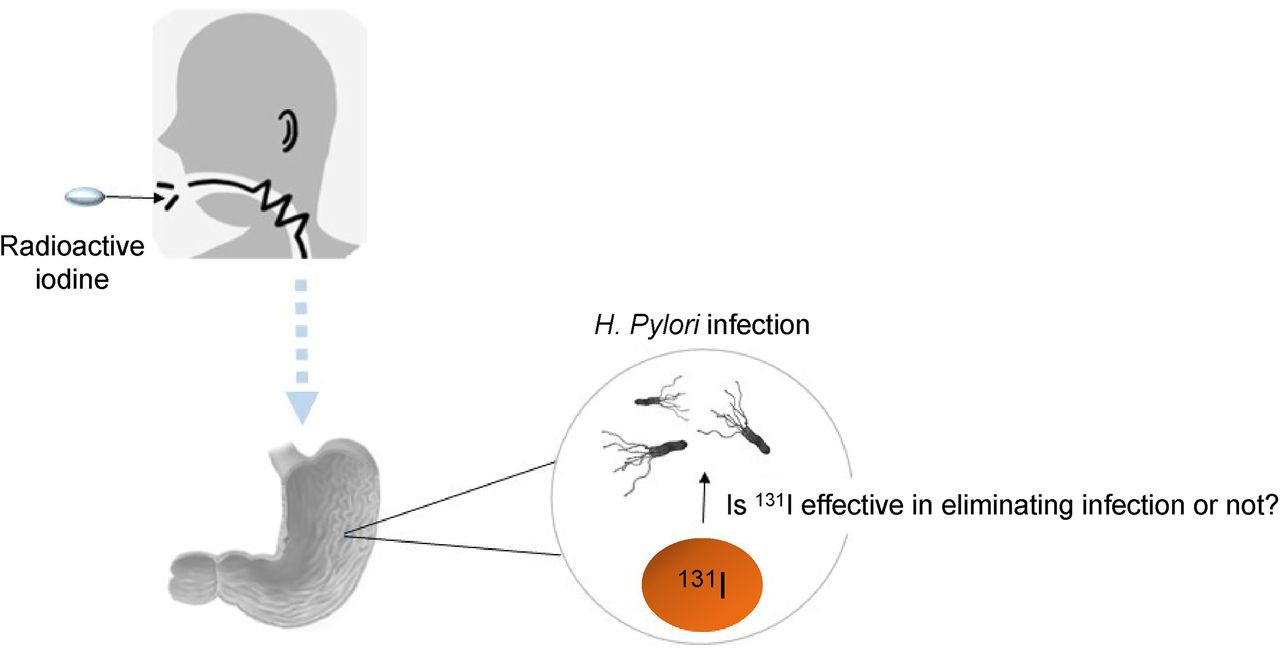
Visual Abstract

Abstract
The leading cause of gastritis and its complications is Helicobacter pylori. Radioactive iodine (131I) accumulates significantly in the stomach after consumption. On this basis, we decided to determine whether different doses of 131I in the stomach would be effective in eradicating the infection. Methods: All patients with hyperthyroidism or differentiated thyroid carcinoma who were referred for 131I treatment were invited to the study. A stool antigen test was conducted before consumption of 131I (0.15–5.5 GBq) and was repeated 2 mo later to detect H. pylori infection. Results: H. pylori positivity was found in 51.8% (14/27) of the patients. At 2 mo after treatment, 13 of the 14 patients with differentiated thyroid carcinoma or hyperthyroidism who had been identified as positive for H. pylori stool antigen before 131I administration were still positive, representing a nonsignificant eradication rate of 7.1%. Conclusion: Administration of 131I to patients with H. pylori did not show potential to eliminate the infection.
The leading cause of chronic gastritis, peptic ulcer, gastric mucosa–associated lymphoid tissue lymphoma, and gastric cancer is Helicobacter pylori, a gram-negative bacteria that infects the human gastric mucosa (1). It has recently been proposed that H. pylori may be related to extraintestinal disorders such as vitamin B12 deficiency, iron-refractory iron-deficiency anemia, and immune thrombocytopenic purpura (2). Worldwide, H. pylori has been considered a group I carcinogen (3), and its eradication leads to healing of peptic ulcers, preventing their recurrence and reducing the risk of gastric cancer (4). In addition, H. pylori–related diseases, including mucosa-associated lymphoid tissue lymphoma, atrophic gastritis, and intestinal dysplasia, are also curable after antibiotic therapy (5).
A triple-therapy regimen comprising a proton pump inhibitor and 2 antimicrobial agents such as amoxicillin, clarithromycin, metronidazole, levofloxacin, and tetracycline is commonly used for eradication. However, the success rate of eradication therapy is dependent on many factors, such as smoking habits and patient compliance. The main factor in reducing therapy efficacy is antibiotic resistance (6). Antibiotic resistance is greater in developing countries than in developed countries (7). Moreover, the frequency of antibiotic use is often a factor in the rate of antibiotic resistance (8). Considering the decrease in the effectiveness of antibiotics against H. pylori strains, the risks caused by antibiotic use, and the need to prevent complications and deaths caused by it, a new therapeutic approach is required.
Remarkably, the stomach and thyroid have a valuable ability to concentrate iodide (9). Thyroid cells phylogenetically originate from iodine-concentrating primary digestive cells. In evolution, these cells move and become specialized in absorbing and storing iodine. Whole-body scans of cancer patients who received high doses of 131I have indicated evidence of 131I uptake in malignant tissue, normal thyroid tissue, the gastric wall, and the salivary glands (10). Gholamrezanezhad et al. (11) showed that radioactive iodine (131I) therapy in patients with differentiated thyroid carcinoma (DTC) and a positive pretreatment urea breath test (UBT) correlated with a significant decrease in the UBT-positive rate. Despite these authors’ acknowledgment that 131I would not be a reasonable therapy for the typical patient with H. pylori, these results could be applied to the use of 131I in eliminating H. pylori in the clinical setting and the food industry. Ionizing radiation directly disturbs the structure of DNA by causing DNA breaks. Secondary effects are the production of reactive oxygen species that oxidize proteins and lipids and cause multiple DNA damages (12). Considering that our geographic region (Ardabil, Iran) has a high prevalence of H. pylori infection, we felt prompted to determine whether different doses of 131I in the stomach are effective in eradicating this infection.
MATERIALS AND METHODS
Patient Selection
The study design was approved by the Ethics Committee of Ardabil University of Medical Sciences. Patients with DTC or hyperthyroidism who had been referred to the Ardabil Nuclear Medicine Center for 131I therapy were asked to participate. Informed consent was obtained from the participants before the research began.
The exclusion criteria comprised previous attempts to eradicate H. pylori using antibiotics or antacids in the previous 1 mo or bismuth in the previous 3 mo, a history of gastrectomy, and pregnancy or lactation.
To evaluate the response to a standard treatment protocol, our research restricted data analysis to patients with DTC or hyperthyroidism who had never previously taken 131I.
Experimental Design
Before therapy, all patients were asked to provide stool samples for H. pylori antigen testing. Stool samples were kept at −20°C until testing. Only patients who were treated with 131I and had a positive H. pylori stool antigen test were eligible for the study. 131I in the range of 0.15–5.5 GBq was administered to patients with DTC or hyperthyroidism. Subsequently, these patients were told not to use any antibiotics, antacids, or bismuth and to return for stool sample testing 8 wk after treatment.
H. pylori Antigen Test
We used a qualitative and immunochromatographic assay to detect H. pylori antigens in stool samples. Each sample was placed into a well and allowed to react with particles coated with anti–H. pylori antibodies. The mixture then moved toward the membrane by capillary action. If H. pylori antigens were present at detectable levels in the sample, a visible colored signal was produced. The appearance of a colored band at the result line and at the control line was considered positive. Complete absence of the control band was considered invalid, regardless of the appearance of the result line (13).
Statistical Analysis
Because of the dichotomous nature of all dependent variables (positive/negative), the McNemar test with the exact method was used to determine any differences before and after the interventions. A P value of 0.05 indicated a statistically significant difference for all compared variables. Statistical analysis was done using SPSS software version 26.0 (IBM).
RESULTS
The ratio of desired changes to undesired changes was 1 to 13 (P = 1). All 14 patients positive for H. pylori antigen had a repeat stool antigen test for the presence of H. pylori 8 wk after 131I therapy. None of the patients had used antibiotics, antacids, or bismuth during the intervention period. H. pylori positivity was seen in 92.8% (13/14) of the patients (Table 1). Different doses (0.15–5.5 GBq) had no significant effect on H. pylori eradication. Of the 14 subjects studied, one (a 41-y-old woman with DTC who received a dose of 5.5 GBq) became negative 2 mo after treatment (Table 1).
Clinical and Therapeutic Details of 18 Patients with DTC or Hyperthyroidism and H. pylori Infection Treated with 131I
DISCUSSION
The prevalence of H. pylori in eastern and southern Europe, South America, and Asia is often higher than 50%, and most infected people are asymptomatic. Currently, a proton pump inhibitor combined with antibiotic therapy is suggested for patients with active H. pylori infection. In the study of Gholamrezanezhad et al. (11), on 71 patients with DTC and a positive pretreatment UBT result, 131I therapy at a dose of 3.7–7.4 GBq was related to a significant decrease in UBT positivity: 32.4% of UBT-positive patients became negative after 2 mo of treatment. These findings provide indirect evidence of H. pylori susceptibility to 131I treatment. In another study, by Xu et al., the mean amount of H. pylori before 131I was 28.36%, whereas it was 18.18% after 131I treatment. A significant decrease in 13C-UBT was observed after 131I treatment compared with before treatment (P < 0.01) (14).
Our results were significantly different from those of these 2 studies (11,14). In the present study, 51.8% (14 of 27) of patients were H. pylori–positive. We found that 131I therapy at different doses, administered to patients with DTC or hyperthyroidism, did not eradicate H. pylori in 92.8% of cases 8 wk after treatment. The conflicting results may be explained by the diversity of the population and the virulence of the gastric mucosal bacteria of treated patients—a factor that was not assessed in this study. A milder degree of H. pylori colonization in the gastric mucosa may result in enhanced sensitivity to 131I, as the infection is less extensive and the gastritis is generally nonatrophic. Meanwhile, our geographic region has the highest rate of H. pylori infection in Iran, and the cytotoxin-associated gene E (cagE)+ genotype of the H. pylori significantly increases the risk of gastric cancer in this high-risk population (15).
In addition, these differences may be due to the experimental techniques used in each study to detect H. pylori infection. Invasive and noninvasive tests are applied to diagnose H. pylori infection. In invasive methods, including cultures, histology, and urea tests, biopsy samples obtained by upper gastrointestinal endoscopy are used. Noninvasive techniques include stool antigen tests, UBT, and serology. UBT is faster and cheaper than the others. Proton pump inhibitors, bismuth, and antimicrobial agents may interfere with this test by inhibiting urea activity. Moreover, other urea-producing microorganisms in the gastric mucosa can create false-positive results (16). Stool antigen testing is an inexpensive way to detect active H. pylori infection. Enzyme immunoassays and immunochromatography are 2 types of this test. Eradication of H. pylori infection is assessed by stool antigen testing. Hence, this test is useful before and after H. pylori therapy (13). In the present study, H. pylori infection was detected by stool samples, whereas Gholamrezanezhad et al. and Xu et al. (11,14) used UBT. In accordance with these findings, a study by Shmuely et al. (17) found that 131I treatment did not eliminate H. pylori infection in Israeli patients. All 18 patients with DTC and H. pylori antigen–positive stool remained positive 3 mo after 131I therapy, representing an eradication rate of 0% with an upper 95% confidence limit of 18.53% (17). Table 2 compares the findings of the present study with previous studies on eradication of H. pylori using 131I.
Comparison of Present Findings with Previous Findings on 131I Effect on H. pylori Eradication
CONCLUSION
In the current study, the effect of different doses of 131I radiation on H. pylori in our local population of DTC or hyperthyroidism patients was investigated. Contrary to previous reports, 131I failed to eradicate H. pylori infection, possibly because of the severe degree of H. pylori colonization in the gastric mucosa. Therefore, further investigation in different populations is needed.
DISCLOSURE
The study was supported and funded by the Deputy of Research and Technology, Ardabil University of Medical Sciences (IR.ARUMS.REC.1399.070). No other potential conflict of interest relevant to this article was reported.
KEY POINTS
QUESTION: Does 131I prescribed in different doses (0.15–5.5 GBq) for DTC or hyperthyroid patients eliminate H. pylori infection?
PERTINENT FINDINGS: Different doses of 131I radiation had no effect on H. pylori in our local population of patients with DTC or hyperthyroidism.
IMPLICATIONS FOR PATIENT CARE: 131I radiation cannot be a rational treatment for the eradication of H. pylori in the people of our geographic region (Ardabil, Iran).
Footnotes
Published online Jan. 9, 2024.
REFERENCES
- Received for publication August 10, 2023.
- Revision received November 6, 2023.
In this issue


Jump to section
Related Articles
Cited By...
- No citing articles found.