


Article Figures & Data
Figures
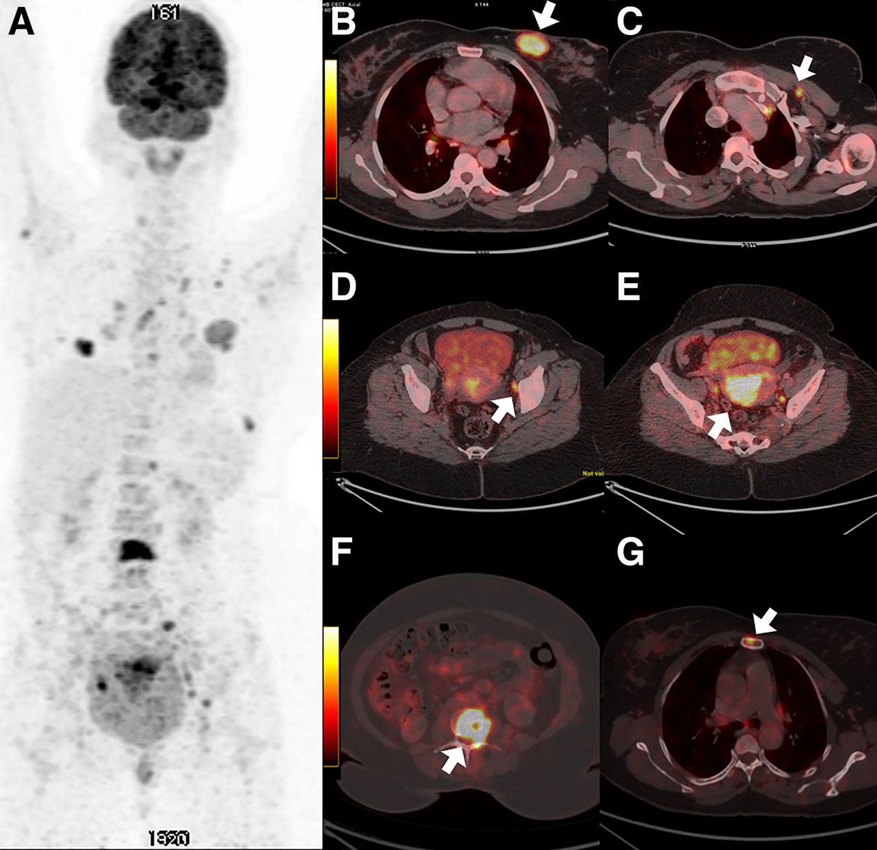
- FIGURE 1.
18F-FDG PET/CT of 42-y-old woman with suspected carcinoma of endometrium. (A) PET maximum-intensity projection showing multiple 18F-FDG–avid lesions. (B) Transaxial images showing lobulated soft-tissue lesion with spiculated margins involving upper inner quadrant of left breast (arrow) and (C) 18F-FDG–avid left interpectoral lymph nodes (arrow), suggestive of breast primary with nodal metastasis. (D and E) 18F-FDG–avid hypodense lesion involving body of uterus (arrow in E) with bilaterally 18F-FDG–avid external iliac lymph nodes (arrow in D), overall favoring breast primary with endometrial metastasis rather than primary endometrial cancer. (F and G) 18F-FDG–avid lytic skeletal lesions involving L3 vertebra (arrow in F) and sternum (arrow in G), suggestive of metastatic involvement.
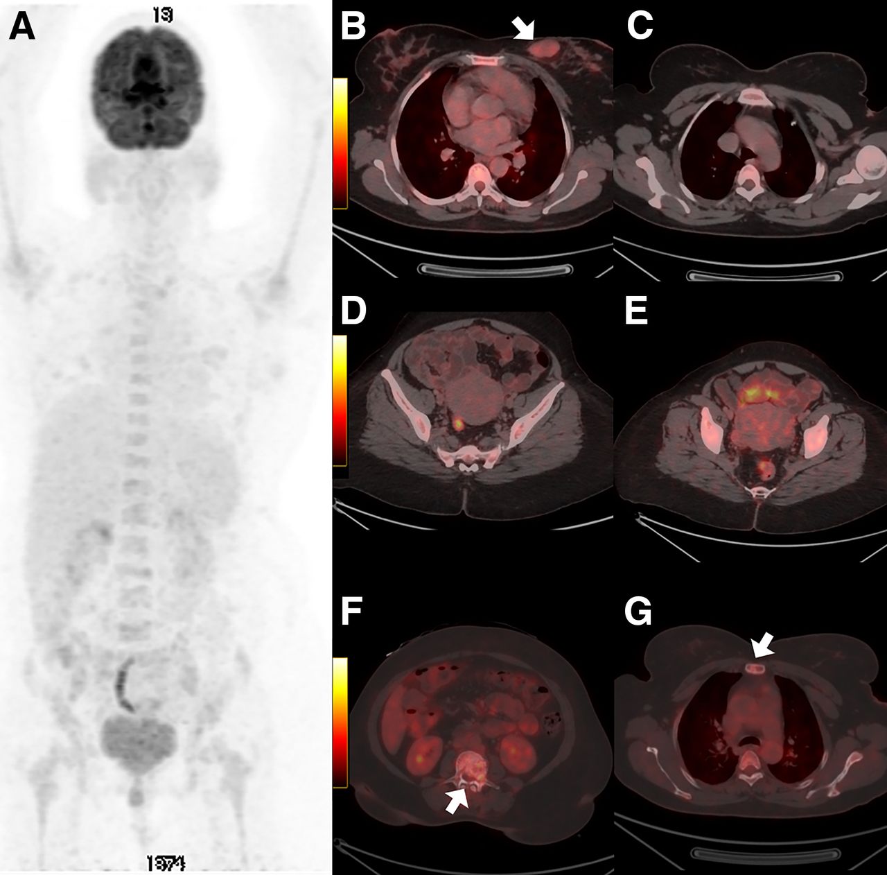
- FIGURE 2.
Follow-up 18F-FDG PET/CT of same patient as in Figure 1, after 5 cycles of chemotherapy. (A) PET maximum-intensity projection showing metabolic activity regression of most of the previous lesions. (B and C) Transaxial images showing regression in size and metabolic activity of left breast mass (arrow) and resolution of left interpectoral lymph node. (D and E) Resolution of hypodense lesion involving body of uterus, and regression in size and metabolic activity of bilateral external iliac lymph nodes. (F and G) Regression in metabolic activity with increase in sclerosis in skeletal lesions (arrow). Scan findings are suggestive of favorable response to therapy.


In this issue


Jump to section
Related Articles
Cited By...
- No citing articles found.