


Article Figures & Data
Figures
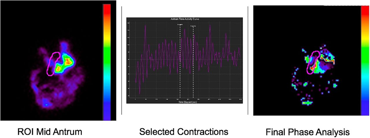
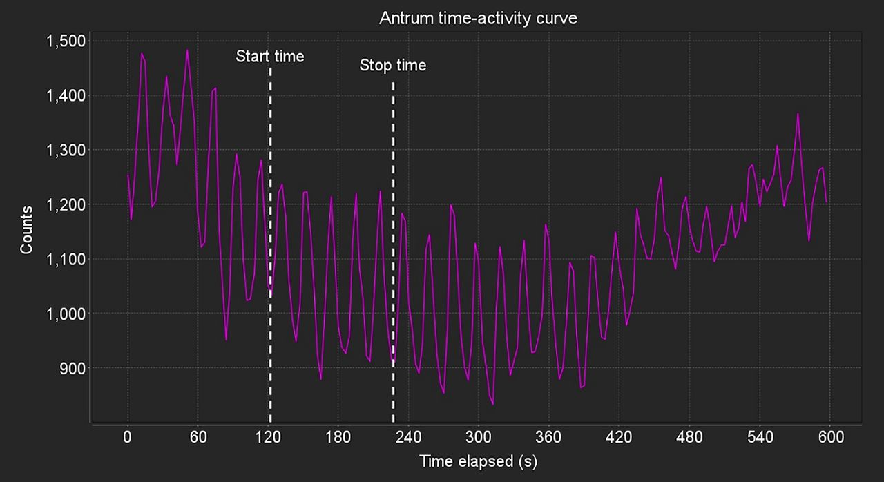
- FIGURE 1.
User selection of set of antral contractions for DACS analysis. This example of time–activity curve from patient study shows that even after use of image motion correction software, patient motion can result in significant motion artifacts in time–activity curve. Software workflow allows user to select optimum subset of image peaks and valleys (as shown between start time and end time), where antral contractions are stable and will be used for DACS processing.
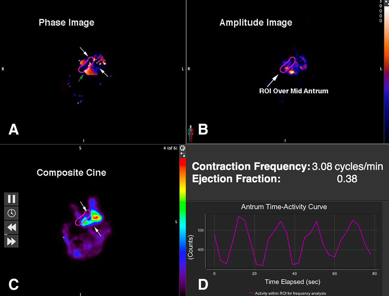
- FIGURE 2.
Healthy volunteer. (A) Fourier phase image showing color-coded pixels of Fourier phase analysis. Two-centimeter-wide region of interest drawn over mid antrum is same as obtained from those pixels in mid antrum with highest amplitude as shown in B. Antral peristaltic wave originates at incisura (white arrows). Resulting phase image shows those pixels that have similar color-coded phase angles clustered in proximal and distal antrum to left and right of mid antral region of interest. Leading edge of in-phase pixels appears as band of pixels (shown here with white color scale or 0° phase angle [green arrow]) in proximal antrum. To left of mid antral region of interest, group of pixels appears (∼180° from leading edge, red/orange color scale) corresponding to retrograde contractions arising in distal antrum. (B) Amplitude image showing color-coded pixels of Fourier amplitude. Image demonstrates cluster of high-amplitude pixels in mid antral region of interest (arrow) and in adjacent proximal antrum. (C) Single frame of composite cine image, with colored pixels representing total counts of radiolabeled solid-food activity in stomach. When viewed as movie display, antral peristaltic wave can be seen to originate at incisura (white arrows) and propagate distally through antrum across mid antral region of interest, followed by retrograde bolus movement back into proximal antrum. (D) Time–activity curve from mid antral region-of-interest–derived gastric counts, which are used to calculate antral contraction frequency and EF. ROI = region of interest.
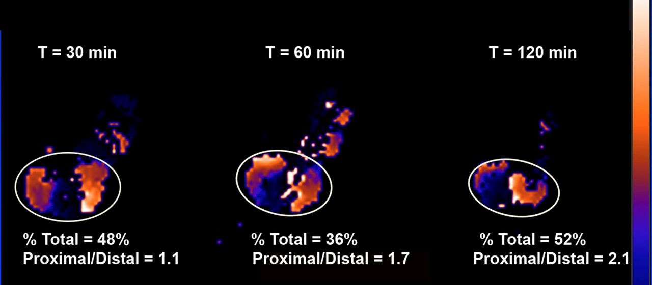
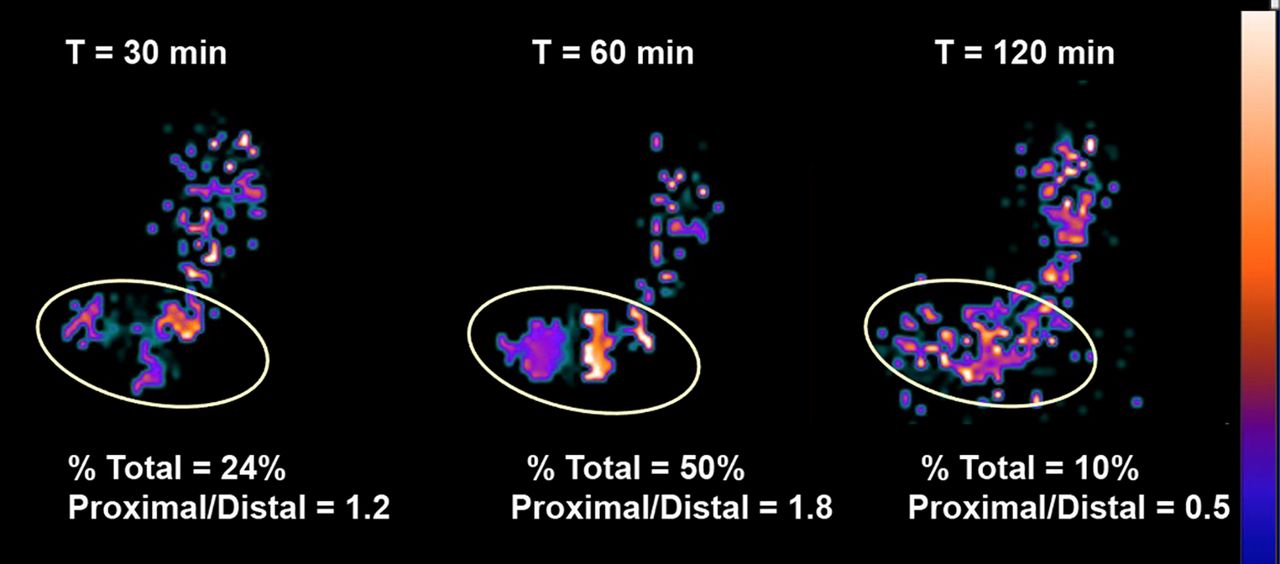
- FIGURE 3.
Patient with normal GE and normal phase analysis. Shown are Fourier phase results at 30, 60, and 120 min. Elliptic region of interest (white) shows total antral area used for analysis. Similarly colored clusters of pixels in proximal and distal antrum are those that have similar phase angles by Fourier analysis. Typically, ratio of proximal-to-distal ratio for in-phase pixels increases from 30 to 120 min.
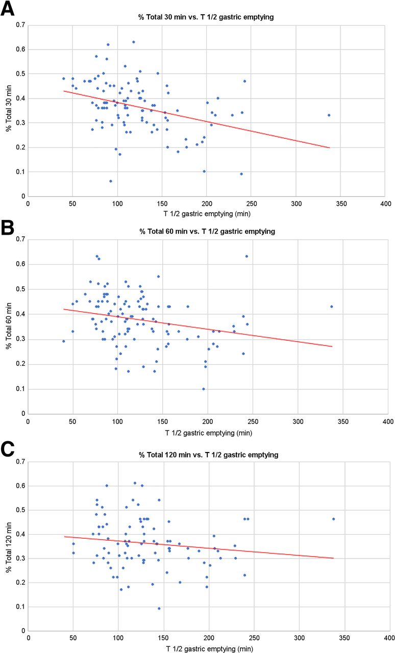
- FIGURE 4.
Linear regressions of percentage total compared with T½ of GE. (A) 30 min (percentage total = 0.4630 + −0.0008 × T½, R = 0.3746, P = 0.0001). (B) 60 min (percentage total = 0.4415 + −0.0005 × T½, R = 0.2559, P = 0.0065). (C) 120 min (percentage total = 0.4024 + −0.0003 × T½, R = 0.1456, P = 0.1680).
- FIGURE 5.
Patient with abnormal GE and abnormal phase analysis. Shown are Fourier phase angle images for patient with delayed GE (T½ = 188 min). Elliptic ROI as in Figure 4 again shows total antral area used for analysis. There is lack of synchronous in-phase proximal and distal antral pixels at 30 and 120 min compared with normal pattern (Fig. 4). At 60 min, there is cluster of proximal antral phasic activity but no coordinated distal phasic contractions.


Tables
- TABLE 1.
Descriptive Summary and 90% Percentile Intervals Based on Healthy Volunteers for Conventional GES Parameters
Variable n Median Range P* 5%, 95% CI Percentage total, 30 min 19 45% 31%–63% 0.11 31%, 63% Percentage total, 60 min 22 40% 17%–63% 19%, 62% Percentage total, 120 min 13 51% 32%–61% 32%, 61% Proximal-to-distal ratio, 30 min 19 1.67 0.36–6.62 0.035 0.36, 6.62 Proximal-to-distal ratio, 60 min 22 1.89 0.71–4.71 0.88, 3.15 Proximal-to-distal ratio, 120 min 13 2.65 1.25–6.38 1.25, 6.38 EF, 30 min 21 23% 8%–44% 0.022 14%, 36% EF, 60 min 22 27% 19%–42% 19%, 40% EF, 120 min 14 32% 11%–41% 11%, 41% Frequency (cycle/min), 30 min 21 3.08 2.58–3.45 0.11 2.67, 3.33 Frequency (cycle/min), 60 min 22 2.86 2.40–3.57 2.76, 3.48 Frequency (cycle/min), 120 min 14 2.91 2.42–3.20 2.42, 3.20 ↵* P value for testing time effect of each variable.
- TABLE 2.
Predictive Capability of DACS Parameters for Abnormal Results Compared with Conventional GES
DACS grouping by… Overall (n = 121) Conventional GES results P* Raw odds ratio† 95% CI Abnormal (n = 53) Normal (patients + healthy volunteers) (n = 68) Percentage total, 30 min 0.001 Abnormal, <31% or >63% 30 (27.5%) 21 (43.8%) 9 (14.8%) 4.49 1.81–11.15 Normal, 31%–63% 79 (72.5%) 27 (56.3%) 52 (85.2%) Reference Percentage total, 60 min 0.17 Abnormal, <19% or >62% 9 (7.6%) 6 (11.8%) 3 (4.5%) 2.84 0.68–11.97 Normal, 19%–62% 109 (92.4%) 45 (88.2%) 64 (95.5%) Reference Percentage total, 120 min 0.21 Abnormal, <32% or >61% 34 (35.1%) 21 (41.2%) 13 (28.3%) 1.78 0.76–4.16 Normal, 32%–61% 63 (64.9%) 30 (58.8%) 33 (71.7%) Reference Proximal-to-distal ratio, 30 min 1.00 Abnormal, <0.36 or >6.62 7 (6.4%) 3 (6.3%) 4 (6.6%) 0.95 0.20–4.46 Normal, 0.36–6.62 102 (93.6%) 45 (93.8%) 57 (93.4%) Reference Proximal-to-distal ratio, 60 min 0.017 Abnormal, <0.88 or >3.15 39 (33.3%) 23 (46.0%) 16 (23.9%) 2.72 1.23–5.99 Normal, 0.88–3.15 78 (66.7%) 27 (54.0%) 51 (76.1%) Reference Proximal-to-distal ratio, 120 min 0.83 Abnormal, <1.25 or >6.38 37 (38.9%) 20 (40.8%) 17 (37.0%) 1.18 0.51–2.69 Normal, 1.25–6.38 58 (61.1%) 29 (59.2%) 29 (63.0%) Reference EF, 30 min 0.48 Abnormal, <14% or >36% 25 (24.8%) 12 (29.3%) 13 (21.7%) 1.50 0.60–3.72 Normal, 14%–36% 76 (75.2%) 29 (70.7%) 47 (78.3%) Reference EF, 60 min 0.011 Abnormal, <19% or >40% 24 (22.6%) 16 (34.8%) 8 (13.3%) 3.47 1.33–9.06 Normal, 19%–40% 82 (77.4%) 30 (65.2%) 52 (86.7%) Reference EF, 120 min 0.26 Abnormal, <11% or >41% 15 (18.3%) 10 (23.3%) 5 (12.8%) 2.06 0.64–6.68 Normal, 11%–41% 67 (81.7%) 33 (76.7%) 34 (87.2%) Reference Frequency, 30 min 0.047 Abnormal, <2.67 or >3.33 11 (10.9%) 8 (19.5%) 3 (5.0%) 4.61 1.14–18.57 Normal, 2.67–3.33 90 (89.1%) 33 (80.5%) 57 (95.0%) Reference Frequency, 60 min 0.32 Abnormal, <2.76 or >3.48 10 (9.4%) 6 (13.0%) 4 (6.7%) 2.10 0.56–7.93 Normal, 2.76–3.48 96 (90.6%) 40 (87.0%) 56 (93.3%) Reference Frequency, 120 min 1.00 Abnormal, <2.42 or >3.20 11 (13.4%) 6 (14.0%) 5 (12.8%) 1.10 0.31–3.95 Normal, 2.42–3.20 71 (86.6%) 37 (86.0%) 34 (87.2%) Reference - TABLE 3.
Multivariable Logistic Regression Identifying Best Subset of DACS Abnormality Parameters Associated with Standard Clinical Diagnosis Using Data from All Subjects (n = 121)*
DACS abnormality variable Adjusted odds ratio 95% CI P By percentage total, 30 min, to <31% or >63% vs. 31%–63% 3.30 1.21–9.00 0.02 By EF, 60 min, to <19% or >40% vs. 19%–40% 2.97 1.08–8.21 0.036 ↵* 24 subjects had missing data on at least 1 variable and hence dropped out of model.


