


Article Figures & Data
Figures
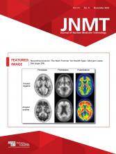
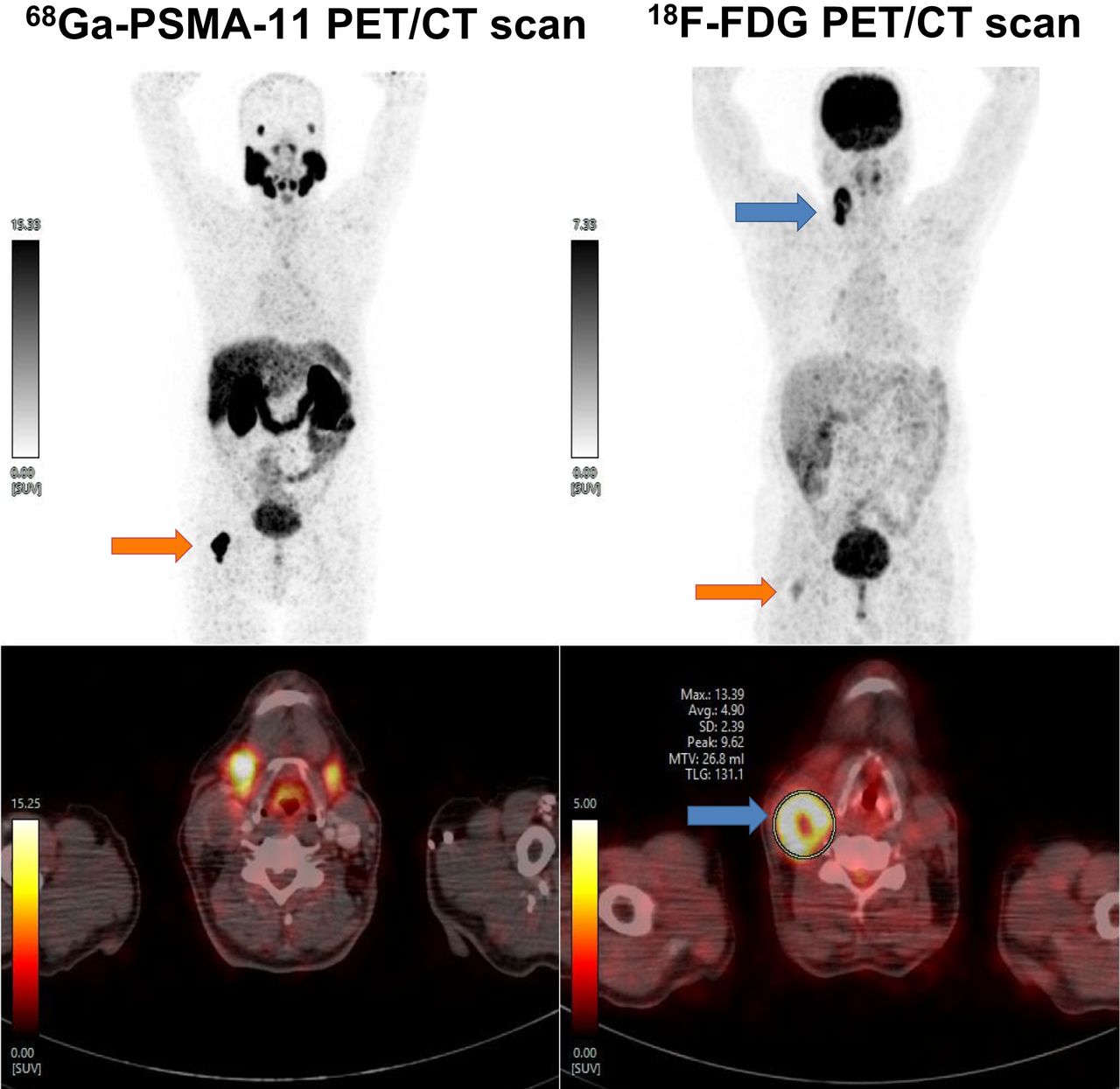
- FIGURE 1.
Whole-body 68Ga-PSMA-11 PET/CT and 18F-FDG PET/CT scans showing 68Ga-PSMA-11 expression (orange arrows) in sclerotic skeletal lesions in body of D4 vertebra (lesion 1), in left internal iliac lymph nodes (lesion 2), and bilaterally in pelvic bones (lesion 3); none of these showed any 18F-FDG uptake. Hypodense lesion in segment VII/VIII of liver showed 18F-FDG uptake (blue arrow) but did not show any significant 68Ga-PSMA-11 expression, raising suspicion and requirement of further confirmation.
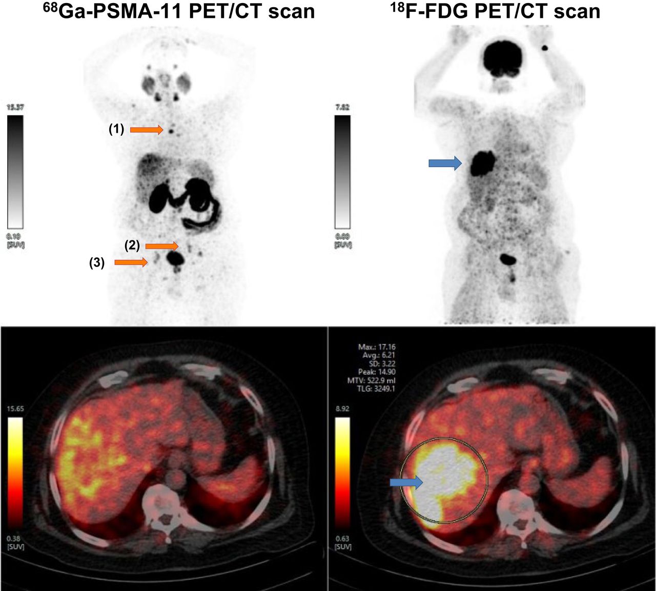
- FIGURE 2.
(A) Scanner view showing tumor with dual morphology. Lower half of left image shows tumor arranged in glandular architecture resembling adenocarcinoma (#), whereas upper half shows tumor arranged in broad trabeculae resembling hepatocellular carcinoma ($). On right, biopsy core shows necrosis (*) (hematoxylin and eosin, ×40). (B) Tumor arranged in glandular architecture, with large cells having moderate nuclear atypia resembling adenocarcinoma (hematoxylin and eosin, ×100). (C) Tumor cells arranged in large nodules and thickened trabeculae with moderate nuclear atypia and eosinophilic cytoplasm suggesting hepatocytic neoplasm (hematoxylin and eosin, ×100). (D) Glandular tumor (adenocarcinoma) component positive for CK7 (immunohistochemistry, ×100). (E and F) Hepatocytic tumor (hepatocellular carcinoma) component positive for glypican-3 (E) and arginase (F) (immunohistochemistry, ×100). Scale bars denote 50 μm.
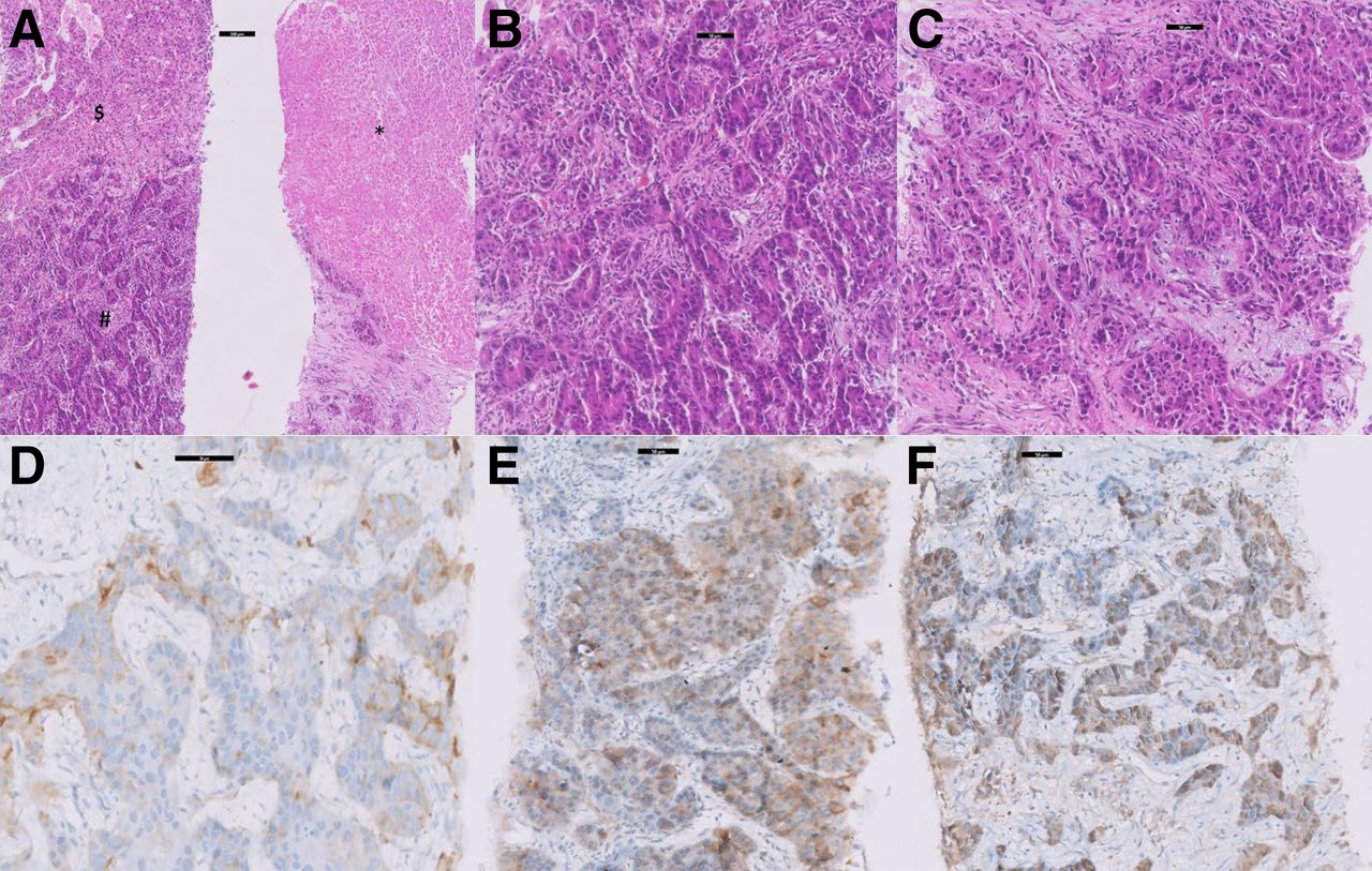
- FIGURE 3.
Coronal (A) and axial (B) posttherapy SPECT/CT images showing 177Lu-PSMA-617–expressing (yellow arrows) and nonexpressing (blue arrows) multiple sclerotic skeletal lesions. Transaxial SPECT/CT (C) and CT (D) posttherapy images showing hypodense lesion in segment VII/VIII of liver (arrow), with absence of 177Lu-PSMA expression.
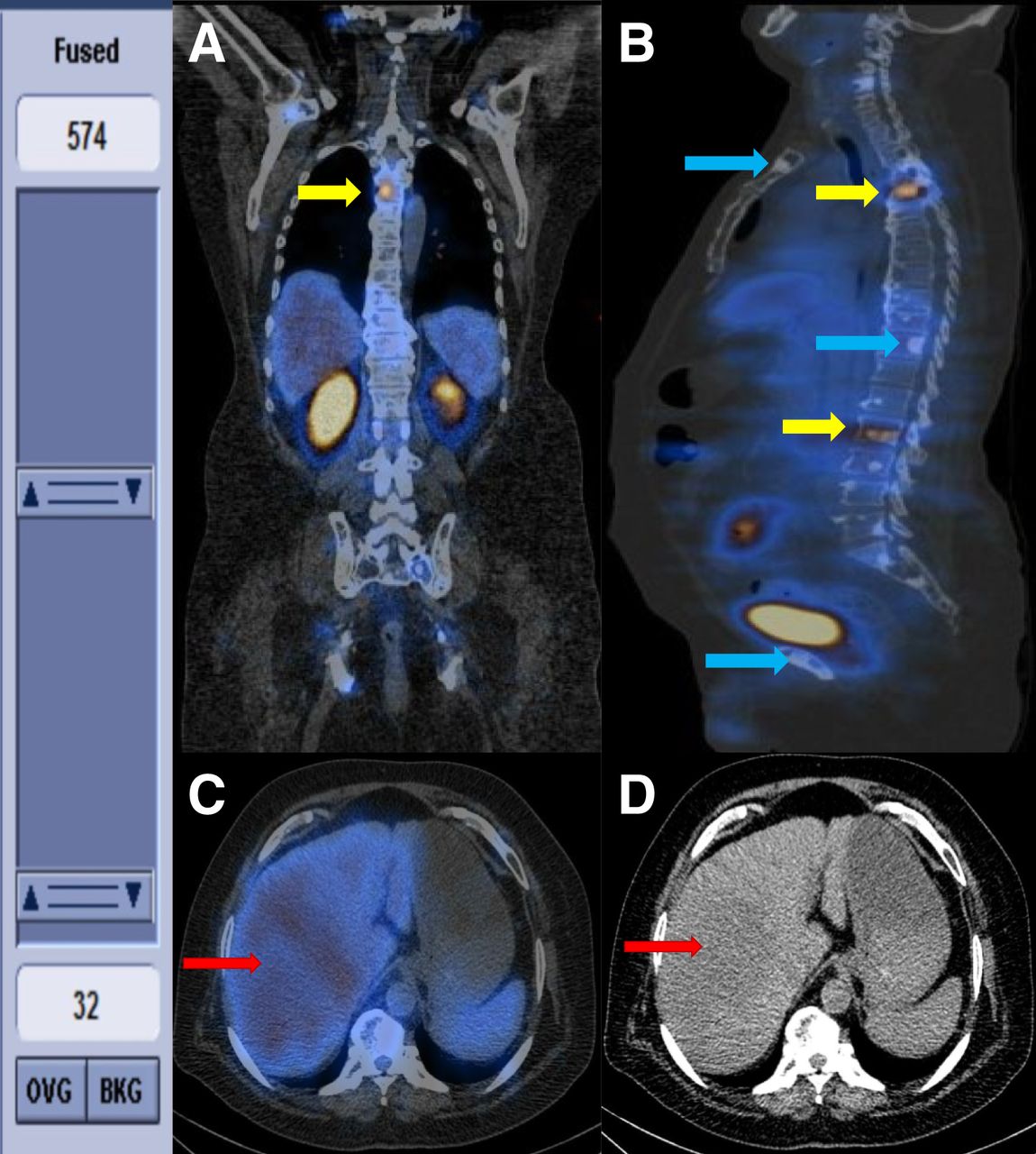
- FIGURE 4.
Whole-body 68Ga-PSMA-11 PET/CT and 18F-FDG PET/CT images showing 68Ga-PSMA-11 expression (orange arrows) in sclerotic lesion involving right neck of femur, and low-grade 18F-FDG uptake, consistent with diagnosis of prostate carcinoma with skeletal metastasis. Right cervical III/IC lymph node shows 18F-FDG uptake (blue arrows) but not 68Ga-PSMA-11 expression, raising suspicion of different pathology.


In this issue


Jump to section
Related Articles
Cited By...
- No citing articles found.