


Article Figures & Data
Figures
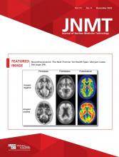
- FIGURE 1.
68Ga-DOTATOC PET/CT images (PET maximum-intensity projection, A; axial view, B; coronal view, C) showing tracer-avid right upper cervical mass.
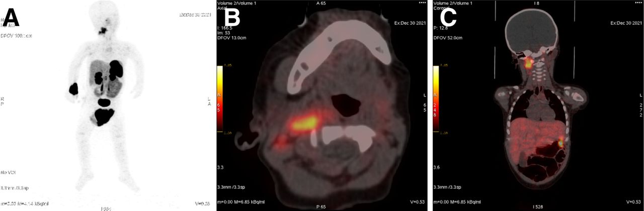
- FIGURE 2.
Contrast-enhanced CT images (coronal view, A; axial view, B) showing slightly enhanced soft-tissue mass in jugulodigastric region (arrow).
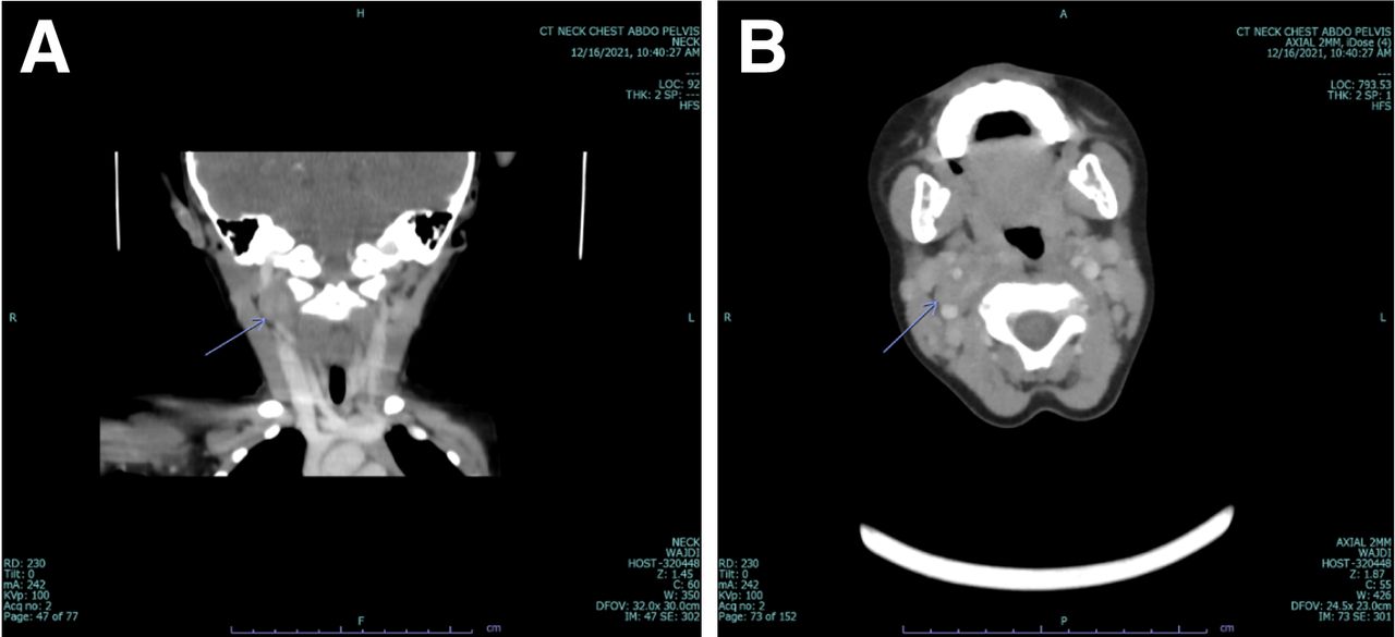
- FIGURE 3.
Neck MRI showing right upper cervical mass (arrows) that is low signal on T1 sequence (coronal view, A), high signal on T2 sequence (coronal view, B), and enhanced on postcontrast T1 sequence (axial views, C and D).
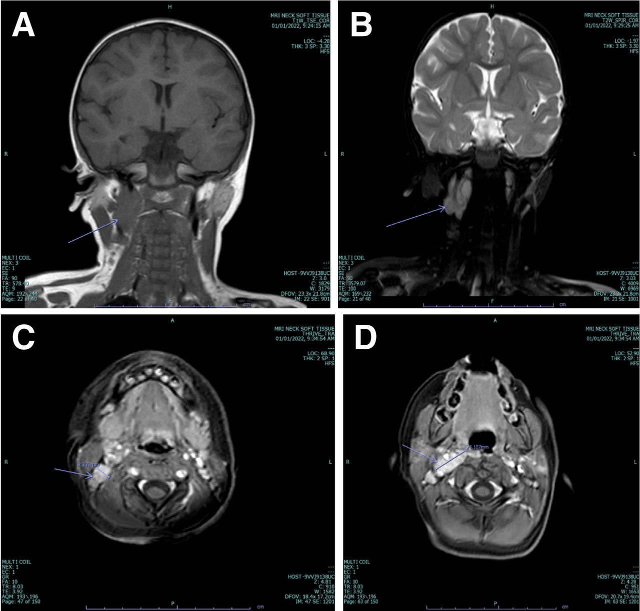
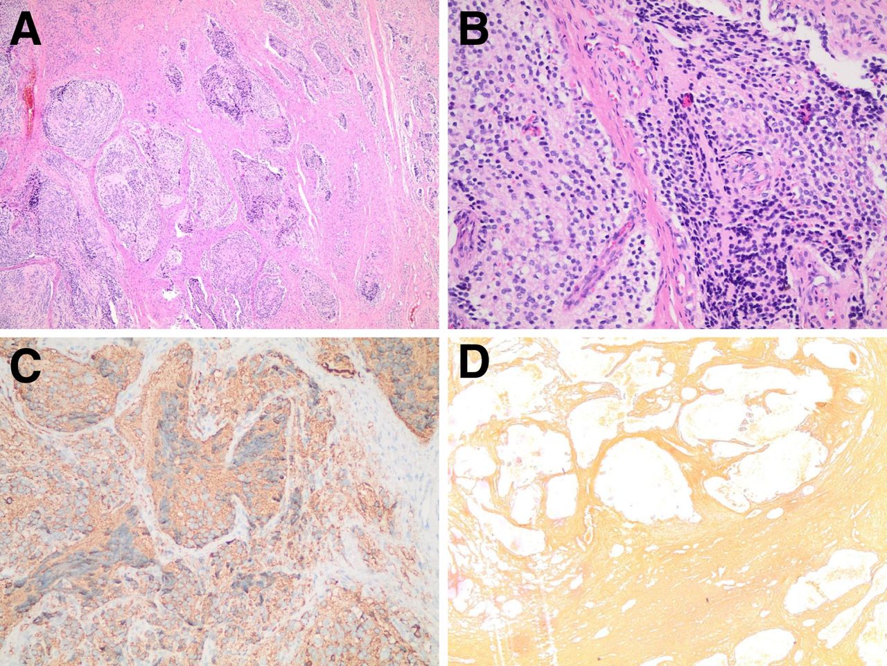
- FIGURE 4.
(A and B) Histopathology showing nodules composed of stroma-poor islands of primitive cells with fibrillary neuropil matrix. (C) Synaptophysin staining in tumor cells. (D) Reticulin stain highlights nested pattern.


