
Main menu

  • Home
  • Content
    • Current
      • JNMT Supplement
    • Ahead of print
    • Past Issues
    • Continuing Education
    • JNMT Podcast
    • SNMMI Annual Meeting Abstracts
  • Subscriptions
    • Subscribers
    • Rates
    • Journal Claims
    • Institutional and Non-member
  • Authors
    • Submit to JNMT
    • Information for Authors
    • Assignment of Copyright
    • AQARA Requirements
  • Info
    • Reviewers
    • Permissions
    • Advertisers
    • Corporate & Special Sales
  • About
    • About Us
    • Editorial Board
    • Contact Information
  • More
    • Alerts
    • Feedback
    • Help
    • SNMMI Journals
  • SNMMI
    • JNMT
    • JNM
    • SNMMI Journals
    • SNMMI

User menu

  • Subscribe
  • My alerts
  • Log in
  • My Cart

Search

  • Advanced search
Journal of Nuclear Medicine Technology
  • SNMMI
    • JNMT
    • JNM
    • SNMMI Journals
    • SNMMI
  • Subscribe
  • My alerts
  • Log in
  • My Cart
Journal of Nuclear Medicine Technology

Advanced Search

  • Home
  • Content
    • Current
    • Ahead of print
    • Past Issues
    • Continuing Education
    • JNMT Podcast
    • SNMMI Annual Meeting Abstracts
  • Subscriptions
    • Subscribers
    • Rates
    • Journal Claims
    • Institutional and Non-member
  • Authors
    • Submit to JNMT
    • Information for Authors
    • Assignment of Copyright
    • AQARA Requirements
  • Info
    • Reviewers
    • Permissions
    • Advertisers
    • Corporate & Special Sales
  • About
    • About Us
    • Editorial Board
    • Contact Information
  • More
    • Alerts
    • Feedback
    • Help
    • SNMMI Journals
  • Watch or Listen to JNMT Podcast
  • Visit SNMMI on Facebook
  • Join SNMMI on LinkedIn
  • Follow SNMMI on Twitter
  • Subscribe to JNMT RSS feeds
Research ArticleTeaching Case Studies

Bone Scanning Findings in a Patient with Heat Stroke–Induced Rhabdomyolysis

Jitesh Dhingra and Yoram Baum
Journal of Nuclear Medicine Technology December 2021, 49 (4) 362-363; DOI: https://doi.org/10.2967/jnmt.121.262501
Jitesh Dhingra
Emory University Hospital, Atlanta, Georgia
  • Find this author on Google Scholar
  • Find this author on PubMed
  • Search for this author on this site
Yoram Baum
Emory University Hospital, Atlanta, Georgia
  • Find this author on Google Scholar
  • Find this author on PubMed
  • Search for this author on this site
  • Article
  • Figures & Data
  • Info & Metrics
  • PDF
Loading

Abstract

We present a case that caused a diagnostic dilemma on a bone scan. We also review the broad spectrum of nonmalignant findings that can impact the interpretation of a bone scan and the value of correlative imaging using SPECT/CT for exact localization and characterization of lesions. The imaging features of important benign pathologies—that is, metastatic mimics—are elaborated so that the reader can avoid misinterpretations when reporting them. We elucidate 4 uncommon benign findings on a bone scan. Rhabdomyolysis is a result of lysis of skeletal muscle with release of cell contents, such as myoglobin and muscle enzymes, and is diagnosed mostly through a combination of clinical appearance and laboratory values. Myositis ossificans is the most common form of heterotopic ossification, usually occurring within large muscles. Its importance stems largely from its ability to mimic more aggressive pathologic processes. Myositis ossificans is one of the skeletal “do not touch” lesions. Such bone lesions are defined by characteristic imaging features, the identification of which precludes the need for additional diagnostic tests or biopsies, thereby avoiding unnecessary interventions. Acute tubular necrosis is kidney injury caused by damage to the kidney tubule cells (kidney cells that reabsorb fluid and minerals from urine as it forms). Common causes are low blood flow to the kidneys, drugs that damage the kidneys, or a severe underlying infection.

  • renal
  • SPECT/CT
  • nonmalignant
  • bone scan
  • renal failure
  • rhabdomyolysis

Bone scanning continues to be the second most commonly used nuclear imaging procedure after nuclear cardiac scans, offering the advantage of total-body examination, low cost, and high sensitivity not only for metastatic lesions but also for benign conditions. Its power rests in the physiologic uptake and pathophysiologic behavior of 99mTc- diphosphonates (1). Radiotracers localize in damaged skeletal muscles because calcium builds up in them and, when combined with phosphate, provides a site for radiotracer deposition (1).

In a case of suspected rhabdomyolysis, not only is bone scintigraphy a sensitive indicator of muscle damage but also the amount of uptake is proportional to the extent of myonecrosis (2–5). Specifically, in sports medicine, bone scintigraphy could be a valuable tool in discriminating bone trauma from muscle trauma (6).

We present an unusual case of a 19-y-old woman who experienced heat stroke and whose bone scan presented several unusual findings; this case demonstrates important teaching points.

CASE REPORT

As per our institute protocol, the patient was injected with 925 MBq of 99mTc-methyldiphosphonate, and after 2.5 h whole-body anterior and posterior images of the entire skeleton were obtained using a γ-camera with gantry motion of 10 cm/min, a matrix size of 256 × 1024, and no magnification. Subsequently, for better visualization of related pathologies, SPECT images of the chest and upper abdomen were obtained using a 360° rotation, noncircular step-and-shoot mode, 128 steps, 20 s per step, a matrix size of 128 × 128, and no magnification.

The whole-body planar and SPECT/CT images demonstrated abnormal radiotracer uptake suggestive of rhabdomyolysis in the muscles of the lower extremity, abnormal bilateral cortical uptake suggestive of acute tubular necrosis within the kidneys, greater abnormal uptake in the right than the left pleural space suggestive of organizing hematoma, and abnormal uptake within the abdominal musculature suggestive of myositis ossificans.

DISCUSSION

Rhabdomyolysis is a complex medical condition leading to disruption of skeletal muscle integrity. This disruption can lead to the direct release of intracellular muscle components such as myoglobin, creatine kinase, aldolase, and lactate dehydrogenase into the bloodstream and extracellular space (2–4).

Bone scans are a sensitive way to visualize muscle injury and have been used with 99mTc-diphosphonate and several similar agents in a wide variety of conditions, including exercise injuries (6), seizures, crush injuries, and polymyositis, and are a good indicator of acute rhabdomyolysis. Myositis ossificans is essentially metaplasia of the intramuscular connective tissue resulting in extraosseous bone formation. Examination of the soft-tissue lesion using 99mTc-diphosphonate bone scans has been helpful in establishing the diagnosis and in determining the full extent of the process early in its evolution (6).

Another important utility of 99mTc-diphosphonate bone scans lies in evaluation of acute renal failure after exercise with flank pain and patchy renal vasoconstriction due to acute tubular necrosis. These findings have been shown in multiple reports, with a prominent example being one written by Watanabe et al. (7). We also found an organizing pleural hematoma with radiotracer uptake. Localization of bone-imaging 99mTc-diphosphonate agents has previously been described in abdominal, chest wall, and pelvic hematomas (8,9).

CONCLUSION

It is extremely important for all 4 benign conditions described above to be recognized on a bone scan. We hope this report educates the reader about the types of uncommon conditions that can be seen on bone scans.

DISCLOSURE

No potential conflict of interest relevant to this article was reported.

FIGURE 1.
  • Download figure
  • Open in new tab
  • Download powerpoint
FIGURE 1.

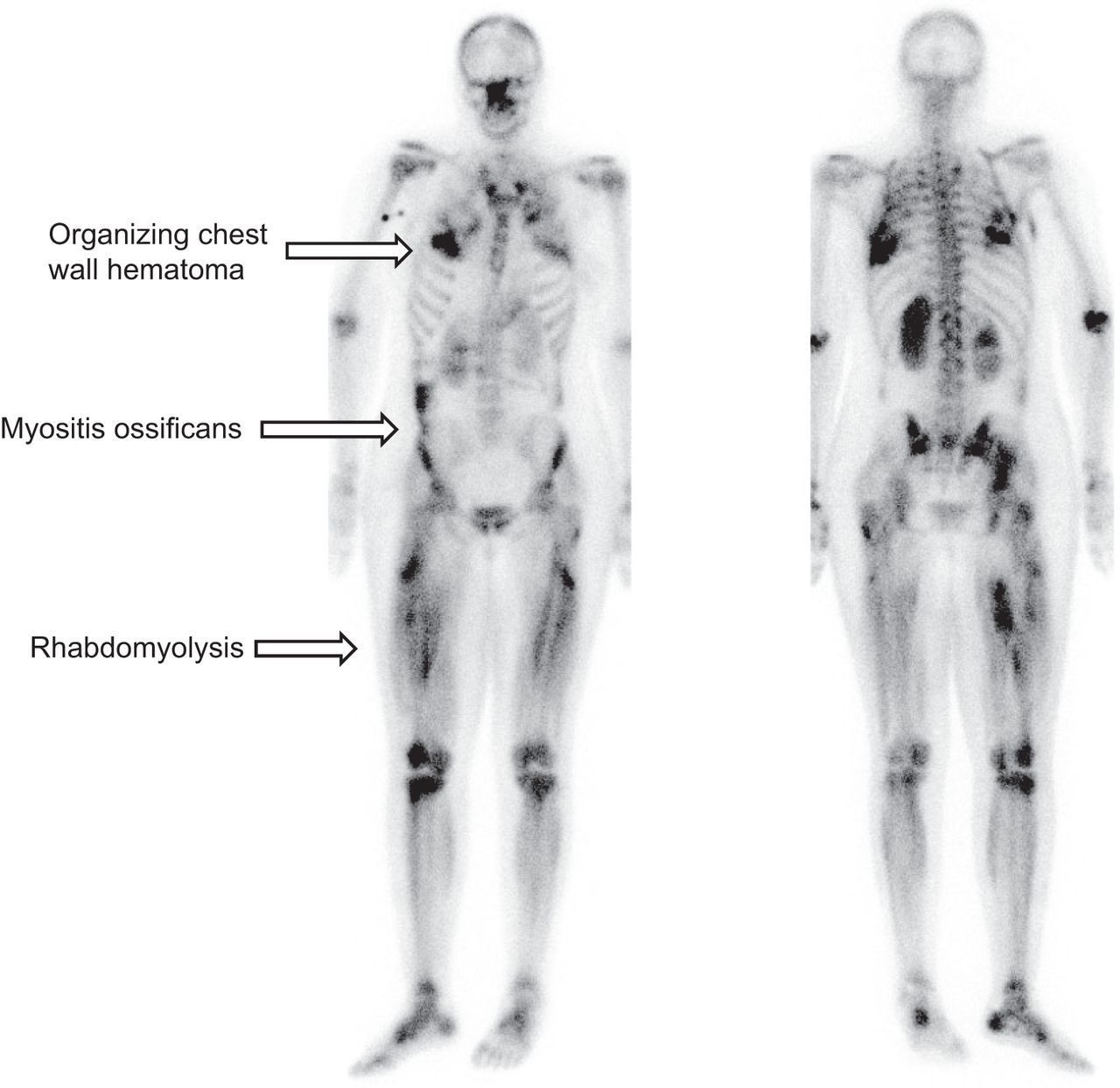
Whole-body 99mTc-methyldiphosphonate SPECT/CT image demonstrating abnormally increased uptake in organizing chest hematoma, myositis ossificans, and rhabdomyolysis. Increased bilateral uptake in several locations in lower extremities along length of muscle corresponds to pattern of rhabdomyolysis.

FIGURE 2.
  • Download figure
  • Open in new tab
  • Download powerpoint
FIGURE 2.

SPECT/CT images demonstrating abnormally increased radiotracer uptake in organizing chest wall hematoma. (Top left) CT image of chest demonstrating chest wall hematoma. (Top right) SPECT image demonstrating abnormally increased uptake within hematoma. (Bottom left) SPECT/CT image demonstrating abnormally increased radiotracer uptake (arrow) corresponding to underlying chest wall hematoma. (Bottom right) Rotating maximum-intensity projection SPECT image demonstrating focal region of uptake in right chest. Also seen is faint focus of uptake in left chest (additional site of hematoma).

FIGURE 3.
  • Download figure
  • Open in new tab
  • Download powerpoint
FIGURE 3.

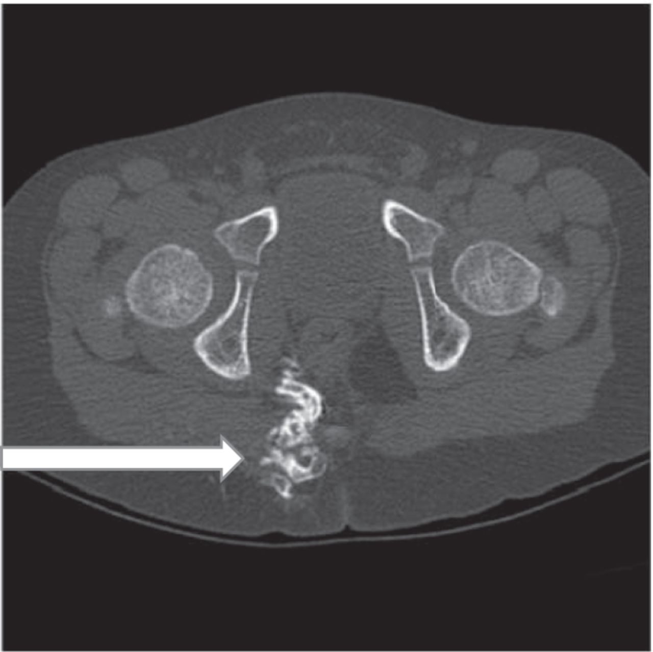
CT images in bone window demonstrating focus of myositis ossificans (new bone formation, arrow) in pelvis.

FIGURE 4.
  • Download figure
  • Open in new tab
  • Download powerpoint
FIGURE 4.

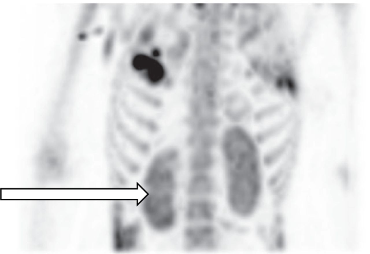
Planar image demonstrating abnormally increased bilateral cortical retention of radiotracer in kidneys in pattern of acute tubular necrosis.

FIGURE 5.
  • Download figure
  • Open in new tab
  • Download powerpoint
FIGURE 5.

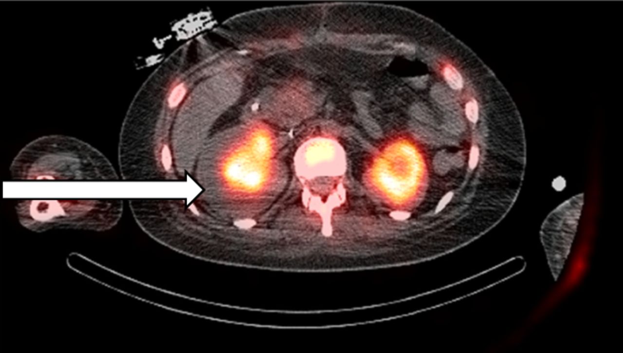
SPECT/CT image demonstrating abnormal bilateral cortical retention of radiotracer in kidneys, corresponding to acute tubular necrosis.

Footnotes

  • Published online July 30, 2021.

REFERENCES

  1.  1.↵
    1. Brenner AI,
    2. Koshy J.
    The bone scan. Semin Nucl Med. 2012;42:11–26.
    OpenUrlCrossRefPubMed
  2.  2.↵
    1. Sanders JA.
    Rhabdomyolysis detected by bone imaging. Clin Nucl Med. 1989;14:431–432.
    OpenUrlPubMed
  3.  3.
    1. Steuart RD,
    2. Morrison RT,
    3. Lot L.
    An incidental finding of rhabdomyolysis on bone scintigraphy: case report. J Nucl Med Technol. 1993;21:63–64.
    OpenUrlAbstract/FREE Full Text
  4.  4.↵
    1. Torres PA,
    2. Helmstetter JA,
    3. Kaye AM,
    4. Kaye AD.
    Rhabdomyolysis: pathogenesis, diagnosis, and treatment. Ochsner J. 2015;15:58–69.
    OpenUrlAbstract/FREE Full Text
  5.  5.↵
    1. Tyler JL,
    2. Derbekyan V,
    3. Lisbona R.
    Early diagnosis of myositis ossificans with 99mTc diphosphonate imaging. Clin Nucl Med. 1984;9:256–258.
    OpenUrlCrossRefPubMed
  6.  6.↵
    1. Williamson MR,
    2. Archibeque F,
    3. Eisenberg B,
    4. Williamson S,
    5. Rosenberg R.
    99mTc pyrophosphate localization in chest wall muscles after bench pressing. Clin Nucl Med. 1989;14:546.
    OpenUrlPubMed
  7.  7.↵
    1. Watanabe N,
    2. Shimizu M,
    3. Kageyama M,
    4. Kamei T,
    5. Seto H,
    6. Kakishita M.
    Diffuse increased renal uptake on bone scintigraphy in acute tubular necrosis. Clin Nucl Med. 1994;19:19–21.
    OpenUrlCrossRefPubMed
  8.  8.↵
    1. Park C,
    2. LaRoy L,
    3. Ali A,
    4. Fordham EW.
    Pelvic hematoma diagnosed on 99mTc methylene diphosphanate bone imaging. Clin Nucl Med. 1989;14:139–140.
    OpenUrlPubMed
  9.  9.↵
    1. Bhattacharya A,
    2. Prasad V,
    3. Mittal BR.
    99mTc MDP uptake in a soft tissue hematoma of the chest wall. Clin Nucl Med. 2004;29:454–455.
    OpenUrlPubMed
  • Received for publication April 27, 2021.
  • Revision received June 10, 2021.
PreviousNext
Back to top

In this issue

Journal of Nuclear Medicine Technology: 49 (4)
Journal of Nuclear Medicine Technology
Vol. 49, Issue 4
December 1, 2021
  • Table of Contents
  • About the Cover
  • Index by author
  • Complete Issue (PDF)
Print
Download PDF
Article Alerts
Sign In to Email Alerts with your Email Address
Email Article

Thank you for your interest in spreading the word on Journal of Nuclear Medicine Technology.

NOTE: We only request your email address so that the person you are recommending the page to knows that you wanted them to see it, and that it is not junk mail. We do not capture any email address.

Enter multiple addresses on separate lines or separate them with commas.
Bone Scanning Findings in a Patient with Heat Stroke–Induced Rhabdomyolysis
(Your Name) has sent you a message from Journal of Nuclear Medicine Technology
(Your Name) thought you would like to see the Journal of Nuclear Medicine Technology web site.
Citation Tools
Bone Scanning Findings in a Patient with Heat Stroke–Induced Rhabdomyolysis
Jitesh Dhingra, Yoram Baum
Journal of Nuclear Medicine Technology Dec 2021, 49 (4) 362-363; DOI: 10.2967/jnmt.121.262501

Citation Manager Formats

  • BibTeX
  • Bookends
  • EasyBib
  • EndNote (tagged)
  • EndNote 8 (xml)
  • Medlars
  • Mendeley
  • Papers
  • RefWorks Tagged
  • Ref Manager
  • RIS
  • Zotero
Share
Bone Scanning Findings in a Patient with Heat Stroke–Induced Rhabdomyolysis
Jitesh Dhingra, Yoram Baum
Journal of Nuclear Medicine Technology Dec 2021, 49 (4) 362-363; DOI: 10.2967/jnmt.121.262501
Twitter logo Facebook logo LinkedIn logo Mendeley logo
  • Tweet Widget
  • Facebook Like
  • Google Plus One
Bookmark this article

Jump to section

  • Article
    • Abstract
    • CASE REPORT
    • DISCUSSION
    • CONCLUSION
    • DISCLOSURE
    • Footnotes
    • REFERENCES
  • Figures & Data
  • Info & Metrics
  • PDF

Related Articles

  • No related articles found.
  • PubMed
  • Google Scholar

Cited By...

  • No citing articles found.
  • Google Scholar

More in this TOC Section

  • High-Sensitivity Troponin Elevation in a Young Woman with Typical Chest Pain: The Heart of the Matter
  • Pulmonary Adenocarcinoma Revealed by Parathyroid Scintigraphy: An Incidental Case to Remember
  • Prominent Right Ventricular Tracer Uptake: A Harbinger of Multivessel Coronary Artery Disease
Show more Teaching Case Studies

Similar Articles

Keywords

  • renal
  • SPECT/CT
  • nonmalignant
  • bone scan
  • renal failure
  • rhabdomyolysis
SNMMI

© 2025 SNMMI

Powered by HighWire