


Article Figures & Data
Figures
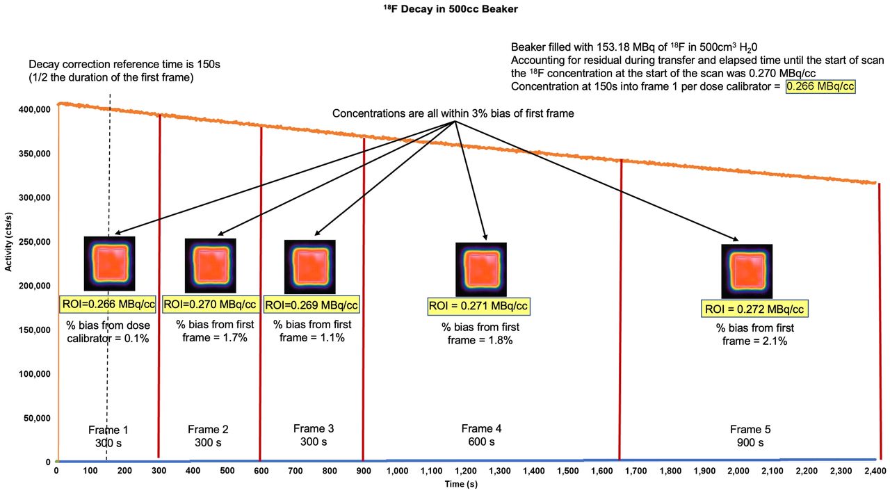
- FIGURE 1.
Decay of 18F in 500-cm3 beaker. A detailed 18F protocol (“Fluorine Decay Correction.docx”) and worksheet (“F18 Decay Correction Worksheet.xlsx”) can be found in the supplemental materials. With syringe, 153.18 MBq of 18F were placed in beaker containing 500 cm3 of H2O. Accounting for residual activity in transfer syringe and elapsed time between dose calibrator measurement and start of scan, concentration of 18F at start of scan was 0.270 MBq/cm3. Scanner performed 2,400-s (40 min) list-mode acquisition. Twenty-four hours later, after all activity decayed, attenuation scanning was performed and 5 serial frames were generated at intervals of 300, 300, 300, 600, and 900 s. ROIs were placed, avoiding beaker boundaries. Activity at time t equaled initial activity × e(−0.693 × t/(half-life of radiotracer). Therefore, with starting activity of 0.270 MBq/cm3, expected activity at 150 s into scan (midpoint of first frame) is 0.266 MBq/cm3. Half-life of 18F is 6,600 s, and 0.270 MBq/cm3 × e (−6.93 × 150 s/6,600s) = 0.266 MBq/cm3. Concentration of ROI in first frame was 0.266 MBq/cm3, which is 0.1% bias from dose calibrator. Concentrations in each subsequent frame were nearly identical to first frame, with biases all within 3% window. There are several conclusions. First, scanner decay-corrects activity to midpoint of first frame. Second, scanner also corrects for duration of each frame, giving activity in MBq/cm3. Third, in biologic systems, only variation in quantitative activity after first frame would be due to physiologic changes and not imaging timing, duration, or decay. Bias from dose calibrator of first frame, also known as efficiency, is inconsequential to measurements of MBF as it cancels out in numerator and denominator of flow equations (6). Bias does inform us on whether test was performed with accurate timing, random, scatter, and dead-time corrections and also on whether camera system was internally calibrated for isotope against standard. If timing of beaker decay test is not precise or camera has not been calibrated, measured concentration could be significantly different from dose calibrator; however, if DC is performed correctly, bias of subsequent frames will be uniform.
- FIGURE 2.
Decay of 13N in 500-cm3 beaker. A detailed 13N protocol (“Nitrogen Decay Correction.docx”) and worksheet (“13N Decay Correction Worksheet.xlsx”) can be found in the supplemental materials. Similar to Figure 1, 348.91 MBq of 13N were placed a beaker containing precisely 500 cm3 of H2O. Scanner performed 600-s (10 min) list-mode acquisition. Two hours later, after all activity decayed, attenuation scanning was performed and 5 serial frames were generated at intervals of 60, 60, 120, 120, and 240 s. Calculations and measurements were as shown in Figure 1. There are several conclusions. First, scanner decay-corrects activity to midpoint of first frame. Second, scanner also corrects for duration of each frame, giving activity in MBq/cm3. Third, in biologic systems, only variation in quantitative activity after first frame would be due to physiologic changes and not imaging timing, duration, or decay.
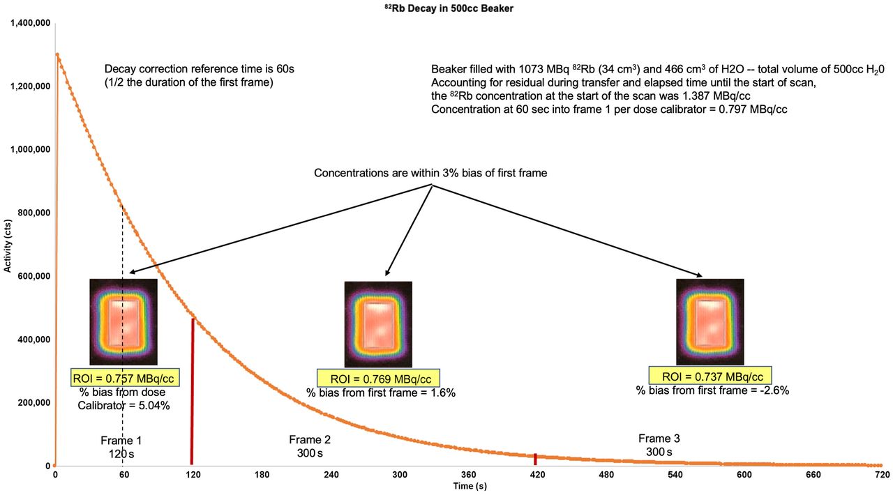
- FIGURE 3.
Decay of 82Rb in 500 cm3 beaker. A detailed 82Rb protocol (“Rubidium Decay Correction.docx”) and worksheet (“82Rb Decay Correction Worksheet.xlsx”) can be found in the supplemental materials. Similar to Figures 1 and 2, 1,073 MBq of 82Rb were placed in a beaker with a total volume precisely 500 cm3 (H2O plus 82Rb eluate). Scanner performed 720-s (12 min) list-mode acquisition. Ten minutes later, after all activity decayed, attenuation scanning was performed and 3 serial frames were generated at intervals of 120, 300, and 300 s. Calculations and measurements were as shown in Figure 1. There are several conclusions. First, scanner decay-corrects activity to midpoint of first frame. Second, scanner also corrects for duration of each frame, giving activity in MBq/cm3. Third, in biologic systems, only variation in quantitative activity after first frame would be due to physiologic changes and not imaging timing, duration, or decay.
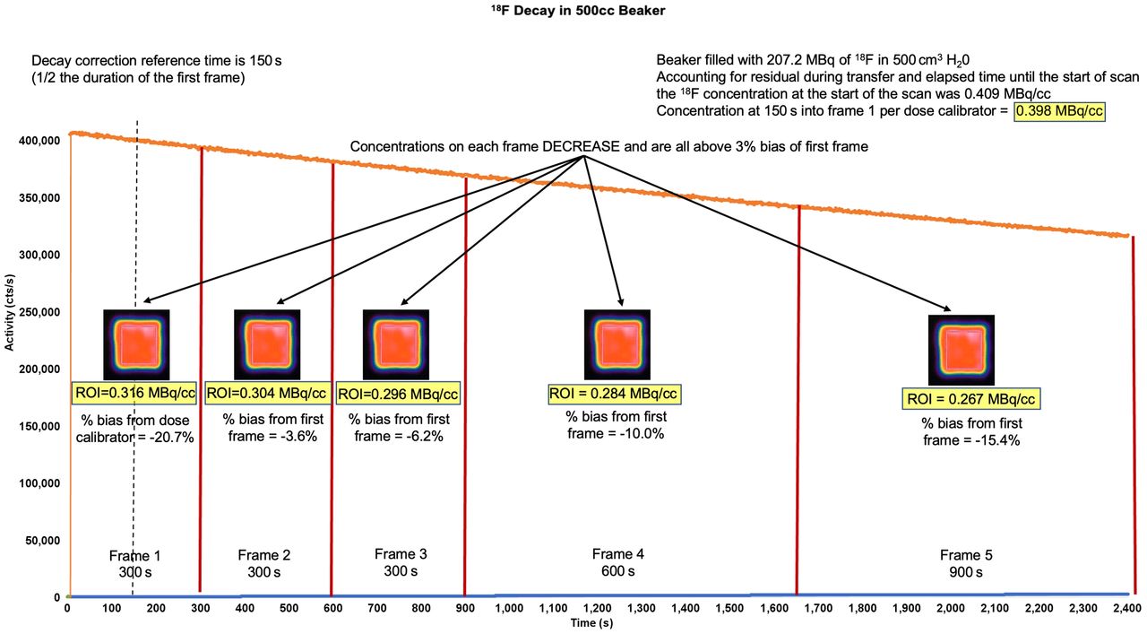
- FIGURE 4.
Similar to Figures 1--3, decay beaker testing, using 18F, was performed on 2D refurbished PET camera when there was concern about accuracy of MBF data. Scanner performed 2,400-s (40 min) list-mode acquisition, and attenuation scans were obtained. Five serial frames were generated. ROI concentration continued to decrease over time and over varying frame durations. There are 2 conclusions. First, scanner does not decay-correct activity to midpoint of first frame or correct for frame duration. Second, in biologic systems, variation in quantitative activity is partly due to inadequate DC or frame duration, which cannot be differentiated from physiologic changes. Therefore, measurement of MBF will not be accurate.
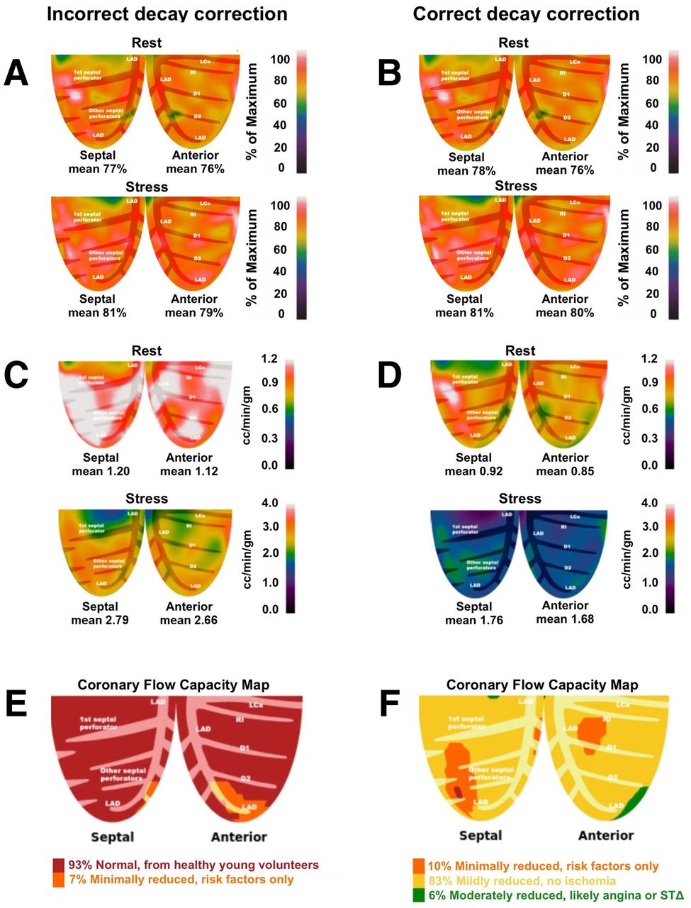
- FIGURE 5.
Figure demonstrates relative and quantitative perfusion data of the left anterior descending coronary artery (LAD) territory obtained from a refurbished 2D PET system where decay correction (DC), as part of the default settings within the camera, was performed incorrectly (left column). After recognition of the error, the DC algorithm was corrected and the study reprocessed. 5A and 5B represent rest and stress relative perfusion images, respectively. Both sets of relative images (incorrect and correct DC) are normal and appear nearly identical. 5C and 5D demonstrate inaccurate and accurate DC of rest and stress absolute perfusion in cc/min/g, respectively. With correct DC, rest and stress MBF are ∼30% and ∼55% lower, respectively. 5E and 5F demonstrate the coronary flow capacity (CFC) maps derived by the integration of absolute flow metrics of the incorrect and correct DC datasets, respectively. With incorrect DC, CFC maps suggest physiology consistent with healthy volunteers without risk factors. However, with correct DC, CFC maps are consistent with mild diffuse epicardial disease for which medical therapy is appropriate. Based on CFC maps, treatment would possibly be different based on the absolute perfusion metrics.


Additional Files
Supplemental Data
Files in this Data Supplement:


