


Article Figures & Data
Figures
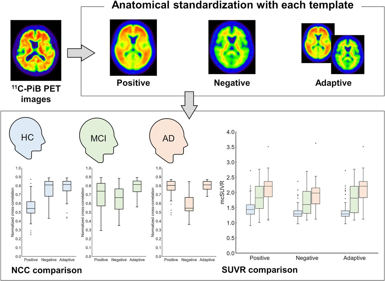
- FIGURE 1.
Workflow of PET-only quantitative evaluation method. First, PET images are anatomically standardized to either template using positive-template method, negative-template method, or adaptive-template method. Second, transformation vector used for standardization is calculated. Third, empirical PiB-prone region of interest (EPP-ROI) is inverse-transformed to individual PET image using transformation vector.
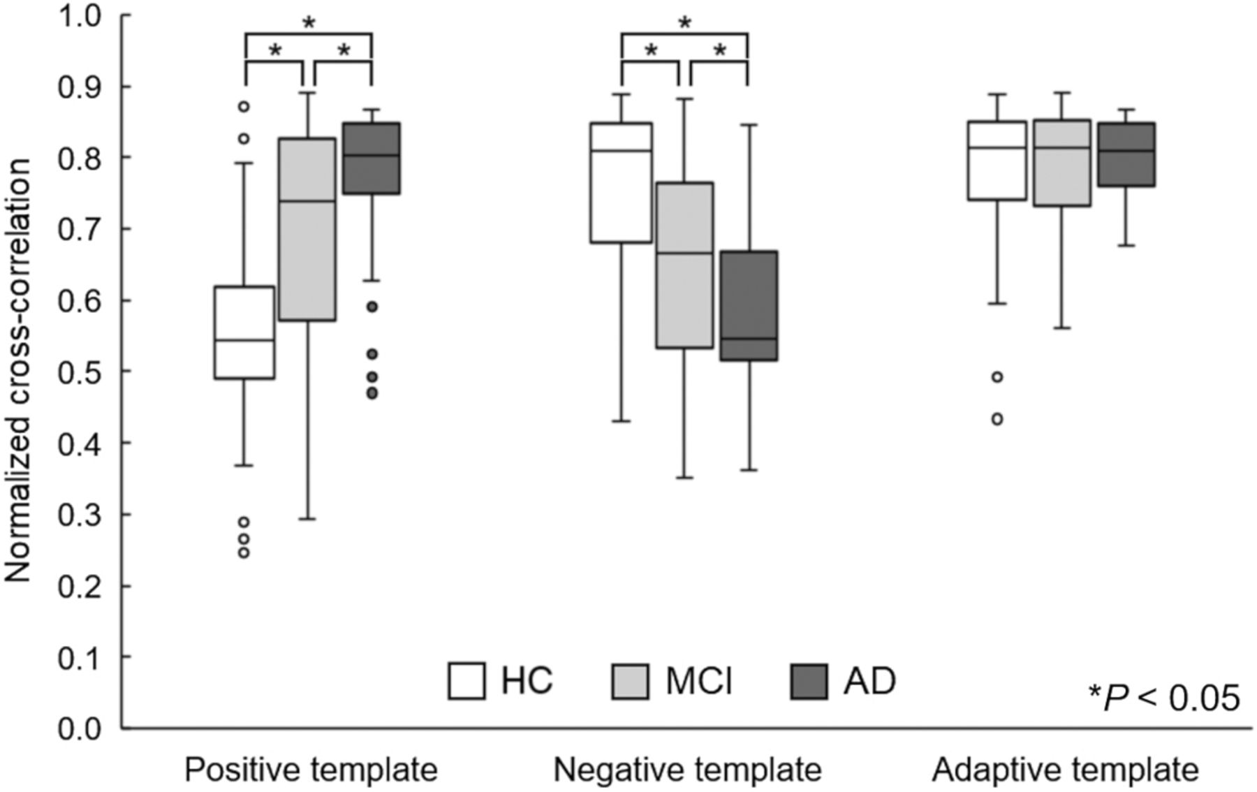
- FIGURE 2.
NCC results. *P < 0.05.
- FIGURE 3.
mcSUVR results. *P < 0.05.
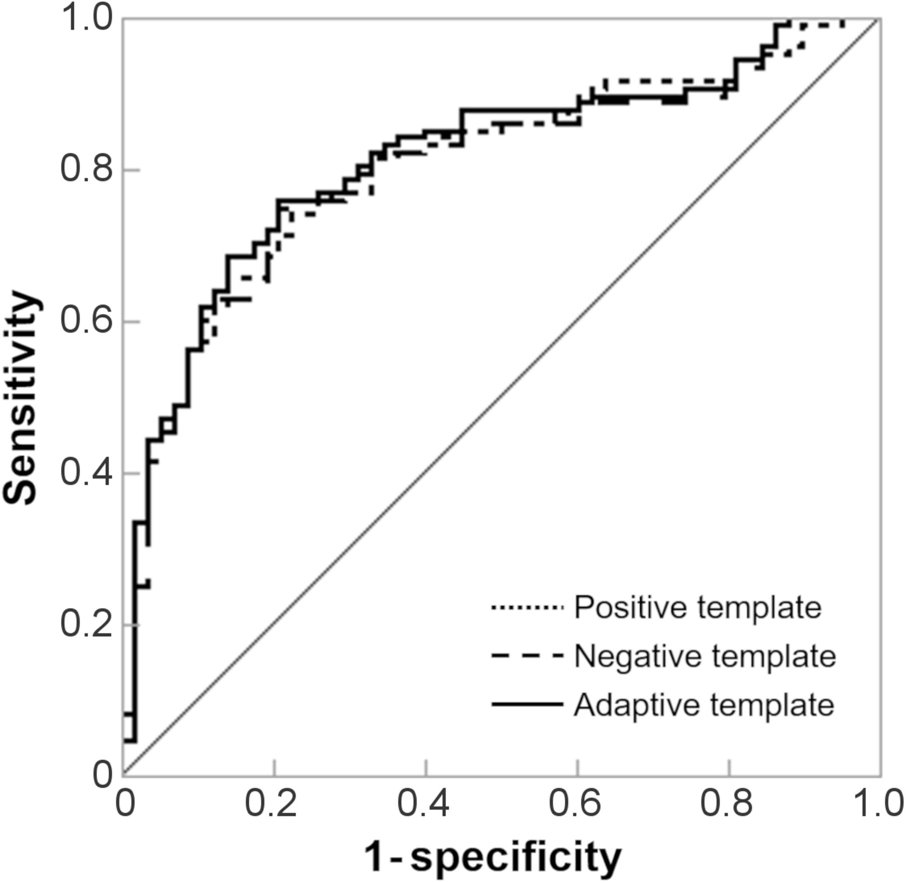
- FIGURE 4.
mcSUVR receiver-operating-characteristic curves for each template. Areas under curve for positive template–based method, negative template–based method, and adaptive template–based method were 0.806, 0.801, and 0.815, respectively.


Tables
Characteristic HC MCI AD Sex (n) Male 30 30 21 Female 28 32 25 Age (y) Mean ± SD 66.4 ± 4.5 71.4 ± 5.5 74.4 ± 6.3 Range 60–80 60–82 62–84 NINCDS-ADRDA — — Probable AD MMSE-J Mean ± SD 29.3 ± 1.1 26.7 ± 1.8 22.2 ± 1.8 Range 24–30 24–30 20–26 CDR-J 0 0.5 0.5 or 1.0 WMS-R Above cutoff Below cutoff Below cutoff Visually positive (n) 14 41 43 Visually negative (n) 44 21 3 NINCDS-ADRDA = National Institute of Neurologic and Communicative Disorders and Alzheimer’s Disease and Related Disorders Association.
Scanner vendor Scanner model Algorithm Iterations Subsets GE Healthcare Advance Iterative (FORE + OSEM) 6 16 Discovery ST Elite Iterative (VUE Point plus) 2 40 Shimadzu Eminence Sophia G/X FORE + DRAMA 4 NA Eminence Sophia B/L FORE + DRAMA 4 NA Eminence G/X FORE + DRAMA 4 NA Headtome V Iterative (FORE + OSEM) 4 16 Siemens ECAT Accel Iterative (FORE + OSEM) 6 16 ECAT Exact HR+ Iterative (FORE + OSEM) 4 16 Biograph 6 Iterative (FORE + OSEM) 4 16 Biograph 16 Iterative (FORE + OSEM) 4 14 FORE = Fourier rebinning; OSEM = ordered-subsets expectation maximization; NA = not available; DRAMA = dynamic row-action maximum-likelihood algorithm.
Visual evaluation Clinical diagnosis No. of participants No. of images Positive template Negative template Adaptive template Positive HC 14 58 0 7 MCI 41 62 0 35 AD 43 46 0 38 Total 98 166 0 80 Negative HC 44 0 58 51 MCI 21 0 62 27 AD 3 0 46 8 Total 68 0 166 86 Concordance rate 59.0% 41.0% 89.2% Coefficient of association not not 0.80 Template AUC Cutoff Sensitivity Specificity Accuracy Positive 0.806 1.80 0.657 0.862 0.729 Negative 0.801 1.40 0.750* 0.793 0.765 Adaptive 0.815 1.40 0.759* 0.793 0.771 *P < 0.05 (difference from positive template).
AUC = area under curve.


